3979
Dynamic contrast-enhanced gastric MRI features are related to adverse histopathological status in advanced gastric cancer1Department of Radiology, the First Affiliated Hospital with Nanjing Medical University, Nanjing, China, 2MR Application Predevelopment, Siemens Healthineers AG, Erlangen, Germany, 3MR Research Collaboration Team, Siemens Healthineers Ltd, Shanghai, China
Synopsis
Keywords: Cancer, Cancer
Motivation: Distinct histopathological types are closely related to the prognosis of advanced gastric cancer. Adverse histopathological status (AHS) is linked to poor gastric cancer outcomes. Extra-dimensional volumetric interpolated breath-hold examination (XD-VIBE) offers high temporal and spatial resolution while allowing free breathing.
Goal(s): To improve the assessment of AHS, we explored the potential for XD-VIBE to improve DCE imaging.
Approach: We evaluated the associations of quantitative DCE MRI parameters (e.g., Ktrans, Ve, and Kep) with AHS.
Results: Using XD-VIBE, quantitative DCE MRI parameters allowed non-invasive assessment of AHS and helped to optimize treatment strategies for advanced gastric cancer.
Impact: In this single-institution study, quantitative DCE MRI features demonstrated promise as markers of AHS in advanced gastric cancer. These features could provide added prognostic value and identify patients who may benefit from adjuvant therapy.
Numerous studies have demonstrated close correlations between histopathological type and gastric cancer prognosis, as well as chemotherapy effectiveness1. We previously showed that overall histopathological score (H-score) offers superior prognostic stratification efficacy, compared with the American Joint Committee on Cancer (AJCC) staging system2. Dynamic contrast-enhanced (DCE) imaging can facilitate the evaluation of gastric cancer by assessing intratumoral vascularization and angiogenesis3. However, few studies have used DCE to explore pathological features of gastric cancer. The most common cause of failure has been patient inability to perform extended breath-hold during gastric MRI. Extra-dimensional volumetric interpolated breath-hold examination (XD-VIBE) is a promising alternative to conventional DCE imaging, which offers high temporal and spatial resolution with free breathing4. We used XD-VIBE to investigate correlations between quantitative DCE MRI parameters and adverse histopathological status (AHS) in gastric cancer .
Methods
From November 2021 to June 2023, we retrospectively enrolled 205 patients with biopsy-proven gastric cancer who underwent MRI scanning on a 3T system (MAGNETOM Skyra, Siemens Healthineers, Erlangen, Germany). For DCE imaging, we used XD-VIBE research sequence with the following parameters: field of view=360´360 mm², voxel size=1.3´1.3´3.5mm, echo time/repetition time=1.26/3.86 ms, flip angle=15°, and temporal resolution=5.68 ms. Quantitative measurements were performed by an experienced radiologist who was blinded to the final pathologic results. Quantitative parameters (e.g., Ktrans, Ve, and Kep) were estimated using the two-compartment Tofts model3; mean and median values for all parameters were calculated for each free-hand region of interest (with area ≥10 mm2). Each region of interest was manually drawn on the images for each tumor layer. Histopathological status was scored as follows: pT2-3 (0 points) vs pT4a (1 point), pN-negative (0 points) vs pN-positive (1 point), intestinal and mixed types (0 points) vs diffuse type (1 point), Borrmann I–III (0 points) vs Borrmann IV (1 point), low-World Health Organization (WHO) histological classification (0 points, papillary or tubular adenocarcinoma) vs high-WHO histological classification (1 point, mucinous adenocarcinoma or signet ring cell carcinoma), low-grade (0 points, well or moderately differentiated) vs high-grade (1 point, poorly differentiated tubular adenocarcinoma), lymphovascular invasion-negative (0 points) vs lymphovascular invasion-positive (1 point), and perineural invasion-negative (0 points) vs perineural invasion-positive (1 point). A histopathological score (H-score) representing overall aggressiveness was determined by summing all status scores for each histopathological subtype. Patients were classified into the low-H-score group (0–4 points) or high-H-score group (5–8 points). Comparisons of normally distributed data were performed with the independent samples t-test; comparisons of non-normally distributed data were performed using the non-parametric Kruskal-Wallis test.
Results
T stage demonstrated a statistically significant relationship with Ve mean (P=0.017); there were no statistically significant relationships for N stage. The WHO classification showed statistically significant relationships with Ktrans mean (P=0.028), Ktrans median (P=0.012), Ve mean (P=0.002), and Kep mean (P=0.027). The Borrmann classification exhibited a statistically significant relationship with Ve mean (P=0.041). The Lauren classification showed statistically significant relationships with Ktrans mean (P=0.004), Ktrans median (P=0.001), and Ve mean (P=0.029). Histological differentiation demonstrated statistically significant relationships with Ktrans mean (P=0.035), Ktrans median (P=0.01), and Ve mean (P=0.009). Lymphovascular invasion displayed statistically significant relationships with Ve mean (P=0.018) and Kep mean (P=0.011). Perineural invasion showed statistically significant relationships with Ve mean (P<0.001), Ve median (P=0.020), Kep mean (P=0.020), and Kep median (P<0.001). The H-score exhibited statistically significant relationships with Ktrans mean (P=0.040), Kep mean (P<0.001), and Ve mean (P=0.004).
Discussion and Conclusion
DCE MRI is an important part of gastric magnetic resonance examinations, which provides valuable insights into the morphological and hemodynamic characteristics of tumors. This study explored the potential for quantitative pharmacokinetic parameters, derived from XD-VIBE DCE MRI, to predict histopathological features of locally advanced gastric cancer. We found that the quantitative parameters Ktrans, Kep, and Ve exhibited statistical significance in terms of distinguishing between low-risk and high-risk gastric cancer. These results suggest strong correlations between these parameters and the biological characteristics of advanced gastric cancer. Such correlations may help to guide treatment decisions and offer insights into patient prognosis. The clinical value of these findings can be enhanced through larger-scale studies that externally validate and corroborate our results. In summary, XD-VIBE DCE offers a reliable method for the imaging-based estimation of quantitative biomarkers, which can facilitate predictions of adverse histopathological status among patients with advanced gastric cancer.
Acknowledgements
No acknowledgement found.References
1. Sano T, Coit DG, Kim HH, et al. Proposal of a new stage grouping of gastric cancer for TNM classification: International Gastric Cancer Association staging project. Gastric Cancer. 2017;20(2):217-225. doi:10.1007/s10120-016-0601-9
2. Li Q, Qi L, Feng QX, et al. Machine Learning-Based Computational Models Derived From Large-Scale Radiographic-Radiomic Images Can Help Predict Adverse Histopathological Status of Gastric Cancer. Clin Transl Gastroenterol. 2019;10(10):e00079. doi:10.14309/ctg.0000000000000079
3. Tofts PS, Brix G, Buckley DL, et al. Estimating kinetic parameters from dynamic contrast-enhanced T(1)-weighted MRI of a diffusable tracer: standardized quantities and symbols. J Magn Reson Imaging. 1999;10(3):223-232. doi:10.1002/(sici)1522-2586(199909)10:3<223::aid-jmri2>3.0.co;2-s
4.Yoon JH, Yu MH, Chang W, et al. Clinical Feasibility of Free-Breathing Dynamic T1-Weighted Imaging With Gadoxetic Acid-Enhanced Liver Magnetic Resonance Imaging Using a Combination of Variable Density Sampling and Compressed Sensing. Invest Radiol. Oct 2017;52(10):596-604. doi:10.1097/RLI.0000000000000385
Figures
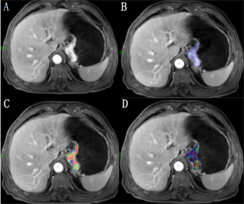
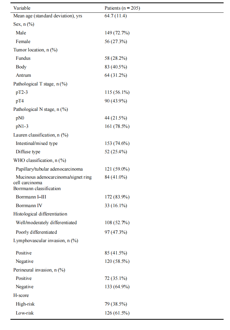
Table 1. Baseline patient characteristics
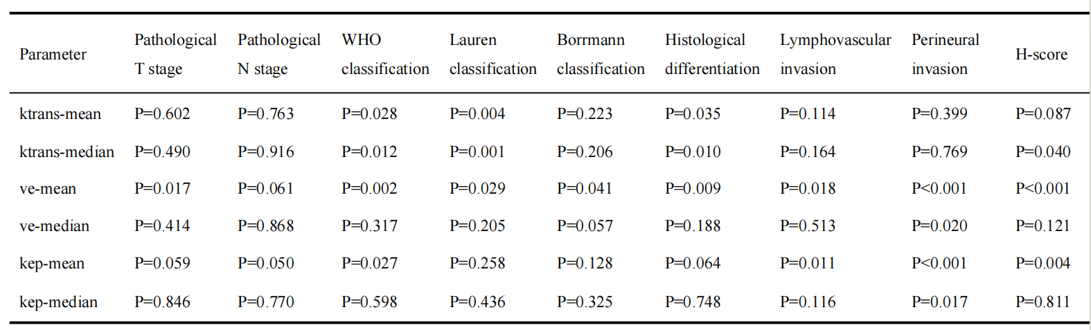
Table 2. P-values for Ktrans, Kep and Ve parameters to identify adverse histopathological status (AHS), according to univariate analysis