3975
Tumour mechanics and vascular fractality quantification via MR-Elastography in the context of liver metastasis from colorectal cancer1INSERM UMRS1149 - Centre de Recherche sur l'Inflammation, University Paris, Paris, France, 2INSERM UMRS1148 - Laboratory for Vascular Translational Science, University Paris, Paris, France, 3siemens-healthineers, Paris, France, 4INSERM UMRS1148 - Laboratory for Vascular Translational Science, University Paris, paris, France, 5Assistance publique - Hôpitaux de Paris, Paris, France, 6Assistance publique - Hôpitaux de Paris, paris, France, 7School of Biomedical Engineering and Imaging Sciences, King’s College London, London, United Kingdom
Synopsis
Keywords: Biomarkers, Vessels, Liver, tumor, biomechanics, Elastography
Motivation: Colorectal cancer is a major global cause of cancer-related deaths, often metastasizing to the liver. Standard treatment includes chemotherapy and anti-angiogenic therapy. Quantifying therapy efficacy remains a clinical challenge.
Goal(s): We explore multifrequency MR-Elastography (MRE) for assessing vascular organization, using a murine liver metastasis model correlated with histopathology.
Approach: The study used MRE imaging of murin liver metastasis model and the corresponding histopathology to analyze vascular organization (fractal dimension), by measuring Hurst index (H-index).
Results: The H-index differs significantly between tumor and healthy liver tissue, with normal vasculature displaying a lower H-index compared to the tumoral tissue.
Impact: Our in vivo elastography study demonstrates the organization of the vascular network by matching with histological findings. This innovative approach paves the way for non-invasive evaluation of treatments targeting tumor vessels, such as bevacizumab or FOLFOX.
Background:
Colorectal cancer is a leading cause of cancer-related deaths globally 1. It tends to metastasize to distant organs, with the liver being a common site of metastasis. Standard treatment for metastatic colorectal cancer typically involves the use of chemotherapy regimens such as FOLFOX 2, along with the recent introduction of BEVACIZUMAB as an anti-angiogenic agent 3. Currently, therapy efficacy quantification is an unmet clinical need because classical criteria for response/resistance such as the RECIST score fail due to the bi-modal therapeutic approach. This combined therapy attacks on the one side cellular integrity, and on the other side the vascular organization. Thus, it is crucial to provide novel imaging biomarkers which allow to quantify on the one hand the impact of the therapy on tissue integrity, and on the other hand the modulation of the vascular organization. It is in this context where we exploit the ability of multifrequency MR-Elastography (MRE) to provide information about the vascular organization stemming from the dispersion analysis of the wave behaviour within the tumour. This analysis yields the so-called Hurst index of the vascular organization. To allow for detailed correlation to histopathology, we propose a murine model of liver metastasis on which multifrequency MRE is performed, and where tumour sections are stained with CD31 to quantify vascular architecture via classical box counting analysis.Methods:
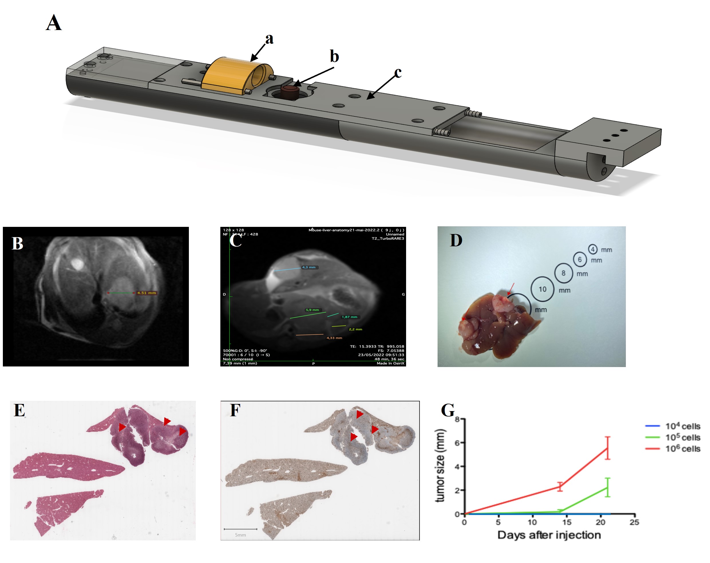
To generate murine hepatic metastases, we injected CT26 tumor cells into the spleens of Balb/C mice 4. The amount of CT26 tumor cells necessary to generate sufficient metastasis in the liver within 4 weeks was optimized (fig 1). Immunostaining was performed using CD31 antibody (Sigma). Imaging was performed using a 7T preclinical MRI scanner from Bruker. A dedicated MRE system was designed to enable the induction of shear waves from underneath the animal (fig1). The system allowed to vary the vibrational frequency from 250-325Hz in order to extract the dispersion properties of the shear waves within the tumors. A multi-slice spin echo MRE sequence was utilized with specific parameters (TR/TE 1000/25ms; FOV 40x40mm; 4 wave phases; 15 slices; isotropic resolution 0.5 mm). MRE data were reconstructed according to 5.Results:
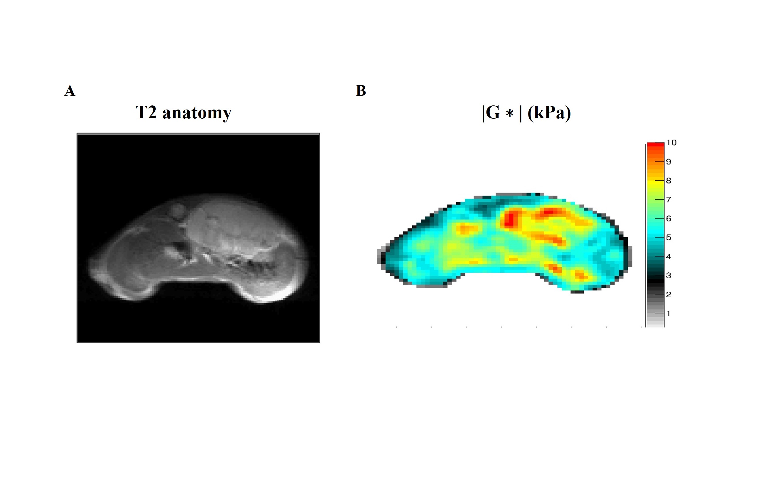
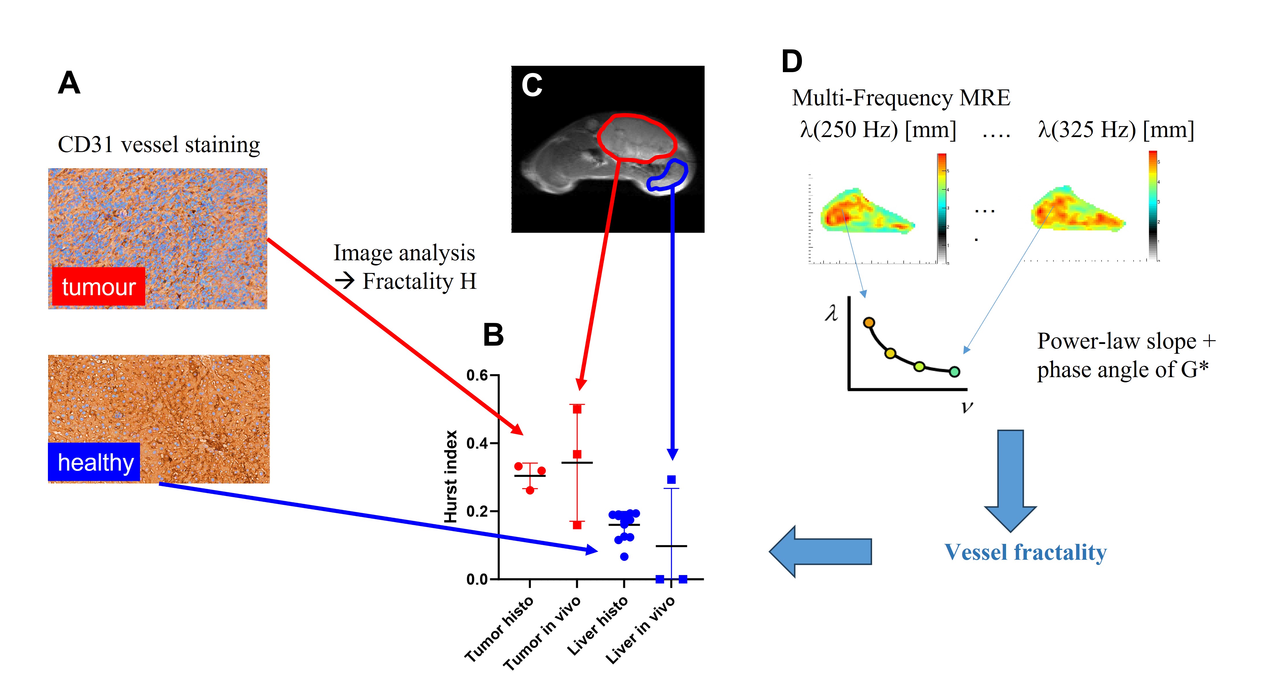
Figure2 shows a transvers section through the abdomen of a mouse which carried a large tumour. The corresponding image of the shear modulus |G*| clearly depicts the tumour as a solid mass which is distinctively different from the liver parenchyma. As such, stiffness itself quantifies the modulation of tissue organization, as published before 6.Fig3 A shows CD31 stained tissue sections from tumours and from normal liver tissue. Clearly, vessel organization is dramatically different within the liver metastasis. This organization can be mathematically quantified via the box-counting approach which characterizes the fractal dimension of the structure. This can further be transformed into the so-called Hurst index, which describes for values between HÎ[0-0.5] the anti-persistent regime 7.Clearly, as shown in Fig.3B, the H-index between tumour and healthy liver tissue differs significantly, with normal vasculature exhibiting a lower H-index, i.e., a fractality closer to 2 which is indicative for a more random organization. The MRE experiment provides, as shown in Fig.3C,D, the wavelength as a function of frequency as well as the corresponding phase angle. In the context of the theory of multiple reflections of waves on vessels which behave as scatterers 8, we can relate the dispersion properties of the shear wavelength together with the phase angle to the Hurst index. This is shown graphically in Fig.3D. The resulting Hurst-indices as quantified from the in-vivo multi-frequency MRE imaging are compared to the one obtained from histopathology in Fig.3B. Although not yet perfectly matching, we observed average values which match, and clearly see the corresponding rise/drop in Hurst-index in the tumour/healthy liver regions, respectively. The main reason for the current imperfections is most likely due to challenging SNR within the MRE data which are impacted by artefacts from residual respiratory motion.
Conclusions:
In this work, we have developed a system for applying multifrequency MRE to rodents within the preclinical setting. Vascular architecture quantified via the Hurst-index and calculated once via histological images stained for vessels, and once extracted from multi-frequency MRE matches on average. More refined imaging protocols are needed to further improve the SNR of the MRE data. In the next step we will investigate the corresponding vascular changes throughout a combined therapy using FOLFOX and BEVACIZUMAB quantified via histology and non-invasively via multi-frequency MRE. These results open up useful new perspectives for assessing the efficacy of anti-tumor treatments targeting vessels, as no satisfactory criteria currently exist for response to therapy to such combined approaches.Acknowledgements
This research was funded by the ITMO Cancer Aviesan / Inserm / Cancer 2020 grant Réf : DESP/PB n°241References
1. Cancer incidence and mortality worldwide: Sources, methods and major patterns in GLOBOCAN 2012 - Ferlay - 2015 - International Journal of Cancer - Wiley Online Library. https://onlinelibrary.wiley.com/doi/10.1002/ijc.29210.
2.Hung, A. & Mullins, C. D. Relative effectiveness and safety of chemotherapy in elderly and nonelderly patients with stage III colon cancer: a systematic review. The Oncologist 18, 54–63 (2013). 3. Thornton, A. D., Ravn, P., Winslet, M. & Chester, K. Angiogenesis inhibition with bevacizumab and the surgical management of colorectal cancer. Br. J. Surg. 93, 1456–1463 (2006).
4. Bhattacharjee, S. et al. Tumor restriction by type I collagen opposes tumor-promoting effects of cancer-associated fibroblasts. J. Clin. Invest. 131, e146987.
5. Sinkus, R. et al. Rheological determinants for simultaneous staging of hepatic fibrosis and inflammation in patients with chronic liver disease. NMR Biomed. 31, e3956 (2018).
6.Garteiser, P. et al. MR elastography of liver tumours: value of viscoelastic properties for tumour characterisation. Eur. Radiol. 22, 2169–2177 (2012).
7.Mandelbrot, B. B. & Hudson, R. L. The (Mis)Behaviour of Markets: A Fractal View of Risk, Ruin and Reward. (Profile Books, 2010).
8.Lambert, S. A. et al. Bridging Three Orders of Magnitude: Multiple Scattered Waves Sense Fractal Microscopic Structures via Dispersion. Phys. Rev. Lett. 115, 094301 (2015).
Figures