3974
Time-dependent response of Silver nanoparticles reinforced biocomposite in Murine fibroblast cells by NMR-based Pharmacometabolomics1Metabolomics Research Facility, Institute of Nuclear Medicine and Allied Sciences (INMAS), DRDO, Delhi, India, 2Department of Chemistry, Indian Institute of Technology Delhi (IITD), Delhi, India
Synopsis
Keywords: Other Preclinical, Toxicity, In vitro Pharmacometabolomics, NMR spectroscopy, Nanocomposite, Nanotoxicology
Motivation: Conventional toxicity assays generally convey the efficiency of nanomaterial, but they lack a mechanistic molecular approach to toxicity.
Goal(s): We aimed to employ NMR-based Pharmacometabolomics to detect early metabolic alterations associated with nanomaterial developed for healthcare.
Approach: The NMR spectroscopy technique was used on Silver nanoparticles reinforced biocomposite exposed murine fibroblast (L929) cells.
Results: Results demonstrated that cellular metabolic profiles reset towards control upon more exposure.
Impact: Our nanomaterial-based study accentuates the role of pharmacometabolomics, which will complement the findings of conventional assays, eventually leading to efficacious clinical translation.
Introduction
With the progress of nanotechnology, new nanomaterials are continually developed for biomedical applications. Nanoparticle-reinforced biocomposites have emerged as a potential candidate for wound management. These composites are better than their bulk counterparts due to various properties, such as the large interfacial area between biopolymers and nanoparticles1, mimicking the native tissue2, and antimicrobial nature3. Sooner or later, human exposure to them is inevitable. Hence, toxicity studies should be carried out meticulously. The current toxicity studies lack molecular information regarding the toxicity mechanism, which may even result in adverse reactions among the population. Pharmacometabolomics can fill this information gap and help translate this material into the commercial world4. In the current work, a time-dependent drug metabolism study was conducted on murine fibroblast cells with the help of Nuclear Magnetic resonance (NMR) spectroscopy. Murine fibroblast (L929) was used to evaluate the metabolic response of Silver nanoparticles (Ag NPs) reinforced biocomposites, as these cells are widely used for in vitro tests, including cell biology studies, cytotoxicity, and biocompatibility testing.Aims and Objectives
To evaluate in vitro metabolic perturbations in murine fibroblast (L929) cells on nanomaterial interaction.Methods
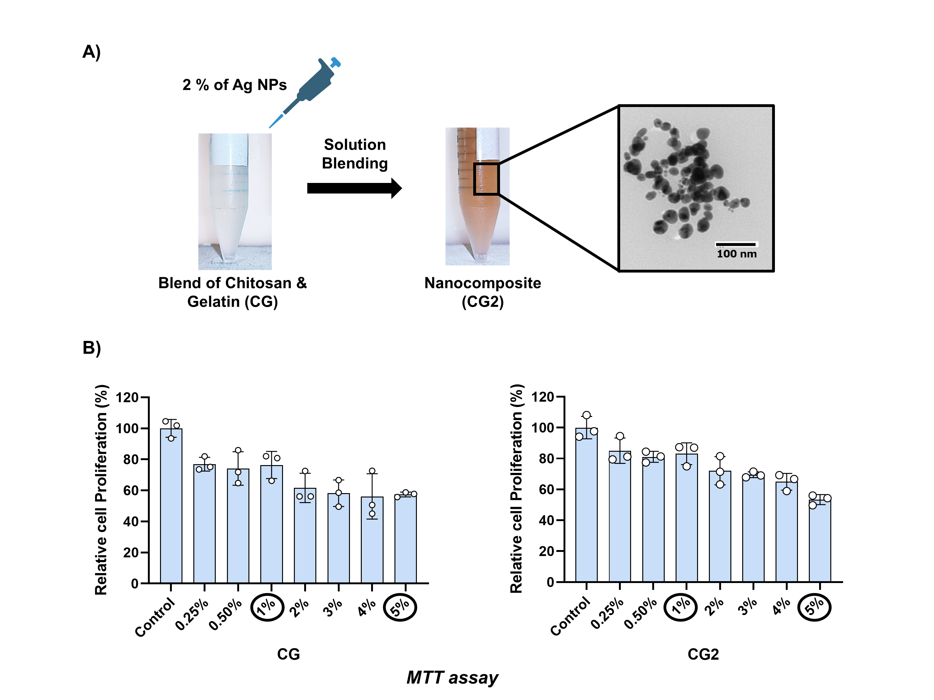
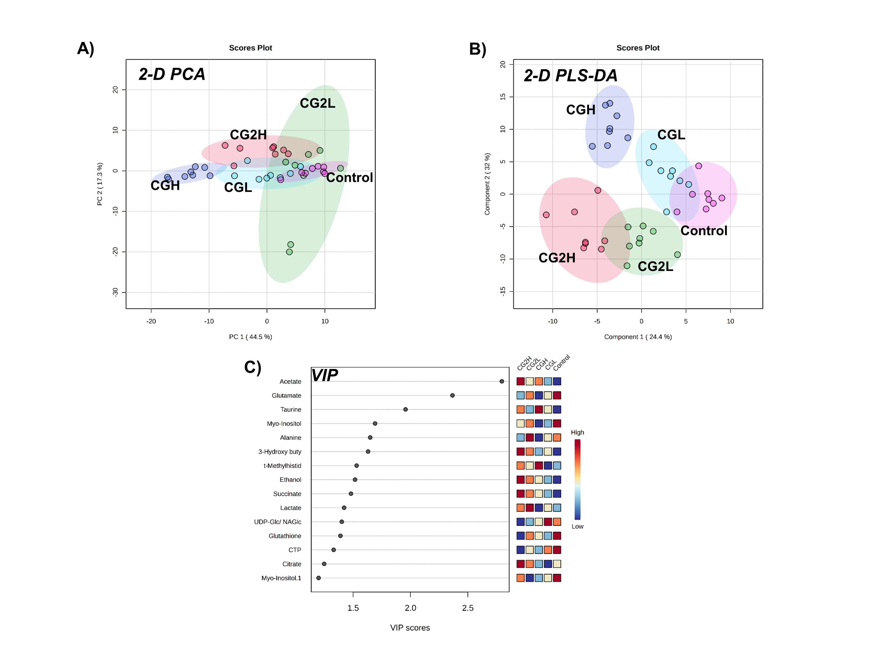
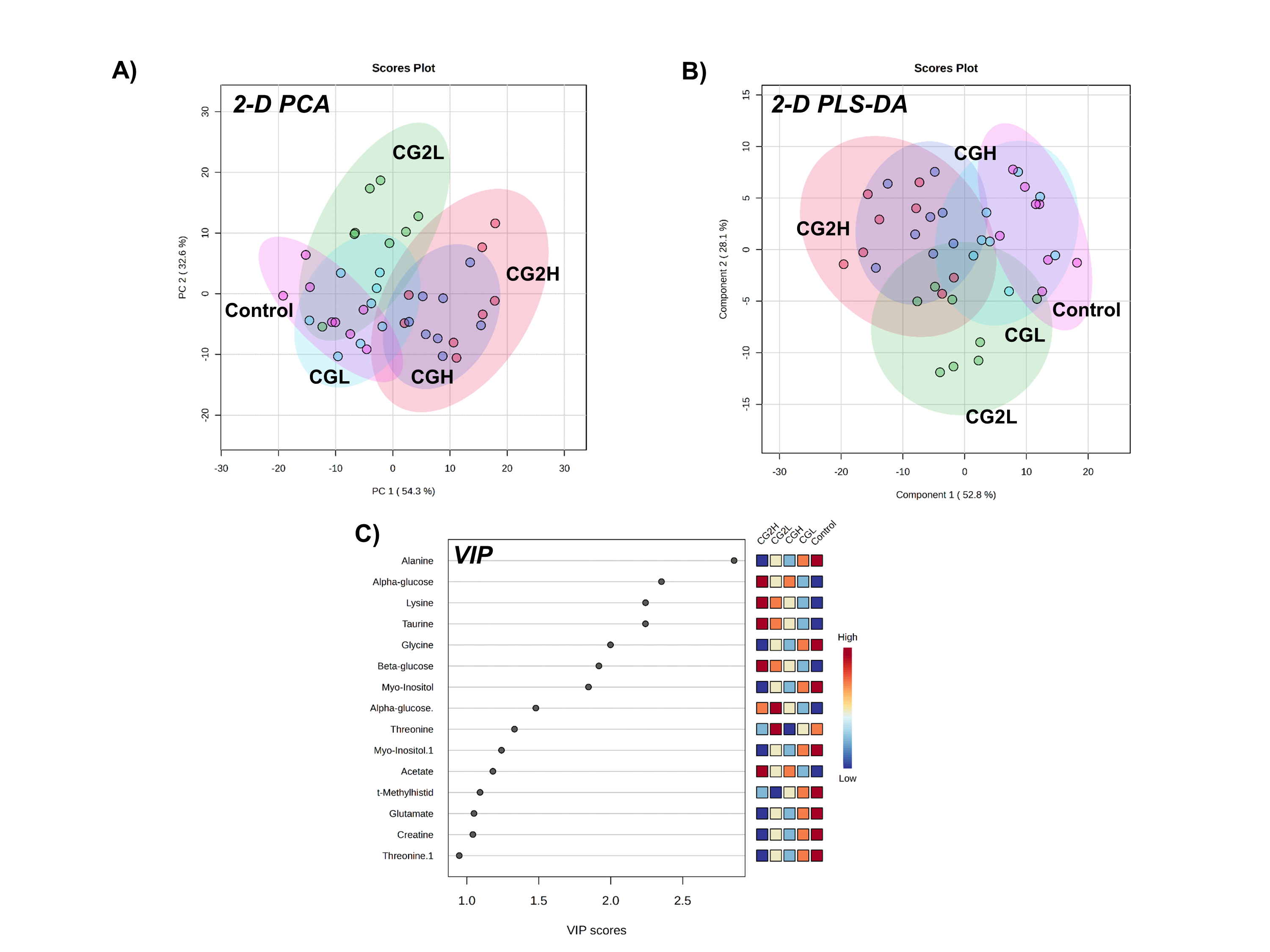
Silver nanoparticles (Ag NPs) were prepared by the green synthesis using Ocimum sanctum (tulsi powder), then dispersed in the blend of chitosan (C) and gelatin (G) biopolymers through the solution blending process to prepare Ag NPs reinforced biocomposite3. The two formulations, CG and CG2, with 0 and 2 % Ag NPs, were used for NMR-based pharmacometabolomics. First, the adherent cells were grown till they reached 90 % confluency. The doses were selected through cellular viability studies (MTT assay). Then, the medium of cells was replaced with the medium containing two doses (L=1% and H=5%) of the two formulations (figure 1(B)). In contrast, fresh medium without formulation was added to the control cells. The cells were collected after incubating for 24 and 48 hours. The polar metabolites were extracted from cells through the dual-phase separation method5. The extracted metabolites were freeze-dried to remove the solvents. The samples were reconstituted in 550 µL of 0.2 M sodium phosphate-buffered deuterium oxide containing 0.1 mM of sodium 3-(trimethylsilyl) propionate-2,2,3,3-d4 (TSP) for spectral acquisition. The 1H Nuclear Magnetic Resonance (NMR) spectra with water suppression (NOESYPR1D) were acquired on a 600 MHz Bruker Avance III spectrometer at 298K with 64 scans collected into 32 K data points with a relaxation delay of 2 s, flip angle of 90° and a mixing period of 100 ms6. Principal component analysis (PCA) and partial least square discriminant analysis (PLS-DA) using Metaboanalyst 5.0 were applied to analyze the clustering of the samples.Results
The dispersion of Ag NPs in the blend of chitosan and gelatin in CG formed reddish brown-coloured nano-biocomposite (CG2), as shown in Figure 1(A). In Figure 2, PCA and PLS-DA showed the separation of high doses (CGH and CG2H) from control after 24 hours of exposure. On increasing this incubation by another 24 hours, the metabolic profiles of high dose treatments were resetting towards control. VIP scores (Figure 2(C) and 3(C)) showed the top 30 important buckets responsible for cluster separation among treatments and control6.Discussion
Exposure of L929 cells to CG and CG2 caused metabolic perturbations in the intracellular metabolome. Amino acids such as alanine, lysine, glycine, glutamate, and threonine profiles were altered, signifying alteration in amino acid metabolism. Taurine and Glutathione levels are altered, implying detoxification pathways in the presence of high doses in the initial 24 hours of exposure. The tricarboxylic acid (TCA) cycle was also hampered due to alteration in acetate concentration at 24 hours of exposure5. High dosages lead to oxidative stress compared to polymer blends due to the leaching of Ag NPs from the composite. On another 24 hours of exposure, creatinine levels decreased, as creatinine is a reserved energy source. After more exposure to biocomposite, the restoration process has been triggered.Conclusion
This is the first time-dependent study for evaluating the material metabolism at the cellular level through NMR Pharmacometabolomics. NMR spectroscopy can help gain the molecular information of Ag NPs reinforced biocomposites toxicity in fibroblast cells, ultimately predicting their biosafety for topical application in wound care.Acknowledgements
The work was supported by Director, Institute of Nuclear Medicine and Allied Sciences (INMAS), Defence Research and Development Organisation (DRDO), Delhi, and Indian Institute of Technology Delhi (IITD), India. Isha Gupta was supported by University Grants Commission, India.References
1. Gupta I, Gandhi S, & Sapra S. Metal/metal oxide nanoparticles reinforced biocomposites for drug delivery. In Fiber and Textile Engineering in Drug Delivery Systems. Woodhead Publishing. 2003:461-485.
2. Wang X, Chang J, & Wu C. Bioactive inorganic/organic nanocomposites for wound healing. Applied Materials Today. 2018;11:308-319.
3. Gupta I, Kumar A, Bhatt AN, et al. Green synthesis-mediated silver nanoparticles based biocomposite films for wound healing application. Journal of Inorganic and Organometallic Polymers and Materials. 2022; 32(8):2994-3011.
4. Awashra M, & Młynarz, P. The toxicity of nanoparticles and their interaction with cells: an in vitro metabolomic perspective. Nanoscale Advances. 2023;5(10):2674-2723.
5. Carrola J, Bastos V, de Oliveira JMPF, et al. Insights into the impact of silver nanoparticles on human keratinocytes metabolism through NMR metabolomics. Archives of Biochemistry and Biophysics. 2016;589:53-61.
6. Gandhi S. Devi M, Pal S, et al. Metabolic regulatory variations in rats due to acute cold stress Tinospora Cordifolia intervention: high resolution 1H NMR approach. Metabolomics. 2012;8(3):444–45.
Figures