3972
Exploring enhanced abscopal effect: Molecular imaging insights into tumor physiologic changes in combined radiation and PD-1 blockade therapy1National Cancer Institute, Bethesda, MD, United States
Synopsis
Keywords: Preclinical Image Analysis, Preclinical
Motivation: Radiation therapy (RT) for primary tumors triggers regression in non-irradiated metastatic lesions via the abscopal effect, can potentiate the effects of immune-checkpoint blockade on the non-treated tumors in the same animal.
Goal(s): To examine physiological alterations of the treated tumor linked to the abscopal effect.
Approach: We used MRI-based imaging techniques, such as EPR oximetry, DCE MRI, and 13C DNP MRI.
Results: After RT and PD-1 blockade, we observed improved oxygenation, permeability, perfusion, and CD8+ T cell infiltration in metastatic (untreated) tumors. Interestingly, primary tumor with increased permeability and perfusion and reduced hypoxic fraction (HF10) before treatment correlated with augmented abscopal effect post-treatment.
Impact: Metastatic tumor conditions improved with RT and ICB, enhancing oxygenation, permeability, perfusion, and CD8+ T cell infiltration. Primary tumor's elevated permeability and lower HF10 correlated with higher AE in metastatic tumors, confirmed through carbogen-enhanced perfusion.
Introduction
Previous reports note that radiation therapy (RT) for the primary tumor rarely leads to regression in non-irradiated metastatic lesions, known as the abscopal effect (AE). AE results from systemic anti-tumor immune responses triggered by localized RT on the primary tumor. These responses are initiated by the activation of tumor-specific CD8+ T cells, primed by antigen-presenting cells capturing tumor-specific antigens from the irradiated tumor. 1 These primed CD8+ T cells can induce apoptosis in tumor cells at distant, non-irradiated sites. 2 However, the overall occurrence rate of the AE due to RT alone is exceptionally low, primarily because anti-tumor effects are hindered by adaptive immune resistance mechanisms, including the PD-1/PD-L1 pathway. 3 Although immunotherapy, especially immune checkpoint blockade (ICB), has been reported to enhance the AE, no investigations into the associated imaging biomarkers have been conducted thus far. 4The tumor microenvironment (TME) has a close relationship with local immune activity. Our research involves assessing pO2 distribution, permeability, perfusion, and glycolytic metabolism in primary and metastatic tumors undergoing combination therapy. Our objective was to investigate the physiological and metabolic alterations in tumors displaying the abscopal effect (AE). Furthermore, we sought to understand the physiological and metabolic features in primary tumors linked to successful AE. This knowledge could enable us to augment the AE during treatment by harnessing advantageous physiological and metabolic conditions.
Methods
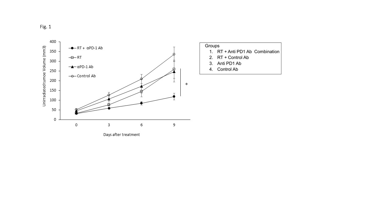
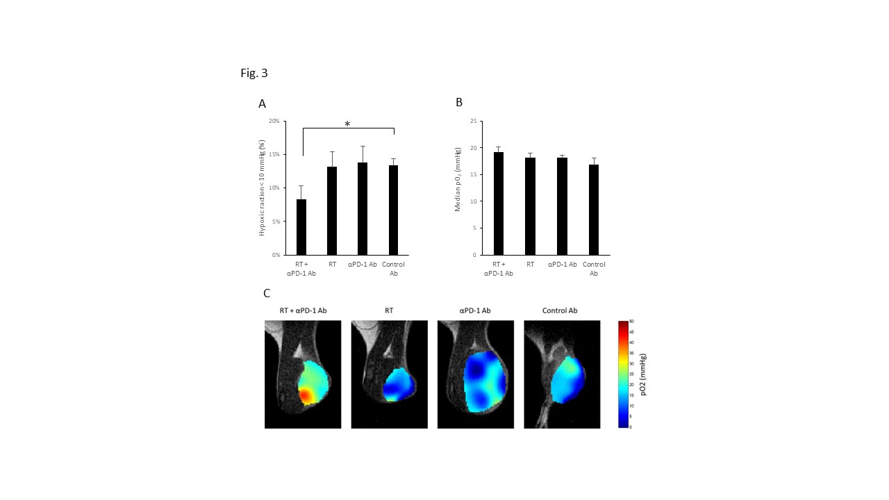
MC38 colon adenocarcinoma treated with RT and a PD-1 inhibitor were employed to assess the AE. In the in vivo treatment model, 1x10⁶ tumor cells were inoculated subcutaneously into the right (primary) hindleg, and 2x10⁵ into the left (metastatic) hindleg of C57BL/6 mice. Four treatment groups were established as outlined in Figure 1.Electron paramagnetic resonance imaging (EPRI) was conducted to map intra-tumor pO2 quantitatively with high resolution (~0.2mm) by observing the linewidth of the exogenously administered trityl radical probe Ox063.
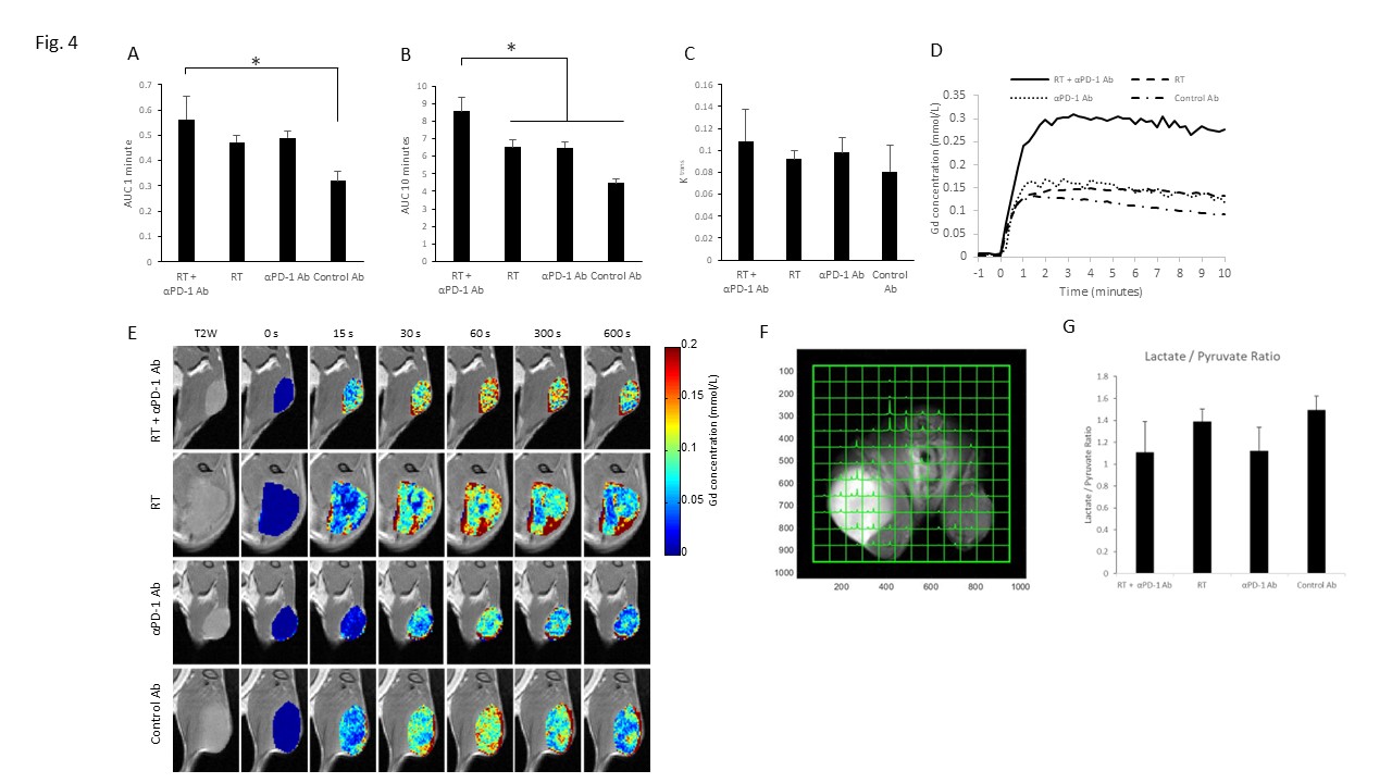
Dynamic contrast-enhanced magnetic resonance imaging (DCE-MRI) was performed on a 3 T scanner. T1-weighted fast low-angle shot (FLASH) images were obtained with TR = 117.2 ms, TE = 6 ms, flip angle = 30˚, two slices, 28 x 28 mm resolution, 15-second acquisition time per image, and 45 repetitions. Gd-DTPA solution was injected through a tail vein cannula. To determine the local concentrations of Gd-DTPA, T1 maps were calculated from three sets of Rapid Imaging with Refocused Echoes (RARE) with the acquisitions being made before running the FLASH sequence.
13C hyperpolarized MRI: Hyperpolarized 13C MRI studies were performed on a BioSpec 3T (Bruker). Hyperpolarized [1–13C] pyruvate was rapidly injected intravenously (20 μL/g). Chemical-shift imaging images were acquired 30 s after the hyperpolarized [1–13C] pyruvate injection with the following parameters: FOV 28×28 mm, slice thickness 8 mm, matrix size 14 × 14, spectral width of 5000 Hz, repetition time 60 ms, and excitation pulse with a flip angle of 5.
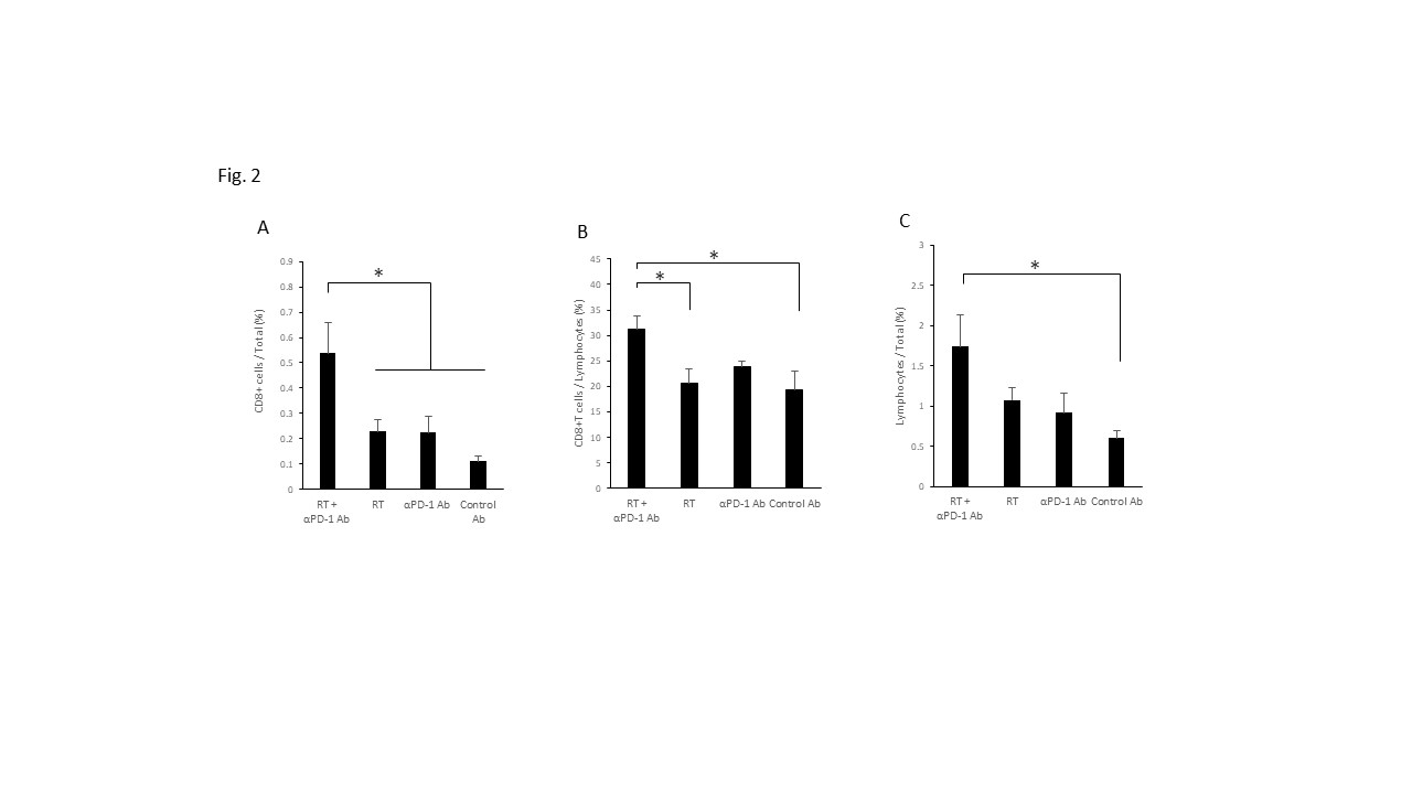
Flow cytometry: To analyze tumor-infiltrating lymphocytes (TILs), on day 9 after treatment, single-cell suspensions were prepared from the left hind leg tumor. The cell surface phenotypes were determined by direct immunofluorescence staining with αCD3 and αCD8 antibodies.
Results and Discussion
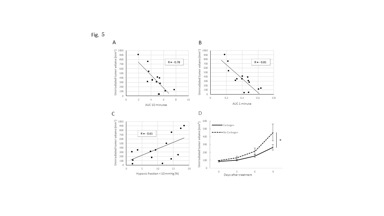
We established a mouse tumor model displaying AE through a combination of RT and PD-1 blockade with MC38 tumors. This combination demonstrated a synergistic effect on metastatic tumors (Fig.1). Flow cytometry confirmed increased CD8+ T cell infiltration in metastatic tumors post RT and PD-1 blockade (Fig.2), implying that the in vivo synergy was driven by increased CD8+ T cell infiltration. The AE corresponded with improvements in imaging biomarkers: lower hypoxic fraction (<10 mmHg) (HF10) and enhanced permeability, perfusion in metastatic tumors (Fig.3 and 4). Additionally, hyperpolarized 1-13C Pyruvate MRI indicated decreased pyruvate to lactate flux with RT and PD-1 blockade in metastatic tumors (Fig.4). Notably, the tumor microenvironmental conditions (high permeability, perfusion, and low HF10) in primary tumor before treatment correlated with the extent of AE induction (Fig.5), underscoring the impact of primary tumor irradiation on AE. Interestingly, the observation was confirmed by enhancing tumor permeability and perfusion during irradiation with carbogen led to superior tumor growth suppression compared to RT and PD-1 blockade alone.Conclusion
Hypoxic fraction pO2<10mmHg, permeability, perfusion and CD8+ T cell infiltration in metastatic tumors improved after the combination of RT and PD-1 blockade. Higher permeability/perfusion and lower HF10 in primary tumor before the treatment were associated with slower metastatic tumor growth after the treatment. These data suggested that the imaging biomarkers potentially predict the successful AE after the combination of RT and PD-1 blockade, and carbogen inhalation during the radiation treatment may be a promising strategy to enhance AE after the combination treatment.Acknowledgements
This research was supported by intramural funds from the Center for Cancer Research of the National Institutes of Health. The authors declare no conflicts of interest.
References
(1) Wang, D.; Zhang, X.; Gao, Y.; Cui, X.; Yang, Y.; Mao, W.; Li, M.; Zhang, B.; Yu, J. Research Progress and Existing Problems for Abscopal Effect. Cancer Manag Res 2020, 12, 6695-6706. DOI: 10.2147/CMAR.S245426.
(2) Henkart, P. A. Lymphocyte-mediated cytotoxicity: two pathways and multiple effector molecules. Immunity 1994, 1 (5), 343-346. DOI: 10.1016/1074-7613(94)90063-9.
(3) Taube, J. M.; Anders, R. A.; Young, G. D.; Xu, H.; Sharma, R.; McMiller, T. L.; Chen, S.; Klein, A. P.; Pardoll, D. M.; Topalian, S. L.; et al. Colocalization of inflammatory response with B7-h1 expression in human melanocytic lesions supports an adaptive resistance mechanism of immune escape. Sci Transl Med 2012, 4 (127), 127ra137. DOI: 10.1126/scitranslmed.3003689.
(4) Liu, Y.; Dong, Y.; Kong, L.; Shi, F.; Zhu, H.; Yu, J. Abscopal effect of radiotherapy combined with immune checkpoint inhibitors. J Hematol Oncol 2018, 11 (1), 104. DOI: 10.1186/s13045-018-0647-8.
Figures