3971
Perfluorocarbon-PLGA particle ultrastructure affects pH sensitivity in 19F NMR and MRI1Wageningen University and Research, Wageningen, Netherlands, 2Leiden University Medical Center, Leiden, The Netherlands, Leiden, Netherlands, 3Institute for Molecules and Materials, Radboud University, Nijmegen, The Netherlands, Nijmegen, Netherlands, 4Institute for Molecular Cardiology, Heinrich Heine University, Düsseldorf, Germany, Dusseldorf, Netherlands, 5Department of Cell Biology and Immunology, Wageningen University and Research, Wageningen, The Netherlands, Wageningen, Netherlands, 6ICMR Equipe Chimie de Coordination, Universite de Reims, Reims, France, 7Wageningen university and research, Wageningen, Netherlands
Synopsis
Keywords: Probes & Targets, Contrast Agent
Motivation: We aim to advance biocompatible 19F-MR contrast agent to enhance imaging diagnostic in cancer.
Goal(s): Nanoparticles (NPs) internal structure impacting Gd-enhanced effect on 19F-MR signal strength, relaxation times and pH-sensitivity.
Approach: Systematic investigation using 1H/19F-MR to understand how NPs structure affects the interaction between co-encapsulated Gd and 19F and focusing on multi-core NPs and their acidic pH-sensitivity on 19F-MR relaxation times.
Results: Our study revealed that 19F-MR signal strength is pH-dependant. After cellular uptake, multi-core NPs co-encapsulating Gd and PFCE exhibited higher T2 values and stronger signal, influenced by lysosomal acidity, highlighting pH role in MR signal modulation.
Impact: The pH-sensitive 19F-MR probe enhance NPs tracking after cell internalization and by that it holds great promise for tissue pathology imaging, as cancer diagnostics.
Body of the abstract
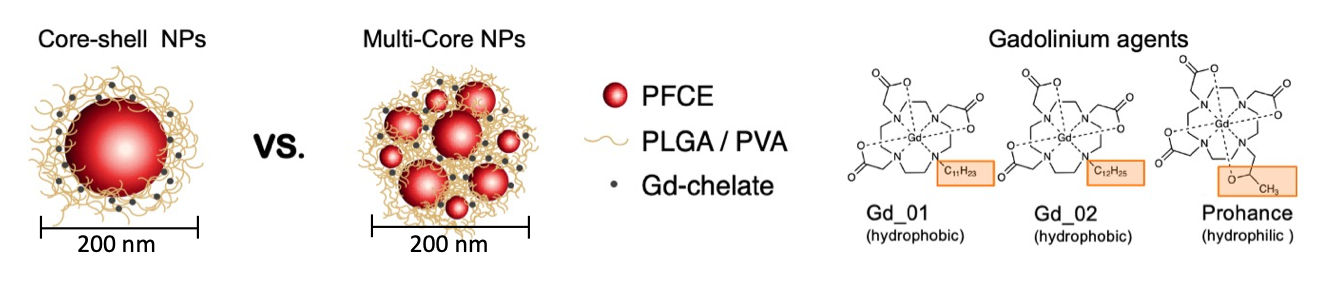
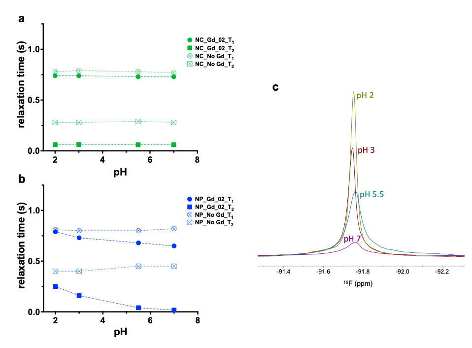
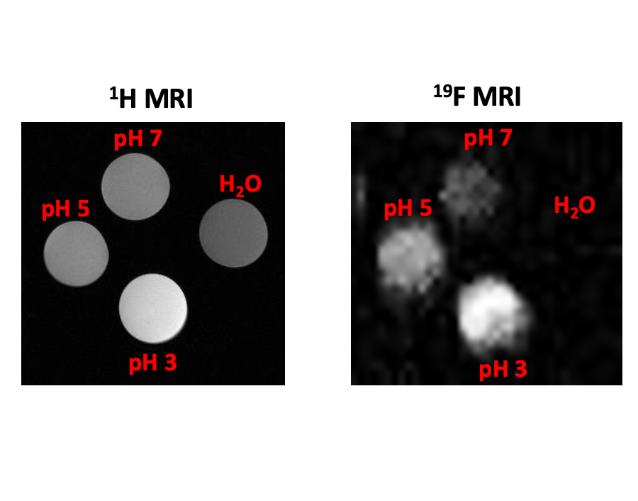
Fluorine-19 magnetic resonance imaging (19F-MRI) holds considerable promise for precise cell tracking. This study investigates the MR relaxation properties of perfluorocarbon-loaded nanoparticles (PFC-NPs) co-encapsulated with a paramagnetic gadolinium (Gd) chelate (as depicted in Figure 1). Paramagnetic relaxation enhancement (PRE) operates at short distances with close proximity between the paramagnetic chelate and the 19F nuclei. We sought to understand the impact of the internal structure, specifically by comparing core-shell and multi-core NPs [1], on the PRE effect of Gd on fluorine MR signal. Our initial findings emphasize the influence of internal structural features on 19F MR relaxation times. In fractal multi-core NPs, Gd chelates affect both longitudinal (T1) and transverse (T2) relaxation times, while core-shell NPs primarily show changes in T2. Furthermore, Gd entrapped within multi-core NPs uniquely enhances 1H MRI, benefiting from their superior water permeability compared to core-shell NPs [2]. Surprisingly, multi-core NPs exhibit pH-sensitive transverse relaxation while maintaining stability, in contrast to core-shell NPs, underscoring the value of the multi-core structure for enhancing 19F signals, as illustrated in Figure 2. The pH sensitivity was established through both 19F NMR (400 MHz) and MRI (14.1T), as shown in Figure 3. The 19F MRI results provide compelling evidence of a progressive increase in signal intensity as the pH transitions from neutral to more acidic conditions, effectively functioning as an 'on-off probe.' Additionally, our findings were corroborated by an in-cell 19F NMR experiment, which unveiled significant variations in T2 relaxation times both within and outside cells. This phenomenon is likely attributable to the acidic environment within lysosomes, offering valuable insights into the cellular microenvironment. Furthermore, preliminary data obtained from cancer cell lines extend the relevance of our probe, showcasing its sensitivity in detecting pH fluctuations. This potential carries significant promise for improving tumor imaging and advancing our understanding of cancer. Notably, the transition of cancer to a metastatic stage often corresponds with increased lysosomal activity, characterized by heightened lysosomal biogenesis and acidification. The ability of our probe to respond to such changes has substantial implications for cancer research and may pave the way for enhanced diagnostic and therapeutic strategies in the future.Acknowledgements
This project has received funding from the European Union’s Horizon 2020 research and innovation programme under the Marie Sklodowska Curie grant agreement No NOVA-MRI (859908). The authors acknowledge the funding from ERC‐2014‐StG‐336454‐CoNQUeST, TTW-NWO open technology grant STW-14716, ERC-2015-PoC-713524-CONQUEST, ERC-2019-PoC-862989-CENYA, and ERA-CVD JTC2017-044.References
1.Koshkina O, Lajoinie G, Baldelli Bombelli F, Swider E, Cruz LJ, White PB, Schweins R, Dolen Y, Van Dinther EAW, Van Riessen NK et al: Multicore Liquid Perfluorocarbon‐Loaded Multimodal Nanoparticles for Stable Ultrasound and19F MRI Applied to In Vivo Cell Tracking. Advanced Functional Materials 2019, 29(19):1806485.
2. Mali, A., Verbeelen, M., White, P. B., Staal, A. H. J., van Riessen, N. K., Cadiou, C., Chuburu, F., Koshkina, O., & Srinivas, M. (2023). The internal structure of gadolinium and perfluorocarbon-loaded polymer nanoparticles affects 19F MRI relaxation times. Nanoscale, 10.1039/d3nr04577c.
Figures