3969
An in-cell NMR strategy optimized for proteins with poor solubility and low thermal stability1Faculty of Life Sciences, Kumamoto University, Kumamoto, Japan, 2Research Institute for Biomedical Sciences, Tokyo University of Science, Noda, Chiba, Japan, 3Nippon Medical School, Tokyo, Japan
Synopsis
Keywords: Preclinical Image Analysis, Drug Development, Protein structure
Motivation: The in-cell NMR method allows direct observations of proteins in cultured human cells, to evaluate protein conformations and interactions in an intracellular environment. Currently, target proteins must be highly soluble and thermally stable.
Goal(s): A technical in-cell NMR strategy is required for proteins with poor solubility and low thermal stability.
Approach: Protein concentration-dependent NMR analyses revealed the self-association sites of proteins. Site-directed mutants gained higher solubility. Cell treatment conditions at lower temperature were established.
Results: We successfully constructed an in-cell NMR protocol that improves protein solubility and cell treatment conditions at 25°C, the lowest temperature established so far.
Impact: By performing experiments according to our strategy, in-cell NMR can be applicable to more types of proteins with poor solubility and low thermal stability. Furthermore, we developed an in-cell system for evaluating therapeutic candidate compounds against target proteins.
Introduction
In-cell NMR has been attracting attention for analyses of the conformations and interactions of proteins in an intracellular environment (Fig. 1). Therefore, in-cell NMR serves to translate in vitro experimental results to in vivo findings. To observe specific protein NMR signals in cells, separately prepared stable isotope-labeled proteins are introduced into cultured mammalian cells by electrical pulse- or toxin-aided cell membrane perforation. Generally, highly concentrated protein solutions (> 1 mM) are required for observations of NMR signals from the introduced intracellular proteins within a reasonable measurement time (< a few hours). Thermal stability at 37°C is also required, as it is the normal growth temperature for cultured human cells. However, only limited numbers of proteins meet these conditions.The aim of this study is to optimize the in-cell NMR method for proteins with poor solubility and low thermal stability. The Chemokine Receptor-Binding Domain (CRBD) of the cytoplasmic chemokine signal-regulator FROUNT [1] (Fig. 2) was utilized.
Methods
All uniformly 15N-labeled CRBD proteins were prepared with an E. coli expression system. Concentrated protein solutions were mixed with HeLa cells, which were dissociated from cell culture dishes, and introduced into the cells by electrical pulses for cell membrane perforation. For the experiment with the wild-type CRBD protein, highly soluble 15N-labeled ubiquitin mutant proteins (Ub3A) were also introduced with the CRBD proteins, as a positive control. These cells were cultured on collagen-coated dishes for three hours, to separate attached live cells from unattached dead cells. The live cells were dissociated, washed with phosphate-buffered saline, and mixed with DMEM medium for in-cell NMR experiments. After the experiments, the supernatants of sonicated cells were subjected to NMR measurements to ensure protein structural integrity. NMR experiments were performed with a 14T spectrometer equipped with a TCI CryoProbe (Bruker).Results and Discussion
Since CRBD protein solutions precipitate at concentrations over 0.8 mM, solutions of 0.6 mM CRBD and an equimolar amount of control Ub3A protein were mixed with HeLa cells, which were subjected to electrical pulses. Collected live cells were used for in-cell NMR experiments. 1H–15N SOFAST-HMQC peaks of CRBD, which were observed in vitro (Fig. 3A), were not detected in the in-cell NMR spectrum (Fig. 3B), while those of Ub3A in vitro (Fig. 3C) were observed. Large, self-associated CRBD oligomers were expected to be prevented from entering the cells through the cell membrane pores induced by electrical pulses.To improve the solubility of CRBD, chemical shift differences of 1H–15N HSQC peaks were analyzed using CRBD solutions between 0.05 and 0.2 mM to identify the CRBD self-association site. Larger chemical shift differences were observed at a hydrophobic patch surrounded by charged amino acids, based on the protein structure that we recently determined by NMR. This protein surface was predicted to be the self-association site, and thus we designed eleven site-directed mutants on this surface to improve the solubility of CRBD. The prepared mutants were evaluated, based on the NMR peak intensity ratios (0.05 mM vs. 0.5 mM) at 25°C. The peak intensity of the wild-type CRBD was increased by only 4-fold (Fig. 4A), while those of most mutants were increased further. Among them, the peak intensity of the Arg-to-Gln (RQ) mutant was the highest and increased by 10-fold (Fig. 4B), showing that the mutation significantly improved the solubility. Unfortunately, its solubility is high at 25°C, but not at 37°C, the temperature at which HeLa cells normally grow.
To overcome this difficult situation, we optimized an in-cell NMR protocol for use at 25°C, from the protein introduction and live cell selection to the in-cell NMR measurement. By using 1.0 mM RQ mutant solutions, 1H–15N SOFAST-HMQC peaks, which were observed in vitro (Fig. 3D), were partially observed in the in-cell NMR spectrum at 25°C (Fig. 3E). The observed peaks were from the mobile part of CRBD, and thus the peaks from the structured part were probably broadened due to interactions with large biomolecules. The spectral pattern of the RQ mutant obtained from sonicated cells (Fig. 3F) was identical to that in vitro (Fig. 3D), showing that the three-dimensional structure of the RQ mutant remained properly folded, even though it was subjected to the intracellular environment for three hours.
Conclusion
We successfully optimized the in-cell NMR method for proteins with poor solubility and low thermal stability. By performing experiments according to our strategy, in-cell NMR can be applicable to more types of proteins. Furthermore, we can develop an in-cell system for evaluating therapeutic candidate compounds against target proteins by monitoring the compound–protein interactions.Acknowledgements
The authors gratefully acknowledge Dr. Kosuke Inomata, Dr. Teppei Ikeya and Prof. Yutaka Ito (Tokyo Metropolitan University) for fruitful discussion.
References
[1] Terashima et al., Nature Commun., 11, 609 (2020)
Figures
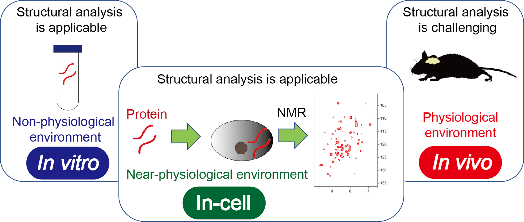
Fig. 1 Role of in-cell NMR
In-cell NMR has been attracting attention to analyze the conformations and interactions of proteins in the intracellular environment. In this method, isotope-labeled proteins are introduced into cultured mammalian cells, and their structures, dynamics and interactions are monitored by NMR. In-cell NMR serves to translate in vitro experimental results to in vivo results.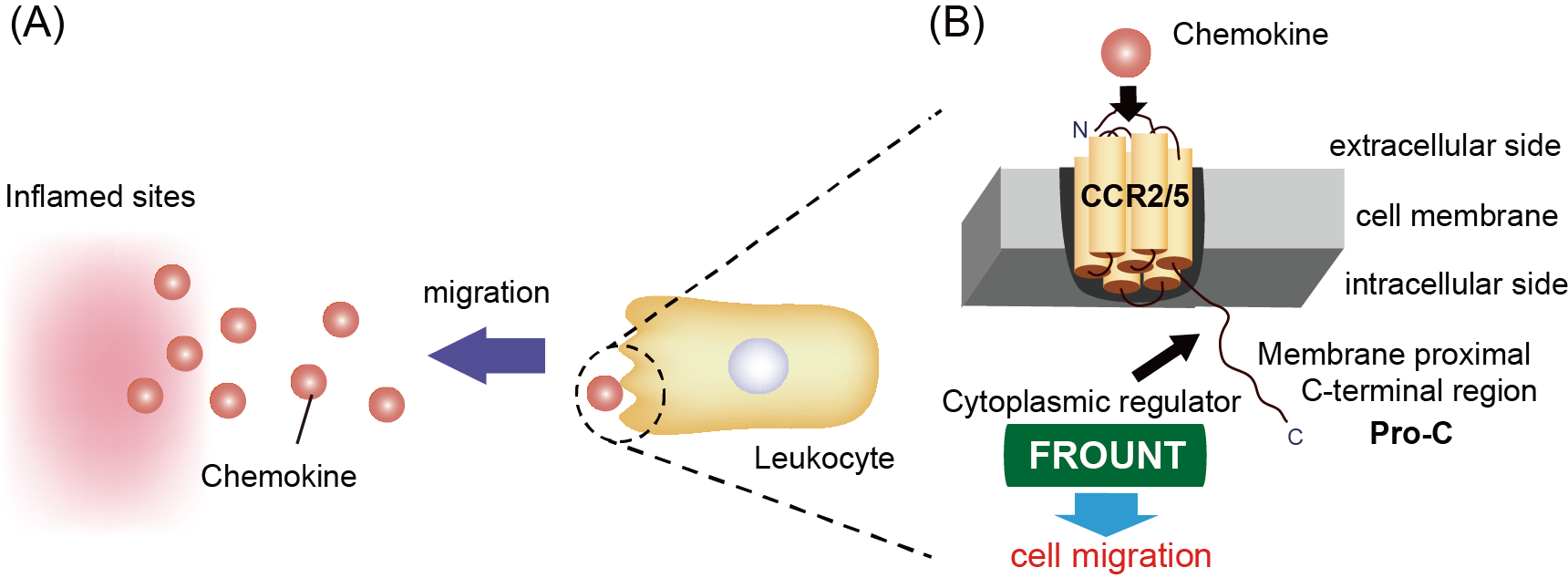
Fig. 2 Cytoplasmic chemokine signal-regulator FROUNT
Chemokines released from inflamed sites are recognized by chemokine receptors on leukocytes, thus triggering leukocyte cell migration (A). Activated chemokine receptors (CCR2/5) promote the binding of the cytoplasmic signaling protein FROUNT to the membrane proximal C-terminal region (Pro-C) for cell migration signaling (B).
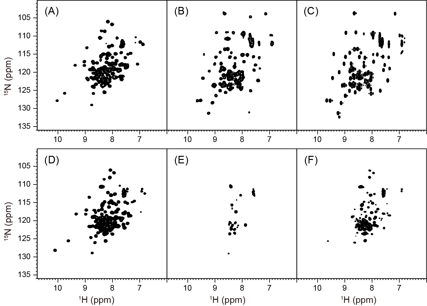
Fig. 3 Comparison of 1H–15N SOFAST-HMQC spectra
In vitro wild-type CRBD spectrum (A), in-cell spectrum of HeLa cells including wild-type CRBD and Ub3A (B), in vitro Ub3A spectrum (C), in vitro RQ mutant spectrum (D), in-cell spectrum of HeLa cells including RQ mutant (E), in vitro spectrum of the RQ mutant from sonicated HeLa cells (F).
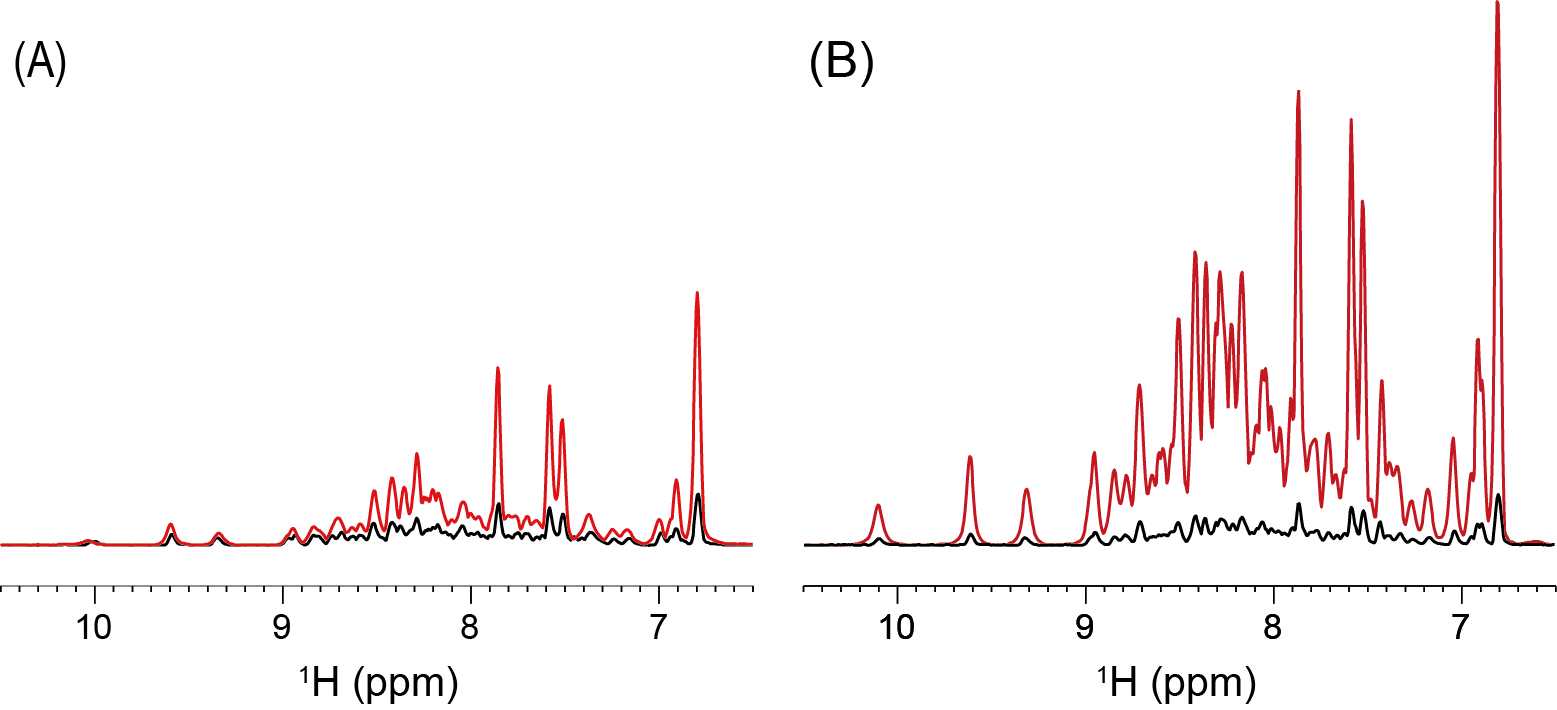
Fig. 4 Comparison of 1D projections of 1H–15N HSQC spectra
Spectra of the wild-type (A) and RQ mutant (B) at 0.05 mM (black) and 0.5 mM (red).