3968
In-vivo Imaging of the adult Zebrafish in an unmodified pre-clinical MRI System at 11.7T1Internal Medicine 2, Ulm University Medical Center, Ulm, Germany, 2Molecular Cardiology, Ulm University Medical Center, Ulm, Germany
Synopsis
Keywords: Biology, Models, Methods, High-Field MRI
Motivation: The zebrafish is an important model organism for the study of vertebrate biology with its genomes showing significant parallels to human genomes.
Goal(s): To show the feasibility of in-vivo imaging at a pre-clinical 11.7T MRI without the need for dedicated hardware components.
Approach: A budget-friendly zebrafish holder was created using a syringe barrel and needle adapters, with a rotating cylinder to maintain water flow. It was positioned on a Bruker Mouse Brain Surface Coil.
Results: The use of standard imaging equipment in combination with a low-cost and easy to built animal holder is sufficient to facility high-resolution imaging of the adult zebrafish.
Impact: This research enables cost-effective, high-resolution MRI for zebrafish, enhancing its potential as a pre-clinical model for vital research in vertebrate biology, ultimately advancing our understanding of human genomics.
Introduction
The zebrafish model (Danio rerio) has emerged as an excellent model organism for studies of vertebrate evolution, diseases, biological pathways as well as toxicologic mechanisms. Another interesting property is its natural tissue regenerative capability, which is especially of interest for studies of cardiovascular development and diseases1. However, due to the opacity of the juvenile and adult stages, traditional optical microscopic methods are not suitable for the study of developmental processes in the adult model.MRI provides a non-invasive imaging technique to overcome limitations of optical microscopy and without the need of sacrificing the animal to allow for longitudinal studies. But the dimensions of the zebrafish and its internals require high imaging resolutions at sufficient SNR which often leads to the construction of dedicated hardware components such as coils2,3 and even gradient systems4.
Methods
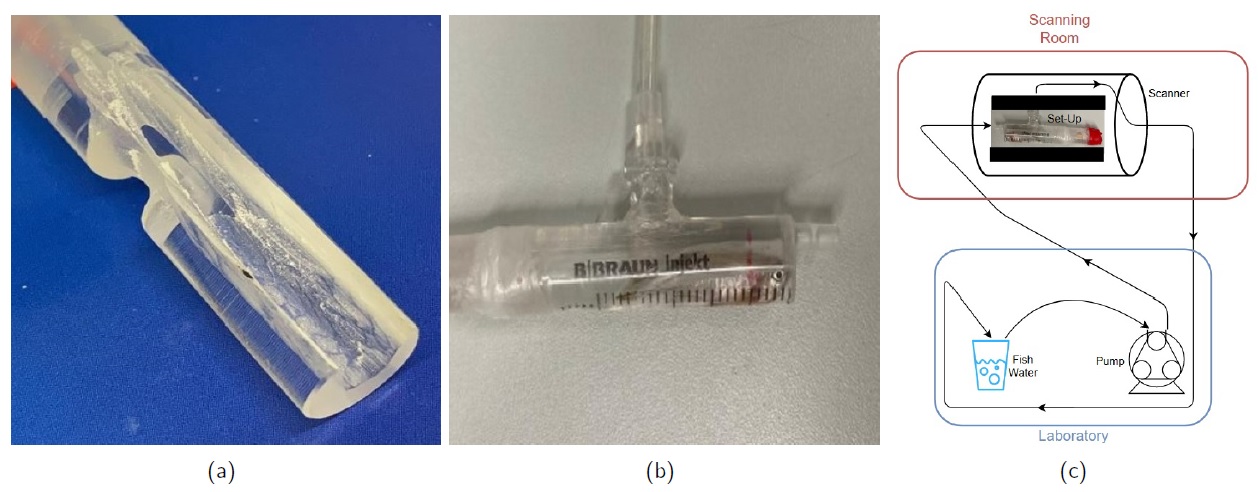
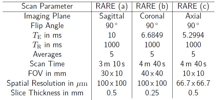
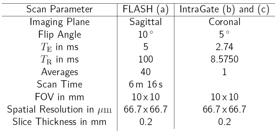
A low-cost animal holder was constructed based on a syringe barrel with the needle adapter functioning as a connector for the inflow tube. For the outflow, a second needle adapter was glued onto a drilled hole to the side of the barrel. A PMMA cylinder was fitted into the syringe with a milled cavity that corresponds to the size of an adult zebrafish. An additional cavity was milled along the circumference to allow for rotation of the cylinder (fish orientation) while maintaining a constant waterflow. All connections were sealed and the in- and outflow tubes were connected to a peristaltic pump (figure 1). The setup was placed on a Bruker Mouse Brain Surface Coil (M.BR LIN RO AD, Bruker, Ettlingen, Germany) such that the fish is placed upright directly on the receive coil. The entire setup was then shuttled into a Bruker BioSpec 117/16-USR pre-clinical MRI system for imaging. Excitation was achieved using a 72 mm inner diameter volume resonator (089/072 Quad to AD, Bruker, Ettlingen, Germany).An anaesthetic concentration of 140 mg/L MS222 was added to pH-controlled water and circulated at a flow rate of approximately 10 mL/min to minimise flow induced motion.
Ex-vivo images were acquired using a RARE (turbo spin-echo) sequence to test the coil sensitivity and overall image quality. In-vivo images were acquired using FLASH and (Cartesian) IntraGate CINE sequences with the aim to resolve respiratory motion. All scan parameters are shown in figure 2 (ex-vivo) and figure 3 (in-vivo).
Results
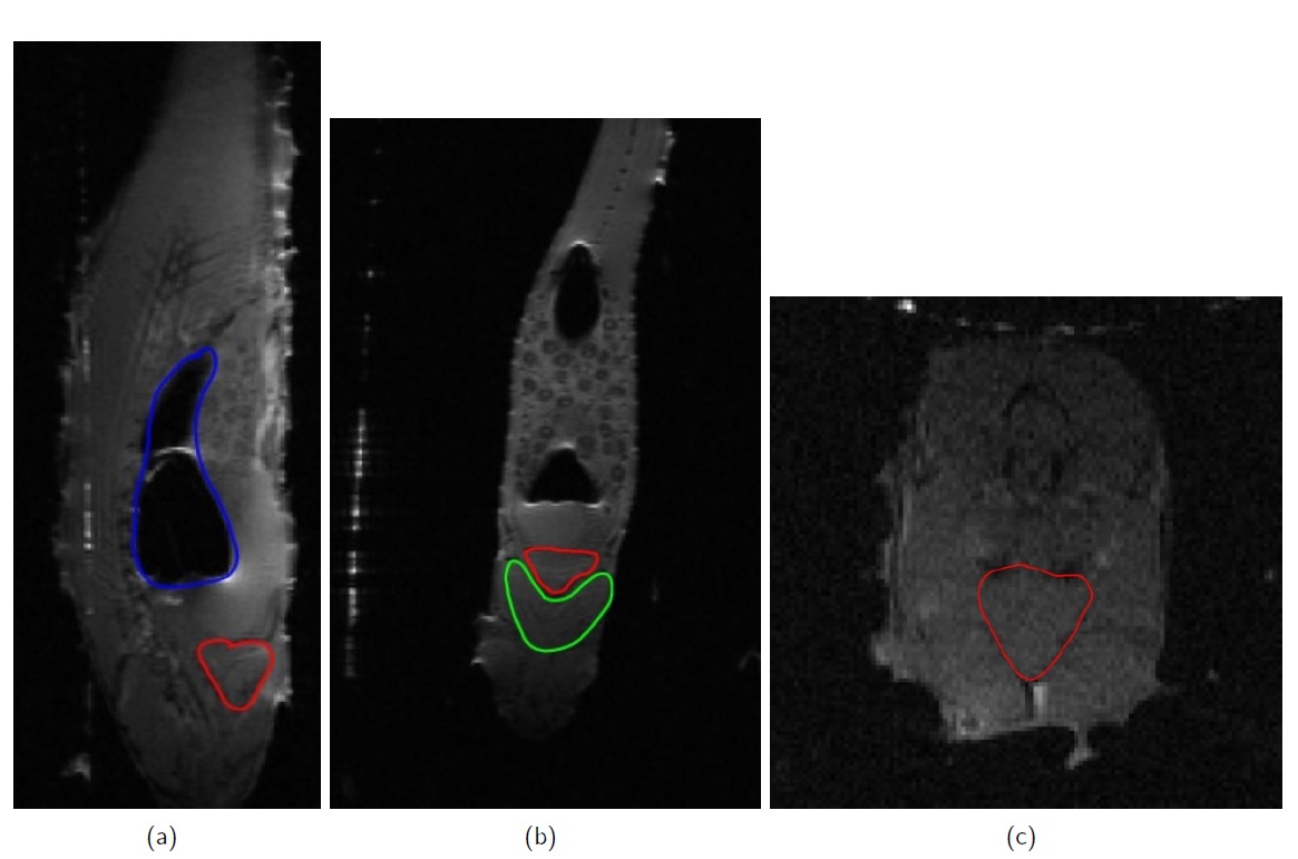
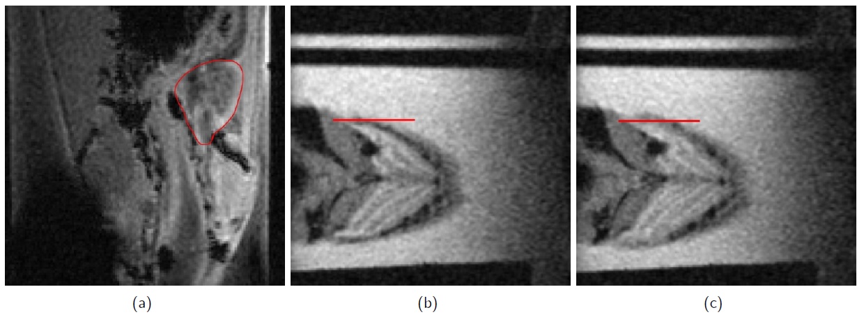
Ex-vivo RARE images with a spatial resolution of 100 μm of all three anatomical planes are shown in figure 4, with the swim bladder highlighted in blue, the heart highlighted in red and the sternohyoid muscle highlighted in green.In-vivo images are shown in figure 5, with a sagittal slice of a FLASH acquisition shown in figure 5a). Again, the heart is highlighted in red. Figure 5b,c) shows two of a total of five stages of respiratory motion, reconstructed from an IntraGate CINE acquisition at a respiratory frequency of 1,33 Hz with an isotropic in-plane resolution of 67 μm. The amplitude of the respiratory motion per gill was measured to be approximately 0.5 mm.
Discussion and Conclusion
The use of standard imaging equipment in combination with a low-cost and easy to built animal holder is sufficient to facility high-resolution imaging of the adult zebrafish.Optimisation of the used imaging sequences led to a clear delineation of cardiac tissue, with enhanced contrasts arising in the in-vivo case. The non-constant water flow restricts in in-vivo imaging the application of spin-echo sequences to the ex-vivo case.
Beneath the resolution of respiratory motion, even the resolution of cardiac motion appears feasible, especially in combination with self-gating techniques such as k0-based gating approaches e.g. in combination with tiny golden angle radial sampling5.
Acknowledgements
The authors thank the Ulm University Centre for Translational Imaging MoMAN for its support.References
1 Koth, Jana, et al. High-resolution magnetic resonance imaging of the regenerating adult zebrafish heart. Scientific reports 7.1 (2017): 1-12.
2 Kabli, Samira, et al. Magnetic resonance microscopy of the adult zebrafish. Zebrafish 3.4 (2006): 431-439.
3 Kabli, Samira, et al. In vivo magnetic resonance imaging to detect malignant melanoma in adult zebrafish. Zebrafish 7.2 (2010): 143-148.
4 Marrifield, Gavin D., et al. Rapid and recoverable in vivo magnetic resonance imaging of the adult zebrafish at 7T. Magnetic resonance imaging 37 (2017): 9-15.
5 Wundrak, Stefan, et al. Golden ratio sparse MRI using tiny golden angles. Magnetic resonance in medicine 75.6 (2016): 2372-2378.
Figures