3967
Pioneering High-Throughput Micro-NMR in Reproductive Biology: Unveiling Metabolic Profiles in Early Bovine Embryos and Oocytes1Annaida Technologies, Lausanne, Switzerland
Synopsis
Keywords: Other Preclinical, Reproductive, Early Embryos
Motivation: Traditional methods for early embryos assessment are often invasive or lack detail, underscoring the necessity for non-invasive, high-resolution techniques.
Goal(s): The primary goal was to test MRS as a predictive tool for embryo viability. By leveraging microchip-based probes, we analyzed the metabolic profile of early-stage bovine embryos and oocytes. This aimed at predicting developmental outcomes with high precision and accuracy.
Approach: In total, we conducted multi-channel high-throughput spectroscopy to perform a minimally invasive analysis of 1 hour on 61 single 8-cell embryos and 84 single oocytes.
Results: We found strong correlation between spectra at the 8-cell stage and subsequent development to blastocyst.
Impact: This work offers new data for MRS and embryology, laying a foundation to improve fertility treatments by selecting viable embryos. By revealing previously inaccessible data it opens to new embryonic research, potentially revolutionizing our understanding of early developmental biology.
INTRODUCTION
Magnetic Resonance Spectroscopy (MRS) has long been recognized as a gold standard, non-invasive technique, invaluable in a range of fields from fundamental research to personalized medicine [1-3]. Recent advances in micro-NMR (a subdivision of MRS focusing on increased sensitivity for volume-limited samples), have unlocked the possibility of performing measurements at volumes in the nanoliter range (i.e., a linear dimension of 100 microns) [4,5]. Using this approach, first data have been published from single 3D human liver cell cultures and early-stage bovine embryos [6,7]. In general, nanoliter MRS could represent a paradigm shift in the context of fertility treatments and developmental biology research, where no alternative method can offer comparable data without harming microscopic cell-cultures such as organoids and early embryos. In this study we leverage an improved micro-NMR probe system to explore the early bovine embryos and oocytes, correlating the signal from mobile lipids to their developmental outcome.METHODS
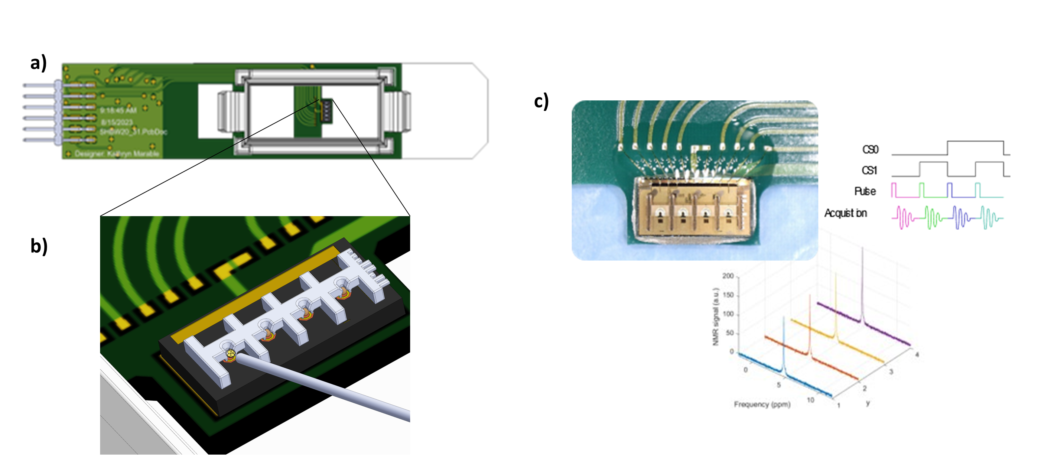
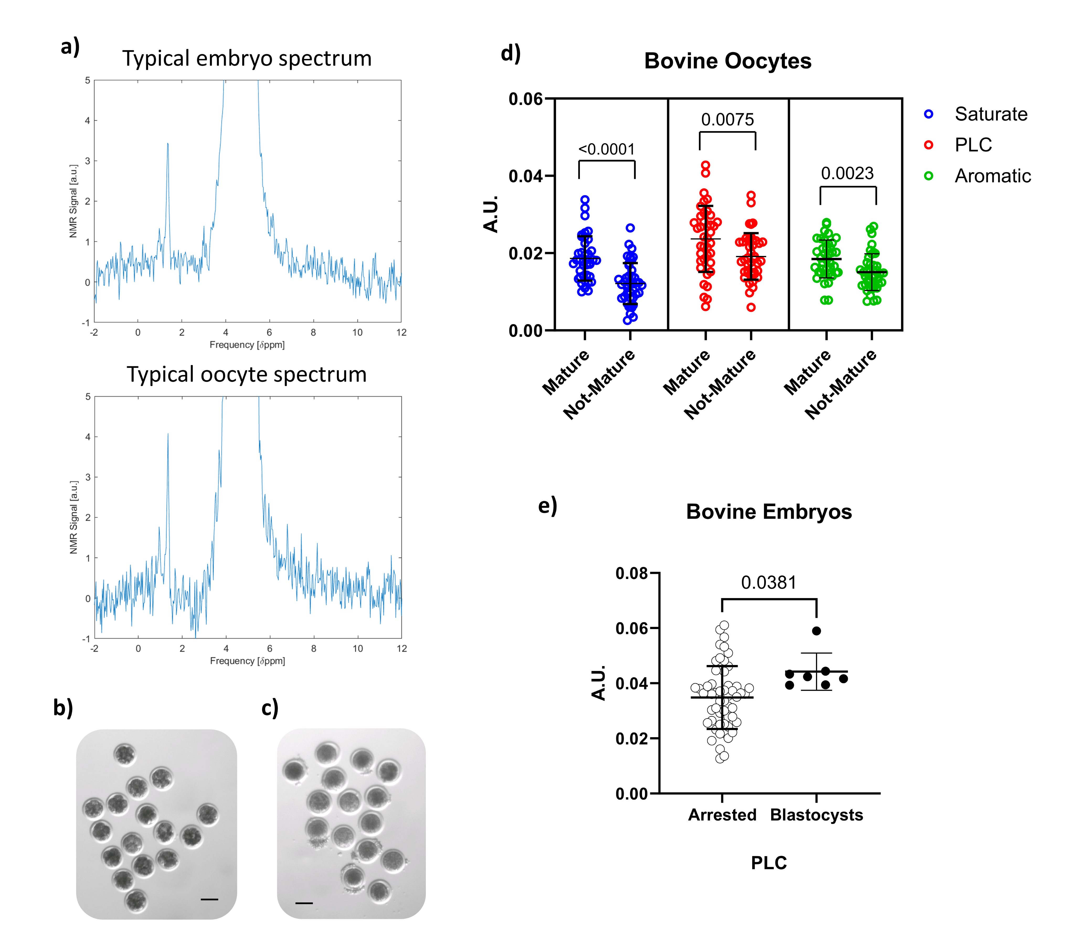
We utilized a cutting-edge scalable and user-friendly micro-NMR probe system, featuring an on-chip transceiver, 4 microcoils for pipeline measurements, 3D micro-printing, and compatible with standard NMR spectrometers (see Fig. 1). This system, operating at frequencies ranging from 150 MHz to 600 MHz, is capable of handling samples between 10 nL and 100 pL in volume. Its CMOS-based design permits simultaneous multi-sample acquisition from 4 sensor coils, making it suitable for high-throughput, detailed studies on embryonic and oocyte samples. In this research, we focused on 61 cow embryos measured at the 8-cell stage. The samples are thawed from cryogenic preservation, their MRS spectrum is recorded at controlled temperature (37 ± 0.5 °C), and they are incubated and monitored with a microscope for the subsequent days to determine how far each embryo develops. We then use these data to draw correlations between MRS profiles and developmental outcomes. We also used the same system to measure 84 oocytes (mature and immature) to evaluate whether MRS profiles capture features correlating with the maturity stage of the samples.RESULTS
The spectra recorded show dominant features attributed to fatty acids. This was expected from previous investigations [4-7]. The embryo’s data analysis revealed a robust 80% precision in predicting the development to the blastocyst stage based on their metabolic profiles at the 8-cell stage. In the oocyte’s data analysis, notable differences (up to p < 0.0001) in the lipid profiles were identified between mature and immature oocytes, with particular emphasis on the saturation levels of lipid molecules. These findings, summarized in Fig. 2, mark the first instance of single mammalian cell NMR spectroscopy, illustrating the unparalleled sensitivity and detail of MRS in this domain.DISCUSSION
By non-invasively deciphering the metabolic signatures of early-stage embryos and oocytes, MRS offers a new lens to observe cellular development, viability, and maturity. The results are a first step towards studying mammalian single cells and embryos, with first data indicating a predictive power of the method in determining embryonic development potential, which demonstrates potential of micro-MRS in embryology and reproductive medicine. A key challenge in Assisted Reproduction Technologies is indeed the identification of those embryos with the highest potential: the average precision in determining embryos potential to pregnancy stands today at about ~40% [8] (see Clinical Data Summary). The differential lipid profiles observed in oocytes further enrich our understanding of oocyte maturity, with potential application for both fundamental biology and clinical practice.Acknowledgements
We thank Prof. Kurt Wüthrich for useful discussions and Dr. Carolina Herrera for providing samples. We thank the CPMA clinic (center of assisted reproduction, Lausanne) for providing culture incubators. This work was partially supported by the European Union's Horizon 2020 research and innovation program under grant agreement N. 681002.References
[1] De Graaf, Robin A. In vivo NMR spectroscopy: principles and techniques. John Wiley & Sons, 2019.
[2] Damadian, Raymond. "Tumor detection by nuclear magnetic resonance." Science 171.3976 (1971): 1151-1153.
[3] Le Bihan, Denis, et al. "MR imaging of intravoxel incoherent motions: application to diffusion and perfusion in neurologic disorders." Radiology 161.2 (1986): 401-407.
[4] Grisi, Marco, et al. "NMR spectroscopy of single sub-nL ova with inductive ultra-compact single-chip probes." Scientific reports 7.1 (2017): 44670.
[5] Montinaro, E., et al. "3D printed microchannels for sub-nL NMR spectroscopy." PloS one 13.5 (2018): e0192780.
[6] Sivelli, Giulia, et al. "NMR spectroscopy of a single mammalian early stage embryo." Journal of Magnetic Resonance 335 (2022): 107142.
[7] Grisi, Marco, et al. "NMR microsystem for label-free characterization of 3D nanoliter microtissues." Scientific reports 10.1 (2020): 1-9.
[8] https://nccd.cdc.gov/drh_art/rdPage.aspx?rdReport=DRH_ART.ClinicInfo&rdRequestForward=True&ClinicId=9999&ShowNational=1
Figures