3966
Synthesis and Evaluation of Porphyrin-Based Gadolinium complex as Multifunctional Theranostic Agent1Division of applied RI, Korea Institute of Radiological & Medical Sciences, Seoul, Korea, Republic of, 2Radiological and Medico-Oncological Sciences, University of Science and Technology, Seoul, Korea, Republic of
Synopsis
Keywords: Probes & Targets, Tumor
Motivation: Currently used Neutron capture therapy agents in clinic have low tumor targeting ability and have different structures from imaging agents that can view bio-distribution, making it difficult to determine the exact distribution of the drug in the human body.
Goal(s): Development of a new MR-image guided drugs for neutron capture therapy
Approach: Two of gadolinium complexes of porphyrin derivative were designed and synthesized. Cell uptake study was performed using normal cell and glioblastoma cells. In vivo MR images and ex vivo fluorescence images were obtained.
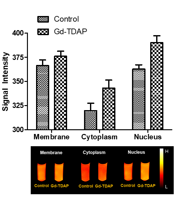
Results: We found that Gd-complexes could enter into the nucleus and be further uptaken by tumor tissues.
Impact: Using the two gadolinium complexes, it is possible to compare the therapeutic efficacy of GdNCT and BNCT and expect a synergistic effect. The development of NCT treatments using small molecule substances may have the potential for clinical application.
Abstract
Neutron capture therapy (NCT) is a precision treatment technology that selectively destroys only tumor cells based on the nuclear reaction that occurs when NCT agents captures thermal neutrons. Representative substances that cause a neutron capture reaction include boron-10 (10B) and gadolinium-157(157Gd).1Boron neutron capture therapy (BNCT) is based on the nuclear reaction between 10B and thermal neutrons (10B[n,α]7Li), resulting in alpha particles (4He) and 7Li nuclei. Because α particles have very short path lengths (5–9 μm), their destructive effect is limited to boron-containing cells. Thus, theoretically, it can selectively destroy tumor cells and adjacent normal cells unaffected. Only two boron compounds, L-4-dihydroxyboryl phenylalanine (BPA) and sodium mercaptoundecahydro-closo-dodecaborate (Na2B12HSH, BSH), have been approved by the Food and Drug Administration (FDA) for clinical use.1,2
Gadolinium neutron capture therapy (GdNCT) is based on a (157Gd[n,γ]158Gd) nuclear reaction, resulting in the emission of prompt γ-rays, internal conversion electrons, and Auger electrons. 157Gd has an extremely large thermal neutron cross section, which is 66 times higher than that of 10B. While BNCT depends primarily on the short fight range of α particles emitted by the boron neutron capture reaction, GdNCT mainly relies on long range gamma rays released by the gadolinium neutron capture reaction.3 Furthermore, because gadolinium agents have been used clinically as magnetic resonance imaging (MRI) agents for a long time, they were directly tested as GdNCT agents.
In developing theranostic drugs, it is important to design a platform that can perform multiple functions simultaneously. This can reduce the side effects caused by unnecessary ingredients from a clinical point of view. In this study, we present the gadolinium complex of 5,10,15,20-(tetra-N,N-dimethyl-4-aminophenyl)porphyrin (TDAP) (Gd-TDAP) and gadolinium complex of 5,10,15,20-(tetra-4-dihydroxyborylphenyl)porphyrin (TDBP) (Gd-TDBP) as multimodal theranostic platforms for brain tumors (optical/MRI and NCT/PDT) (Figure 1).4
Porphyrin, a heterocyclic macrocycle compound, is a promising theranostic agent owing to its selective tumor uptake, good blood brain barrier (BBB) penetration, low toxicity, potential for conjugation with biological moieties, and strong affinity for metallic species. It is used as a radiopharmaceutical not only for single-photon emission computed tomography (SPECT) or PET by chelation with metallic radioisotopes, such as 99mTc, 111In, 68Ga, 89Zr, and 64Cu, but also for magnetic resonance imaging (MRI) or neutron capture therapy (NCT) agents in combination with Gd(III). In addition, porphyrin emits fluorescence; therefore, it can be used as both an optical imaging and a photodynamic therapy (PDT).5
To verify the functions of Gd-TDAP and Gd-TDBP, we here report the photophysical characterization, magnetic properties, brain tumor targeting ability, in vivo MRI contrast enhancement and optical imaging capability.4
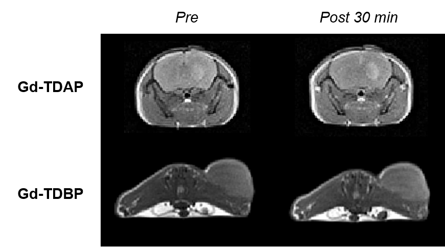
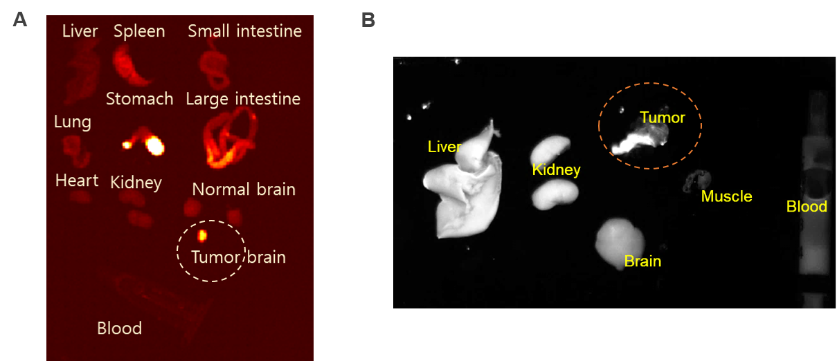
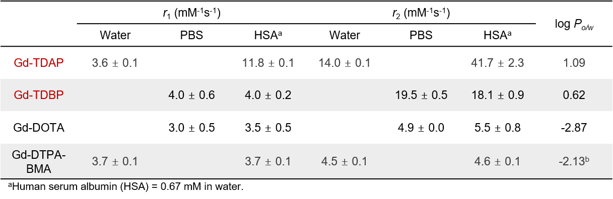
The porphyrin-based gadolinium complexes Gd-TDAP and Gd-TDBP have shown high relaxivities, lipophilicity (Table 1). T1-weighted MR imaging showed that brain tumors could be detected within 20 min post injection (p.i.) of Gd-TDAP and subcutaneous tumor could be detected within 30 min p.i. of Gd-TDBP. (Figure 2). In addition, after injection, the fluorescence images showed a clear difference between the extracted tumor and normal brain tissues (Figure 3). Furthermore, the accumulation of Gd-TDAP in the nucleus after cell fractionation demonstrated potential application for Gd-NCT (Figure 4).
In summary, we have prepared and evaluated two gadolinium complexes. Based on the research so far, we will be able to perform MRI-guided neutron capture therapy and compare the effects of GdNCT and combination of Gd and BNCT.
Acknowledgements
This work was supported by a grant of the Korea Institute of Radiological and Medical Sciences(KIRAMS), funded by MSIT, Republic of Korea (No. 50462-2023).References
1. Jalilian AR, Shahi A, Swainson IP, et al. Potential theranostic boron neutron capture therapy agents as multimodal radiopharmaceuticals. Cancer Biother. Radiopharm. 2022;37(5):342-354.
2. Sauerwein WA, Sancey L, Hey-Hawkins E, et al. Theranostics in boron neutron capture therapy. Life. 2021;11(4):330.
3. Shanmugam M, Kuthala N, Kong W, et al. Combined gadolinium and boron neutron capture therapies for eradication of head-and-neck tumor using Gd10B6 nanoparticles under MRI/CT image guidance. JACS Au. 2023;3(8):2192-2205.
4. Kim S, Yang J-u, Ahn JH, et al. Porphyrin-based tumor-targeting theranostic agent: Gd-TDAP. ACS Med Chem Lett. 2021;12(9):1459-1463
5. Tsolekile N, Nelana S, Oluwafemi OS. Porphyrin as diagnostic and therapeutic agent. Molecules. 2019;24(14):2669.
Figures