3962
A Preclinical Co-Registration Pipeline for MRI and PET/CT: Enabling Multi-tracer Multiparametric PET-CT/MRI in Solid Tumors1Department of Biomedical Engineering, University of Alabama at Birmingham, Birmingham, AL, United States, 2Department of Radiology, University of Alabama at Birmingham, Birmingham, AL, United States, 3O'Neal Comprehensive Cancer Center, University of Alabama at Birmingham, Birmingham, AL, United States
Synopsis
Keywords: Preclinical Image Analysis, Quantitative Imaging, Multi-tracer, Multiparameteric, Registration
Motivation: Robust preclinical multimodal image registration must be developed to provide rich multiparametric data to noninvasively reveal biological treatment-induced tumor alterations.
Goal(s): Establish a pipeline to enable multimodal registration of semi-rigid, subcutaneous tumors in preclinical models to evaluate multi-tracer multiparametric PET/CT-MR metric alterations.
Approach: Utilizing a breast cancer murine model, a custom-designed mouse couch, augmented with an injectable liquid fiducial marker, was assessed for MR-CT co-registration, facilitating the development of a registration pipeline.
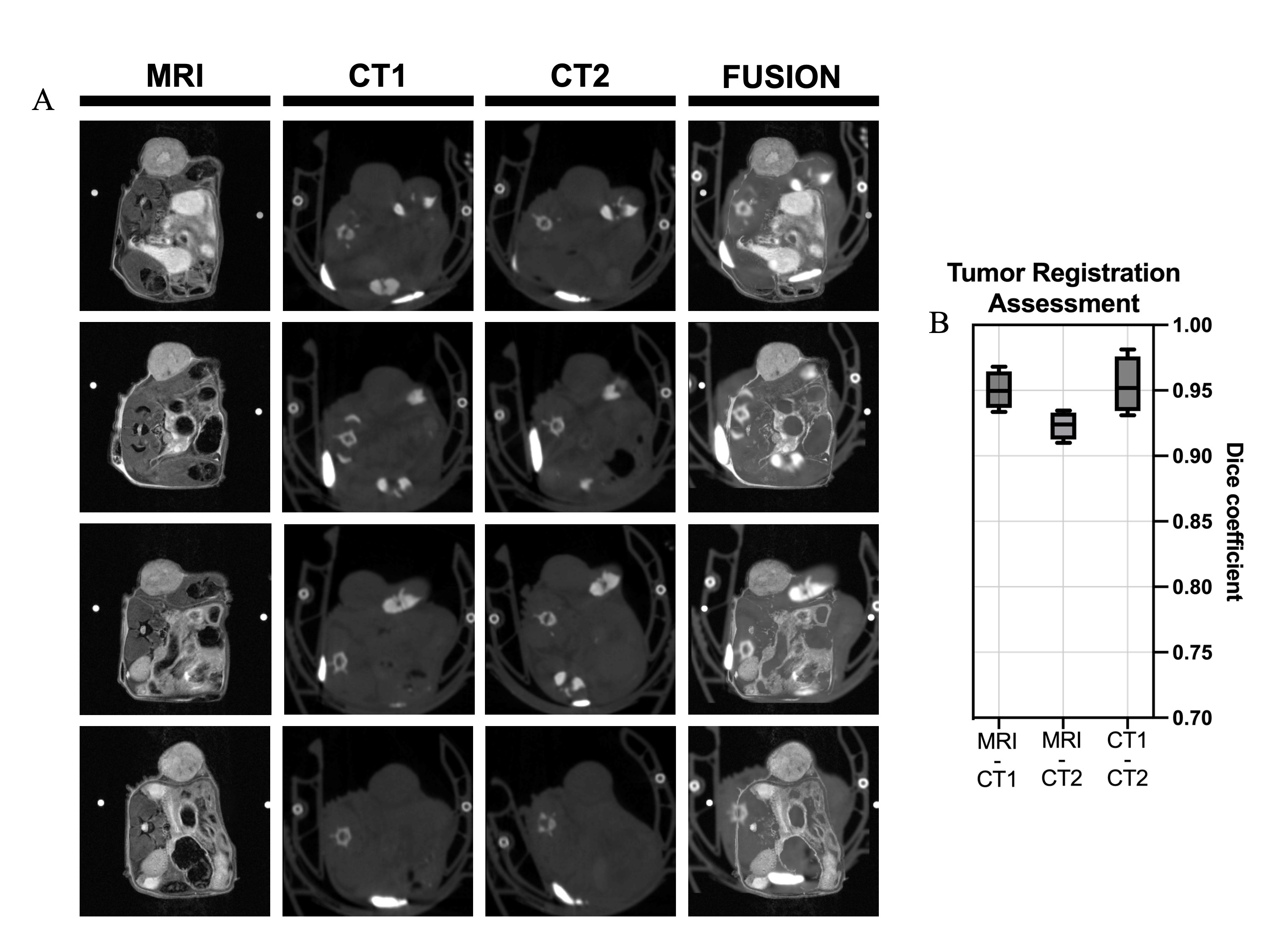
Results: Post-registration tumor segmentation performed independently across modalities yielded high Dice Similarity Coefficient scores (0.92-0.95), indicating accurate tumor alignment, and enabling preliminary multiparametric voxel-wise comparisons of multimodal imaging metrics.
Impact: Preclinical imaging with our registration pipeline enables for biological treatment-induced characterization of subcutaneous tumors through sequential multi-tracer multiparametric PET/CT-MR.
Introduction
Biological characterization of tumors through multimodal imaging is essential for cancer prognosis and treatment strategy optimizations. While clinical PET/MR studies have demonstrated the benefits of integrating quantitative DW- and DCE-MRI with PET compared to molecular imaging alone1–3, preclinical multiparametric and multimodal PET/MR evaluations present technical and logistical challenges. Predominantly brain-centric registration pipelines4 and limitations in MRI-based attenuation correction5 hinder the application in preclinical subcutaneous cancer models which critically serve as first pass evaluation of therapy response. To address this gap, an acquisition and processing pipeline was developed to aid in semi-rigid registration of multimodal images of solid subcutaneous-implanted tumors. The pipeline was evaluated for the synchronization of sequential multi-tracer preclinical PET/CT and multiparametric MR data to enable precise multimodal registration, facilitating the generation of voxel-wise multi-tracer multiparametric maps.Methods
Flank tumor-bearing breast cancer murine models were established. The imaging protocol included multiparametric MRI + two PET imaging tracers: [18F]FDG and [18F]FLT PET/CT. Diffusion-weighted imaging (DWI), T2-weighted, and dynamic contrast enhanced (DCE)- MRI sequences were obtained using a 40mm volumetric coil (9.4T Bruker BioSpec MRI). Subsequent to MRI, each mouse received 100 µCi of [18F]FDG intravenously and was imaged, referenced as CT1 (SOFIE GNEXT PET/CT). Post-[18F]FDG decay, a second [18F]FLT PET/CT scan was acquired (CT2).Custom mouse couches were designed, and 3D printed in polylactic acid chosen for MR-compatibility and CT-inert properties (Figure 1A). Two additional methods to aid in registration were incorporated and evaluated, saline-filled capillary tubes and injectable liquid radiopaque fiducial markers discernable on MR and CT imaging.
Imaging acquisition parameters:
| Imaging Sequence | TE/TR (ms) | FA | Voxel size (mm) | Acquisition matrix | Additional parameters | Generated Maps |
| Axial multi-slice diffusion-weighted (DWI) | 22.73/4500 | 90˚ | [0.53, 0.53, 1] | 64x64x11 | b-vals = (150, 500, 800) | Apparent Diffusion Coefficient (ADC) |
| Axial multi-sliceT2-weighted rapid acquisition and relaxation enhancement (T2RARE) | 24/2000 | 180˚ | [0.13, 0.13, 1] | 256x256x11 | - | High-resolution anatomical reference |
| T1 map variable repetition time (VTR) | 10/ - | 180˚ | [0.53, 0.53, 1] | 64x64x11 | TR = [255,400, 800, 1500, 3000, 5500] | T1 map |
| Axial dynamic contrast enhanced fast low angle shot (DCEFLASH) | 1.65/100 | 30˚ | [0.53, 0.53, 1] | 64x64x11 | 200 x 6.4s frames | Ktrans,ve,vp,kep |
| PET/CT | - | - | [0.2, 0.2, 0.2] | 161x161x600 | CT-based AC, 80kVp | SUV [18F] FDG and [18F] FLT –PET maps |
Image processing techniques:
Reframing: PET/CT slices were averaged adjacently to match the slice thickness of MR acquisitions.
Resampling: PET/CT voxel size was upsampled to match the voxel size of MR acquisitions (0.13 x 0.13) to facilitate accurate overlay and ensure that each voxel represents matching physical space.
Registration: An affine 3D transform was applied to register each of the CT volumes to the T2-weighted MR scan. Parameters include: Optimizer – regular step gradient descent, Iterations – 100, Metric – mattes mutual information, Weighting – rough tumor crop range.
The PET/CT images were reframed, resampled, and registered to the T2-weighted MR scans, summarized in Figure 1B-C. Tumors were manually annotated separately on each anatomical modality post-registration. Dice Similarity Coefficient (DSC) was utilized to gauge correspondence of segmentations. Multiparametric maps were generated through voxel-wise hierarchical agglomerative clustering of maps and multiregional spatial interaction (MSI) was used to quantify cluster spatial colocalization of clusters.
Results
Qualitative assessment of tumor alignment demonstrated correspondence (Figure 2). Saline-filled capillary tubes and liquid fiducial markers confirmed consistent murine positioning with minimal contribution to the enhancement of image registration. The custom mouse couch enabled reproducible positioning across imaging sessions. Quantitative assessments, employing DSC, revealed a score of 0.95 ± 0.01 (mean±std) for MR tumor segmentations compared to CT1, and a slightly lower score with the subsequent CT scan (CT2), 0.92 ± 0.01. Inter-CT comparison yielded a high concordance in tumor annotations, denoted by a DSC of 0.95 ± 0.02 (Figure 2B). The mean metrics of each cluster demonstrated varying intratumoral habitats graphically represented in Figure 1D. Moreover, MSI analysis confirmed substantial colocalization amongst identified clusters, with a value of 0.90 vs random 0.61.Discussion
The proposed acquisition and processing pipeline for registration of multimodal imaging of subcutaneous preclinical tumors has demonstrated its utility in preliminary multiparametric PET/CT-MR map generation. Regions characterized with high FDG, FLT, Ktrans, and kep and low ADC indicate aggressive growing regions (clusters 3 and 4), while clusters with lower vascularity metrics and higher ADC (clusters 1 and 2) may exhibit better treatment response. High DSC and MSI scores confirm the accuracy of tumor segmentation and the effectiveness of cluster matching across modalities, allowing for appropriate voxel-wise multiparametric analysis. These findings support the potential utility of this pipeline in preclinical intratumoral habitat classification.Acknowledgements
This project was in part supported by: NIH NCI R01CA240589, and NIH NCI R01CA276540. Imaging and computational resources for this research were supported by the O’Neal Comprehensive Cancer Center’s Preclinical Imaging Shared Facility P30CA013148. We gratefully acknowledge the support of Sharmila Sridhar and Luke Sligh for their image acquisition contributions.References
1. Furtado FS, Suarez-Weiss KE, Vangel M, et al. Clinical impact of PET/MRI in oligometastatic colorectal cancer. Br J Cancer. 2021;125(7):975-982. doi:10.1038/s41416-021-01494-82. Park J, Pak K, Yun TJ, et al. Diagnostic Accuracy and Confidence of [18F] FDG PET/MRI in comparison with PET or MRI alone in Head and Neck Cancer. Sci Rep. 2020;10(1):9490. doi:10.1038/s41598-020-66506-8
3. Furtado FS, Hesami M, Mcdermott S, Kulkarni H, Herold A, Catalano OA. The synergistic effect of PET/MRI in whole-body oncologic imaging: an Expert Review. Clin Transl Imaging. 2023;11(4):351-364. doi:10.1007/s40336-023-00577-1
4. Presotto L, Bettinardi V, Mercatelli D, et al. Development of a new toolbox for mouse PET–CT brain image analysis fully based on CT images and validation in a PD mouse model. Sci Rep. 2022;12(1):15822. doi:10.1038/s41598-022-19872-4
5. Hu Z, Yang W, Liu H, et al. From PET/CT to PET/MRI: Advances in Instrumentation and Clinical Applications. Mol Pharm. 2014;11(11):3798-3809. doi:10.1021/mp500321h
Figures
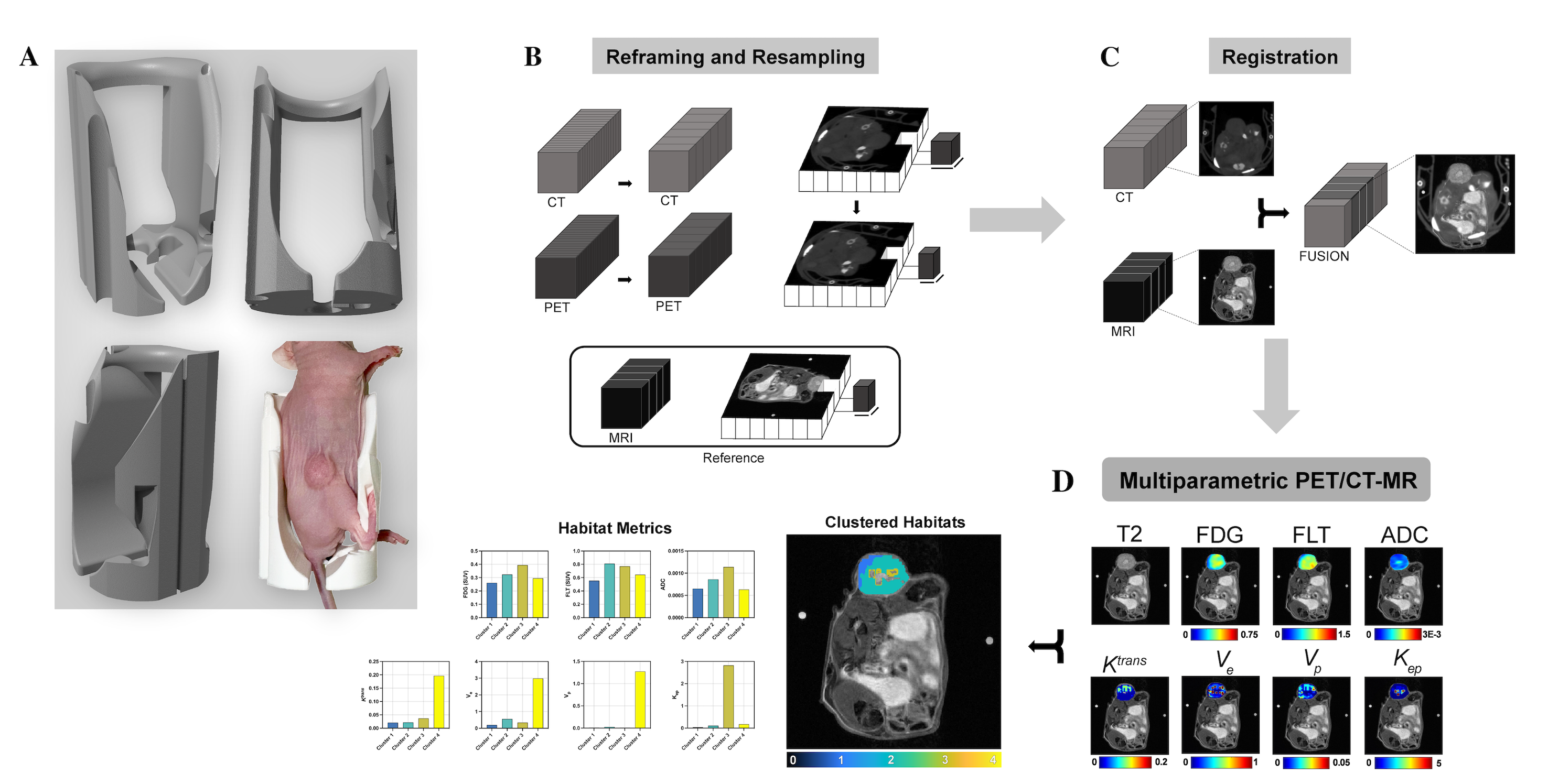
Figure 1: CT-MR tumor registration pipeline scheme. Mice are positioned in the custom designed couch (A). Scans are reframed and resampled to match the MRI reference (B). Registration is performed (C). Multi-tracer multiparametric PET/CT registered to quantitative MR metrics as a preclinical application of this pipeline (D).