3961
A novel ADAMTS-specific MR peptide probe for characterization of atherosclerotic plaques in a murine model1Radiology, Charite Universitaetsmedizin - Berlin, Berlin, Germany, 2Division 1.5 Protein Analysis, Federal Institute for Materials Research and Testing (BAM), Berlin, Germany, 3Institute of Inorganic and Analytical Chemistry, Westfaelische Wilhelms-Universität Muenster, Muenster, Germany, 4Diagnostic and Interventional Radiology, Technische Universitaet Muenchen, Munich, Germany, 5Department of Cardiology and Angiology, Charite Universitaetsmedizin - Berlin, Berlin, Germany
Synopsis
Keywords: Probes & Targets, Atherosclerosis, ADAMTS4
Motivation: A precise diagnosis of atherosclerosis is of clinical importance, since cardiovascular disease remain one of the leading causes of death worldwide.
Goal(s): The goal of this study was to evaluate the feasibility of characterizing a disintegrin and metalloproteinase with thrombospondin motifs (ADAMTS4) using molecular magnetic resonance imaging in a preclnical model of atherosclerosis.
Approach: Molecular magnetic resonance imaging using a novel probe targeted against ADAMTS4 was used in a high-fat murine model.
Results: It was possible to image atherosclerotic lesions in a mouse model using an ADAMTS4- specific probe for magnetic resonance imaging.
Impact: Using molecular MRI targeting ADAMTS4 is a promising method for characterization of plaque composition and could possibly impact plaque vulnerability assessment in the diagnosis and treatment of atherosclerosis in patients.
Background
Atherosclerosis represents a systemic disease, characterized by fatty streaks, inflammatory mediators and fibrous scar tissue within the arterial walls. It is the underlying cause of various cardiovascular pathologies with high rates of morbidity and mortality1. The present-day clinical imaging modalities such as ultrasound, computer tomography and magnetic resonance imaging (MRI) focus on the anatomical landmarks of atheromas such as size and degree of obstruction2-3. However, histological data suggests significant differences in the composition of plaques of similar size4. Therefore, there is an unmet need for non-invasive, non-ionizing imaging techniques that provide information about the plaque structure on the molecular level. A disintegrin and metalloproteinase with thrombospondin motifs 4 (ADAMTS4) is a strongly upregulated proteoglycan-cleaving enzyme in atherosclerosis5.Purpose
The purpose of this study is to evaluate the feasibility of a molecular MRI peptide probe against ADAMTS4 for characterizing atherosclerosis in a murine model.Materials and methods
Male 8-week old apolipoprotein E-deficient mice (ApoE−/−; strain: B6.129P2-Apoetm1Unc/J) were fed a high-fat diet (Altromin Spezialfutter, Lage, Germany) for two (n=11) or four months (n=11), respectively. Additionally, a treatment group (n=11) receiving pravastatin was included. The animals in the treatment group received pravastatin (40 mg/kg) dissolved in the drinking water for four months alongside the high-fat diet. The control group consisted of C57BL/6J male mice (n=10) on standard lab chow. Mice were anesthetized with medetomidine (500 µg/kg), midazolam (5 mg/kg), and fentanyl (50 µg/kg) intraperitoneally. Molecular MRI was performed on a 3T clinical scanner (Biograph, Siemens Healthcare, Erlangen, Germany) using a 4 cm single loop coil with the mice in prone position. MRI was acquired prior to and after intravenous administration of the ADAMTS4-specific probe (0.013 mmol/kg), followed by euthanasia and ex vivo analyses of the aortic arch, brachiocephalic and carotid arteries.Horos (Version 7.1, OsiriX Foundation, Geneva, Switzerland) was used to quantify the MR signal. Morphometric measurements were performed on high–resolution MR images and regions of interest (ROIs) were co–localized with the atherosclerotic-plaque (highest signal within the arterial wall) and defined as areas of enhancement. The contrast-to-noise ratio (CNR) was calculated as follows: CNR = (Combined signal of vessel wall and atherosclerotic plaque − blood signal)/noise. The noise was defined as the standard deviation in pixel intensity from a ROI in the background air. Microsoft Excel (Version 2016, Microsoft, Redmond, USA) and Origin (Version 2023, OriginLab Corporation, Northampton, MA, USA) were used for the statistical analysis and a p-value < 0.05 was considered statistically significant.
Results
The increase in fat intake resulted in the development of atherosclerotic plaques in the aortic arch, thoracic aorta, brachiocephalic and carotid arteries and was confirmed by histology (Fig. 1). Using immunofluorescence staining, ongoing expression of ADAMTS4 was seen in the experimental groups, while no ADAMTS4 was detected in the control group (Fig. 1) . The treatment group showed a decrease in ADAMTS4-expression (Fig. 1). With advancing disease progression, a significant increase in CNR was measured after intravenous application of the novel molecular probe (mean pre-contrast=2.25; mean post-contrast=11.47, p<0.001 for the four-month group) (Fig. 2). A strong correlation between ADAMTS4 content measured via immunofluorescence stain and increase in contrast-to-noise ratio was measured (R2=0.69) (Fig. 2).Conclusions
Molecular imaging using a novel ADAMTS4-specific agent for MRI is a promising method for characterization of atherosclerosis and could possibly improve plaque vulnerability assessment in the diagnosis and treatment in patients.Acknowledgements
This research project was funded by the Deutsche Forschungsgemeinschaft (DFG, German Research Foundation) - Project-ID 372486779 - SFB 1340, B01, B02.
References
- E.J. Benjamin, M.J. Blaha, S.E. Chiuve, M. Cushman, S.R. Das, R. Deo, S.D. de Ferranti, J. Floyd, M. Fornage, C. Gillespie, C.R. Isasi, M.C. Jimenez, L.C. Jordan, S.E. Judd, D. Lackland, J.H. Lichtman, L. Lisabeth, S. Liu, C.T. Longenecker, R.H. Mackey, K. Matsushita, D. Mozaffarian, M.E. Mussolino, K. Nasir, R.W. Neumar, L. Palaniappan, D.K. Pandey, R.R. Thiagarajan, M.J. Reeves, M. Ritchey, C.J. Rodriguez, G.A. Roth, W.D. Rosamond, C. Sasson, A. Towfighi, C.W. Tsao, M.B. Turner, S.S. Virani, J.H. Voeks, J.Z. Willey, J.T. Wilkins, J.H. Wu, H.M. Alger, S.S. Wong, P. Muntner, C. American Heart Association Statistics, S. Stroke Statistics, Heart Disease and Stroke Statistics-2017 Update: A Report From the American Heart Association, Circulation 135(10) (2017) e146-e603.
- R. Virmani, A.P. Burke, A. Farb, F.D. Kolodgie, Pathology of the vulnerable plaque, J Am Coll Cardiol 47(8 Suppl) (2006) C13-8. [3] M.I. Cybulsky, M.A. Gimbrone, Jr., Endothelial expression of a mononuclear leukocyte adhesion molecule during atherogenesis, Science 251(4995) (1991) 788-91.
- P. Libby, J.E. Buring, L. Badimon, G.K. Hansson, J. Deanfield, M.S. Bittencourt, L. Tokgozoglu, E.F. Lewis, Atherosclerosis, Nat Rev Dis Primers 5(1) (2019) 56.
- T.N. Wight, M.J. Merrilees, Proteoglycans in atherosclerosis and restenosis: key roles for versican, Circ Res 94(9) (2004) 1158-67
- D. Wagsater, H. Bjork, C. Zhu, J. Bjorkegren, G. Valen, A. Hamsten, P. Eriksson, ADAMTS-4 and -8 are inflammatory regulated enzymes expressed in macrophage-rich areas of human atherosclerotic plaques, Atherosclerosis 196(2) (2008) 514-22.
Figures
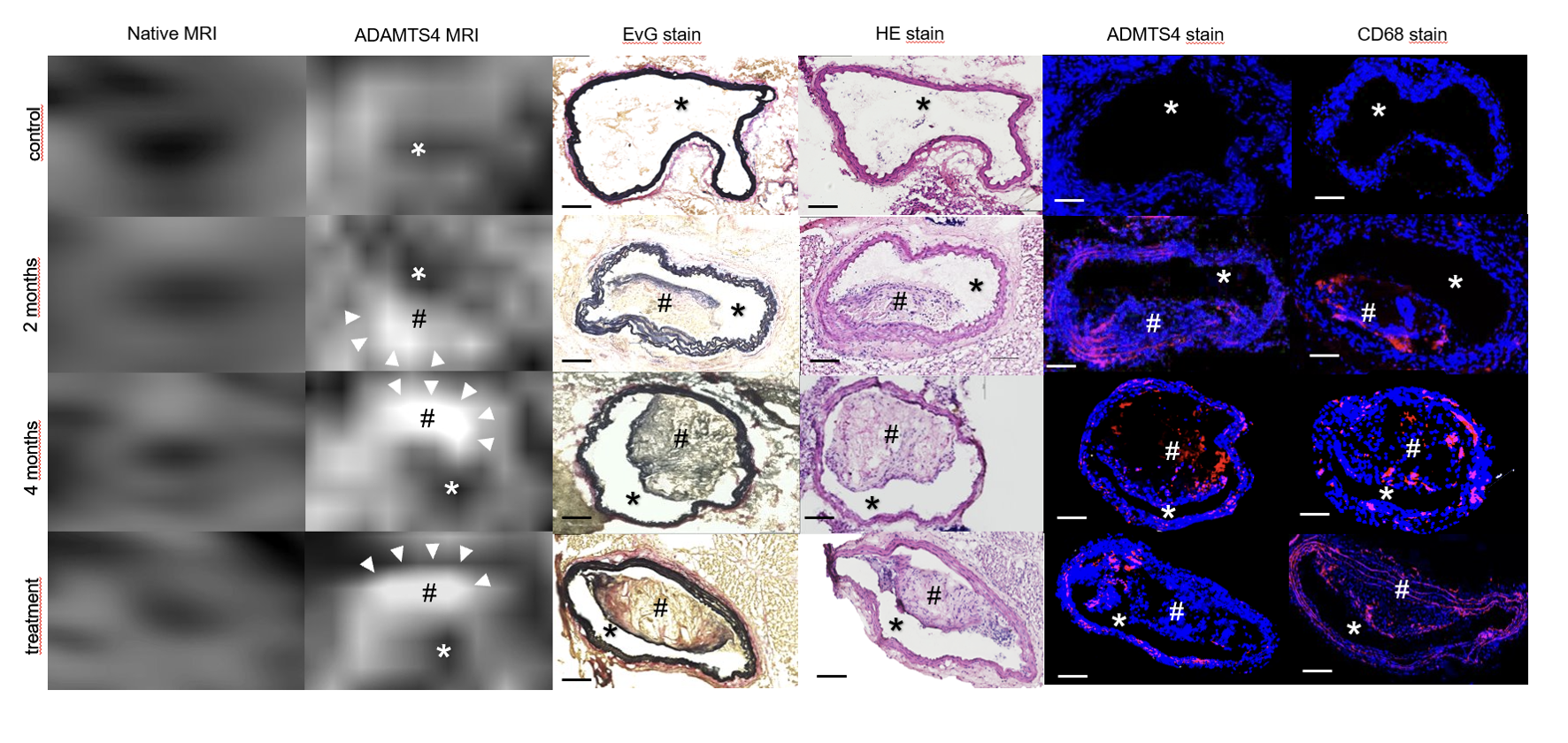
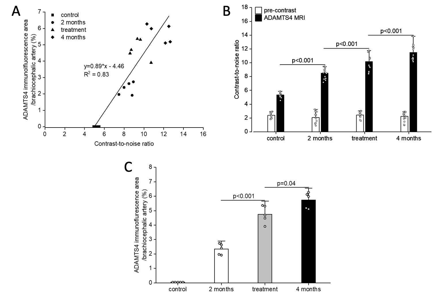
Figure 2. A. The histological measurements of ADAMTS4 were in good correlation with the MRI contrast-to-noise ratio (CNR). B. With increasing duration of the high-fat diet, a significantly stronger enhancement in the CNR was measured after intravenous administration of the novel probe. C. An increasing ADAMTS4-expression was observed after 2 and 4 months of high fat diet in immunofluorescence staining, respectively. No ADAMTS4 expression was seen in the control group. The treatment with pravastatin did not significantly decrease the ADAMTS4 expression.