3960
PET/MRI dual-model Granzyme B targeted probe for non-invasive early diagnosis of acute heart allograft rejection1MR Center, Fuwai Hospital, State Key Laboratory of Cardiovascular Disease, Beijing, China
Synopsis
Keywords: Probes & Targets, Preclinical
Motivation: Early diagnosis of transplant rejection can help to improve the immune-related management of transplant recipients.
Goal(s): Developing the new methods for early and non-invasive diagnosis of acute cellular rejection to unmet clinical needs.
Approach: Develop a PET/MRI dual-model granzyme B (GzmB) targeted probe for non-invasively detection of transplant acute cellular rejection.
Results: In preclinical heart graft models of rejection, our PET/MRI dual-model GzmB targeted probe allow noninvasive discrimination of early acute cellular rejection mediated by recipient cytotoxic CD8+ T cells.
Impact: In the future, this technology developed a PET/MRI dual-model probe for imaging GzmB produced by cytotoxic CD8+ T cells, enabling early non-invasive diagnosis of allograft rejection with high sensitivity and high spatial resolution.
Introduction
Cardiac transplantation remains the single most effective treatment for end-stage heart failure [1]. Despite the routine use of anti-rejection drugs in clinical practice, approximately 40% of cardiac transplant patients still experience unavoidable rejection [2]. Timely diagnosis of early acute cellular rejection is essential to prevent further tissue damage through effective immunosuppressive therapy. Endocardial biopsy is still the currently used gold standard for diagnosing tissue transplant rejection, but this procedure is invasive, subject to sampling error [3]. During acute cellular rejection, transplant rejection and graft damage is primarily mediated by recipient cytotoxic CD8+ T cells, which attack allografts by releasing perforin and granzyme B (GzmB) [4, 5]. It has been demonstrated that GzmB is highly expressed in early-onset acute cellular rejection [2, 6]. This finding demonstrates opportunities for targeting GzmB as an early indicator of acute cellular rejection and highlight the need to develop novel techniques for visualizing GzmB in vivo, which could aid in the diagnosis and monitoring of organ transplant rejection.Methods
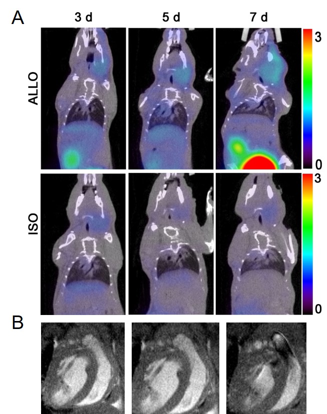
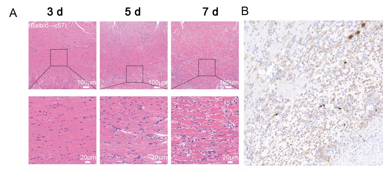
PET/MRI dual-model Granzyme B targeted probe (grazytracer) was synthesized and radiolabeling with gallium-68. The GzmB inhibitory activities and water proton longitudinal relaxation rate (1/T1) of grazytracer were tested. The mouse cervical heterotopic heart transplantation model was established. BALB/c and C57BL/6 mice were used as donor and recipient to establish a mouse allogeneic heart transplantation model. The donor and recipient of the syngeneic mouse heart transplantation model are C57 mice. Mice were subjected to GzmB-targeted PET/MRI dual model imaging on post-operative days 3, 5, and 7. After imaging monitoring, transplantation hearts were removed from recipients, specimens were formalin-fixed, paraffin-embedded. Then harvest tissues were sliced and stain with haematoxylin and eosin or immunohistochemistry with specific antibodies like GzmB (ab283315, abcam) and CD8+.Results
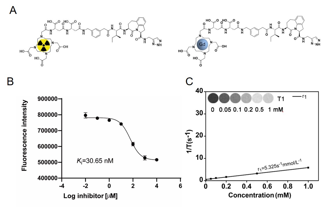
The efficiency of GzmB was determined and the enzymatic Michaelis Menten constants (Km) were calculated to be 30.65 nM. The linear regression fitting determined longitudinal relaxation rates depicted that shows that the r1 value of grazytracer are 5.325 s-1·mmol/L-1 (Fig.1). The uptake of 68Ga-grazytracer was observed increased with the extension of rejection time compared with syngeneic mouse heart transplantation (SUVmax:0.54 ± 0.07 vs. 0.19 ± 0.05 at 7 days, P<0.001) (Fig.2a). The MRI imaging can detect the heart transplant rejection (Fig.2b). Hematoxylin and eosin staining of the myocardium indicated massive inflammatory cell infiltration and disruption of the myocardial integrity in the allograft myocardium (Fig.3a). Immunohistochemistry showed significantly high expression of GzmB and CD8 (Fig.3b).Discussion
Our study focused on GzmB activity as an early biomarker for acute heart allograft rejection. The superior performance of the probe enables dual PET and MRI imaging with higher sensitivity and spatial resolution. It can be used to monitor the acute rejection of heart transplantation noninvasively.Conclusion
In summary, we developed a GzmB-targeted PET/MRI dual mode probe for imaging GzmB produced by cytotoxic T lymphocytes, enabling early non-invasive diagnosis of heart allograft rejection. This study provides an alternative method for monitoring allograft status without biopsy. In the future, the development of imaging of GzmB may be more readily translated into clinical applications.Acknowledgements
This study was supported by the National Key Research and Development Program of China (No. 2021YFF0501400 and 2021YFF0501404) and National Natural Science Foundation of China (No. 22277002 and 92059101).References
1. Siren EMJ, Luo HD, Tam F, Montgomery A, Enns W, Moon H, et al. Prevention of vascular-allograft rejection by protecting the endothelial glycocalyx with immunosuppressive polymers. Nature Biomedical Engineering. 2021;5:1202-16. doi:10.1038/s41551-021-00777-y.
2. Gao T, Yi L, Wang Y, Wang W, Zhao Q, Song Y, et al. Granzyme B-responsive fluorescent probe for non-invasive early diagnosis of transplant rejection. Biosensors and Bioelectronics. 2023;232. doi:10.1016/j.bios.2023.115303.
3. Mac QD, Mathews DV, Kahla JA, Stoffers CM, Delmas OM, Holt BA, et al. Non-invasive early detection of acute transplant rejection via nanosensors of granzyme B activity. Nature Biomedical Engineering. 2019;3:281-91. doi:10.1038/s41551-019-0358-7.
4. Nankivell BJ, Schwartz RS, Alexander SI. Rejection of the Kidney Allograft. New England Journal of Medicine. 2010;363:1451-62. doi:10.1056/NEJMra0902927.
5. Kołt S, Janiszewski T, Kaiserman D, Modrzycka S, Snipas SJ, Salvesen G, et al. Detection of Active Granzyme A in NK92 Cells with Fluorescent Activity-Based Probe. Journal of Medicinal Chemistry. 2020;63:3359-69. doi:10.1021/acs.jmedchem.9b02042.
6. Choy JC. Granzymes and perforin in solid organ transplant rejection. Cell Death & Differentiation. 2009;17:567-76. doi:10.1038/cdd.2009.161.
Figures