3955
Development of an Ex-Vivo Porcine Model of the Bladder for 3D Dynamic MRI Validation1University of Wisconsin - Madison, Madison, WI, United States, 2University of Wisconsin SMHS, Madison, WI, United States, 3Urology, University of Wisconsin - Madison, Madison, WI, United States
Synopsis
Keywords: Biology, Models, Methods, Bladder, Uro-dynamic MRI
Motivation: Recently developed MRI-Urodynamics methods require rigorous validation. In vitro approaches have been implemented, however lack of tissue-like mechanical properties of the in models limits their applicability.
Goal(s): Develop and implement an ex-vivo bladder model for validation of MRI-Urodynamics.
Approach: Porcine bladder was obtained and connected to a syringe pump to conduct MRI experiments during filling and voiding at various flow-rates. Simultaneous pressure measurements were performed during MRI.
Results: Ex-vivo bladder MRI experiments were successfully conducted. MRI-derived flow-rates agree within 10% when compared to the flow-rates imposed by the syringe pump. Pressure-volume analysis provided means of comparison of bladder performance between filling and voiding.
Impact: 3D dynamic MRI can assess anatomical changes in the bladder during voiding and filling. Systematic validation of this technique can enhance its clinical use.
Introduction
Lowery urinary tract symptoms (LUTS) and changes in bladder function occur with age 1-3. Patients with LUTS are evaluated with multi-channel urodynamics studies (UDS) to determine bladder flow and pressure during voiding and filling to assess the severity of symptoms 4. These tests, however, are invasive and do not fully characterize the bladder biomechanics5. Recently, MRI-based techniques have been developed that allow comprehensive assessment of the bladder during voiding and filling4. However, it largely remains unvalidated. Recently, a highly controlled experimental validation using an in vitro, synthetic, bladder mimicking phantom was investigated, however, a major limitation is the lack of tissue-specific behavior of the bladder model. The goal of this study was to perform highly controlled experimental studies to validate our recently developed MRI-based urodynamics method in an ex-vivo porcine model.Methods
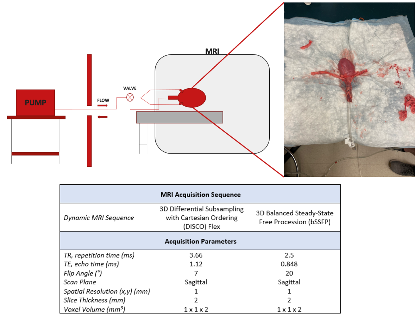
Ex Vivo Bladder Model Preparation Porcine bladder was obtained from pigs euthanized for unrelated purposes. In preparation, the ureters were dissected from the renal hilum, the urethra was dissected as distally as possible given anatomical restrictions. The ureters, urethra and bladder were removed en bloc. After excision, the ureters and urethra were isolated. Two infusion catheters were placed inside each ureter while a drain catheter was placed in the urethra at the level of the bladder neck. These were sutured in place and were capable of being hooked up to a programmable, two-channel syringe pump (Chemyx, Inc.) to simulate bladder voiding and filling. Two 300ml syringes were utilized in the pump, filled to 250cc, and facilitated flow. For filling and voiding studies, water was injected through the ureters and withdrawn from the urethra, respectively. Ex-vivo bladder was placed in a 3.0T clinical MRI scanner (GE Healthcare) and volumetric data was acquired using two different sequences: (1) 3D Differential Subsampling with Cartesian Ordering (DISCO) Flex and (2) 3D Balanced Steady-State Free Procession (bSSFP). Three different flow-rates for both filling and voiding were used (100, 200 and 300cc/min) resulting in 12 total volumetric acquisitions. bSSFP acquisitions were carried out followed by DISCO Flex. This is because DISCO-Flex requires the use of gadolinium-based contrast for signal enhancement. Prior to DISCO Flex acquisitions, 5 ml of gadolinium-based contrast (0.1mmol/kg) was hand-injected into a 1L volume of DI water used to fill the syringes in the flow loop. A summary of acquisition parameters for each sequence are shown in Figure 1. Pressure measurements 1cm inside the bladder were acquired using MRI-compatible, fiber-optic pressure transducers (OPP-M200, Opsens Inc). Pressure measurements were acquired for the entirety of each flow test at a frequency of 1000Hz. Pressure and MRI-volumetric data were post-processed using a custom MATLAB script.Results
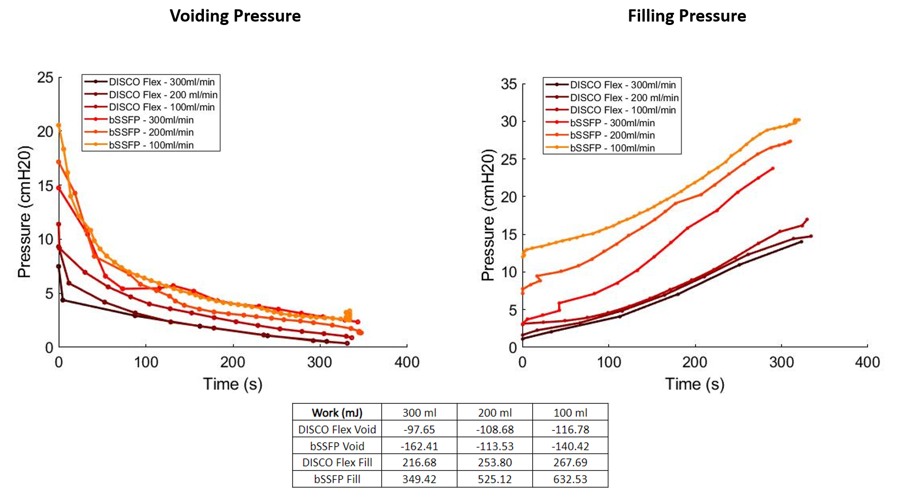
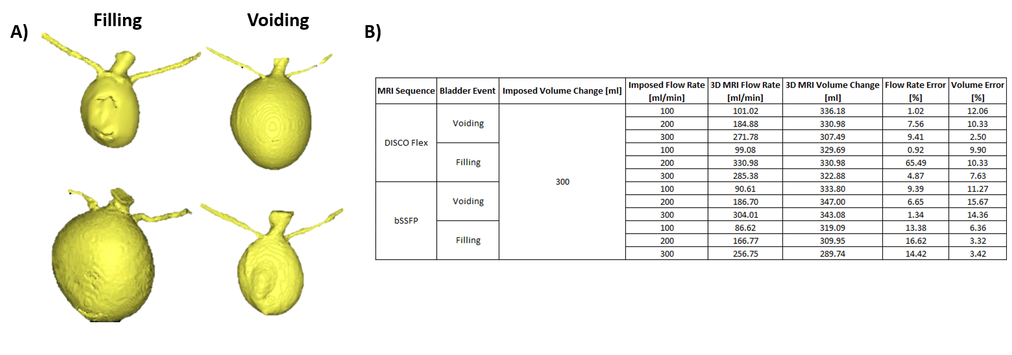
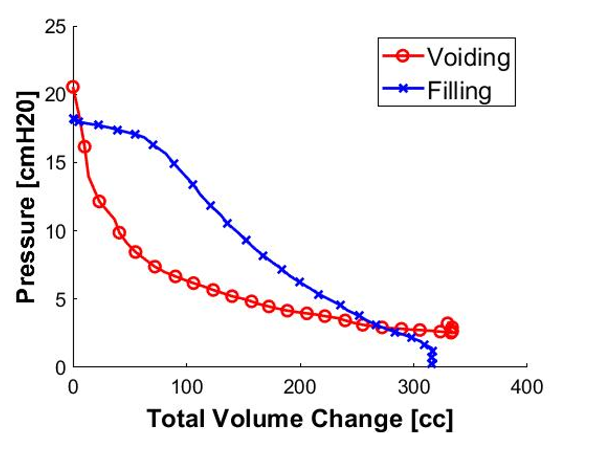
Figure 2 shows ex-vivo bladder volumes over time during a voiding and filling event generated at a flow-rate of 300ml/min. MRI-derived flow-rates agree within 10% when compared to the flow-rates imposed by the syringe pump with average errors of 8.9%. Figure 3 shows pressure-volume (PV) loops obtained for each voiding and filling test. A pressure drop is observed for all voiding events while pressure increases are seen during filling. The work associated with bladder emptying and filling is computed as the integral of the PV-loop. More work output was associated with increased flow rates during voiding and filling. A comparison of PV-loops for voiding and filling events allows for the generation of hysteresis-like curves (Figure 4) suggesting the ex-vivo bladder may behave differently during filling and voiding.Discussion
Bladder volume during 12 total voiding and filling tests were successfully calculated from 3D MRI data and used to estimate volumetric flow-rate. MRI-derived flow-rates agree with imposed flow-rate within 10%. As flow rate decreased, errors tended to increase, likely due to the difficulty in segmentation as there are smaller volume changes. Both voiding and filling pressure maps were qualitatively similar in profile. Peak pressures in both voiding and filling were higher for slower flow-rates. Differences between voiding and filling behavior at the same flow-rate are shown in Figure 4. This represents what can be thought of as a hysteresis curve, displaying the difference in voiding and filling profiles for the same flow-rate and suggesting the bladder may deform differently during emptying and filling. This modeling pipeline represents a novel approach to systematically assess the validity of MRI-urodynamics that better replicates the tissue-specific behavior compared to in vitro bladders made of synthetic material. Future efforts will focus on fine-tuning this model to better understand how the bladder shape changes during experimentation.Acknowledgements
We acknowledge support from GE Healthcare, who provide research support to the University of Wisconsin - Madison as well as R01 DK126850-01 and WPP AAM3497 -Wisconsin Partnership program. Additionally, this investigation was supported by the National Institutes of Health, under Ruth L. Kirschstein National Research Service Award T32 HL 007936 from the National Heart Lung and Blood Institute to the University of Wisconsin-Madison Cardiovascular Research Center.
References
[1] Wei, J. T., Calhoun, E., & Jacobsen, S. J. (2005). Urologic diseases in America project: benign prostatic hyperplasia. The Journal of urology, 173(4), 1256-1261.
[2] Yao M, Simoes A. Urodynamic Testing and Interpretation. [Updated 2023 Aug 14]. In: StatPearls [Internet]. Treasure Island (FL): StatPearls Publishing; 2023 Jan-. Available from: https://www.ncbi.nlm.nih.gov/books/NBK562310/
[3] Pewowaruk R, Rutkowski D, Hernando D, Kumapayi BB, Bushman W, Roldán-Alzate A. A pilot study of bladder voiding with real-time MRI and computational fluid dynamics. PLoS One. 2020;15(11 November). doi:10.1371/journal.pone.0238404
[4] Rutkowski, D. R., Medero, R., Ruesink, T. A., & Roldán-Alzate, A. (2019). Modeling Physiological Flow in Fontan Models With Four-Dimensional Flow Magnetic Resonance Imaging, Particle Image Velocimetry, and Arterial Spin Labeling. Journal of biomechanical engineering, 141(12), 1210041–1210049. https://doi.org/10.1115/1.4045110
[5] Glass Clark, S., Nagle, A. S., Bernardo, R., Vinod, N., Carucci, L., Carroll, A., Speich, J., & Klausner, A. P. (2020). Use of Ultrasound Urodynamics to Identify Differences in Bladder Shape Between Individuals With and Without Overactive Bladder. Female pelvic medicine & reconstructive surgery, 26(10), 635–639. https://doi.org/10.1097/SPV.0000000000000638
Figures