3953
Reference ranges for functional and volumetric CMR in pigs as pre-clinical model1Center for Preclinical Development, University of Zurich and University Hospital Zurich, Zurich, Switzerland, 2Advanced Clinical Imaging Technology, Siemens Healthineers International AG, Zurich, Switzerland, 3Institute for Biomedical Engineering, University and ETH Zurich, Zurich, Switzerland
Synopsis
Keywords: Large Animals, Nonhuman Primates, Animals, CMR, pig
Motivation: Domestic pigs are frequently used as experimental animals for cardiovascular research but are subject to somatic growth.
Goal(s): To investigate the relationship of cardiac functional and volumetric parameters with animal somatic growth.
Approach: In this retrospective CMR study in 58 female Swiss large white pigs, we correlate functional volumetric and dimensional CMR parameters to animal weight.
Results: : Left ventricular mass, left and right ventricular volumes and stroke volumes correlate with animal weight. Weight-independent parameters were left and right ejection fractions. Our findings match values found in humans.
Impact: We provide a regression analysis of clinical functional, volumetric and dimensional CMR parameters for experimental planning and refinement of animal experiments in pigs and illustrate healthy parameter ranges.
Introduction
Cardiovascular diseases (CVD) are the leading cause of human deaths worldwide, with increasing incidence of events. For the development of novel diagnostic tools, and surgical, pharmacological and interventional treatment options, animal models play a key role. Although most of basic research has been conducted in rodents, it is known that there are some crucial differences when comparing their cardiovascular system to the human [1], [2]. Animal models of choice for CVD are therefore large animal models like pigs. With the important aspects of 3R in animal experiments, it is crucial to properly plan and refine experimental design before using animal models. In this abstract, we present a retrospective study on clinical CMR values as a function of animal weight in Swiss large white pigs to guide the refinement of study design and provide healthy control data to reduce overall animal usage.Methods
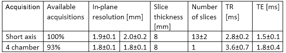
We retrospectively included 58 healthy female Pigs (Swiss large white, 23-109kg) used in cardiovascular studies at our center. All experiments were approved by the cantonal veterinary office. The inclusion criteria were: animals were part of the healthy control group, no manipulation to the cardiovascular system, and availability of clinical routine bSSFP short axis multi-slice cine imaging at 1.5T (N=33) or 3T (N=25) clinical MR scanners (Achieva/Ingenia, Philips Healthcare, Best, The Netherlands). Imaging data was collected during free breathing and either electrocardiogram or pulse oximetry gating. The imaging parameters for short-axis and complementary four-chamber views are summarized in Table 1. Data analysis was performed semi-automatically using the Segment Toolbox (Medviso AB, Lund, Sweden) and left/right ventricular ejection fractions (LVEF/RVEF), stroke volumes (LVSV/RVSV), end-systolic and end-diastolic volumes (LVESV/RVESV, LVEDV/RVEDV) as well as LV end-systolic and end-diastolic masses (LVESM, LVEDM) were estimated. Further, we report the dimensions of left and right atria (LA/RA) and the LV itself from four-chamber view imaging when available. All data analysis was performed according to the 2020 update of the SCMR Task Force on Standardized Post-Processing [3]. The measured CMR parameters were plotted against animal weight and linear as well as non-linear (two parameter: logarithm, square root, potential function) regression was performed. Root mean squared error (RMSE), best fitting function and coefficient of determination R2 of the fit parameters are reported. RMSE was used to determine the best fitting function.Results
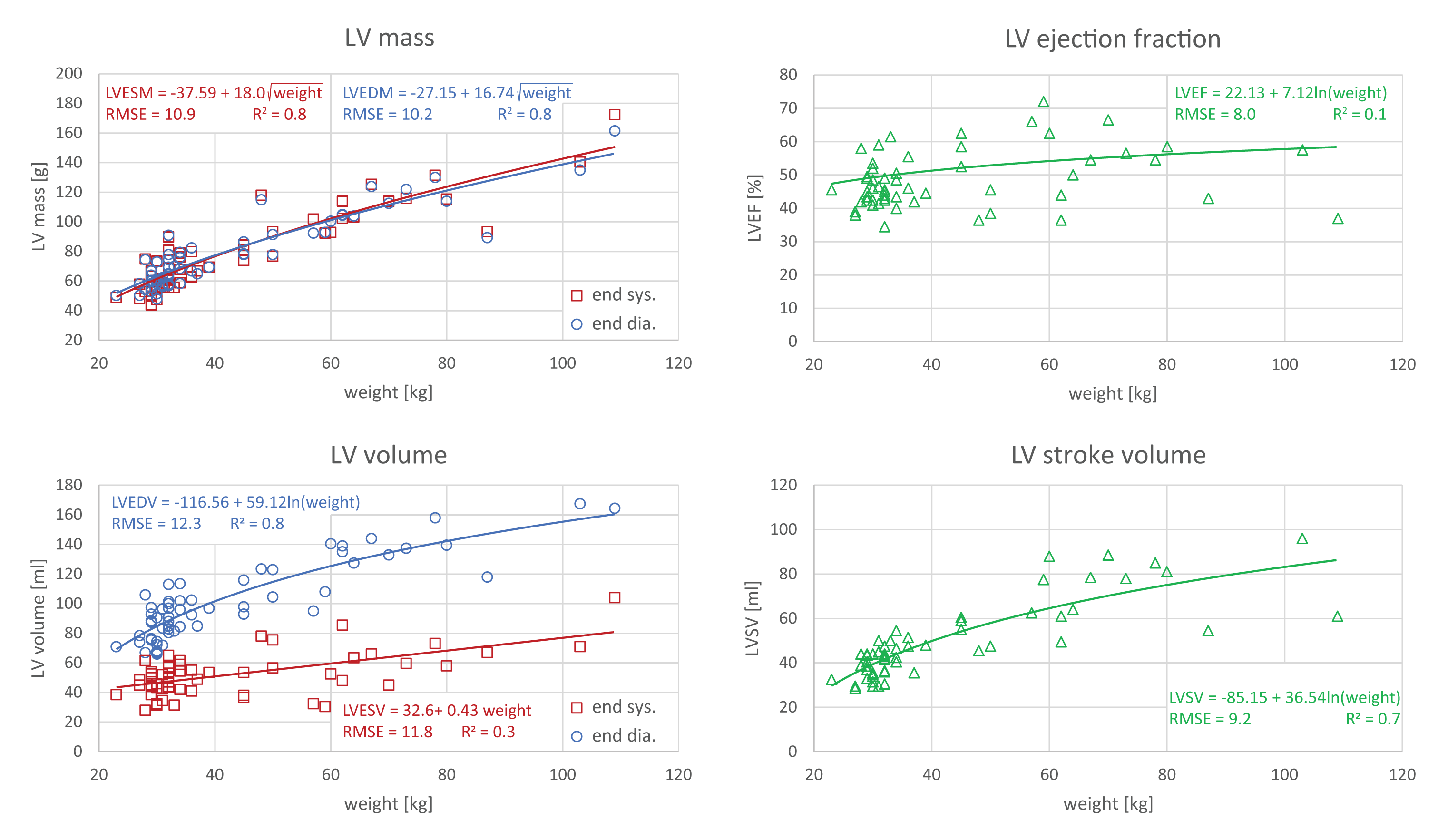
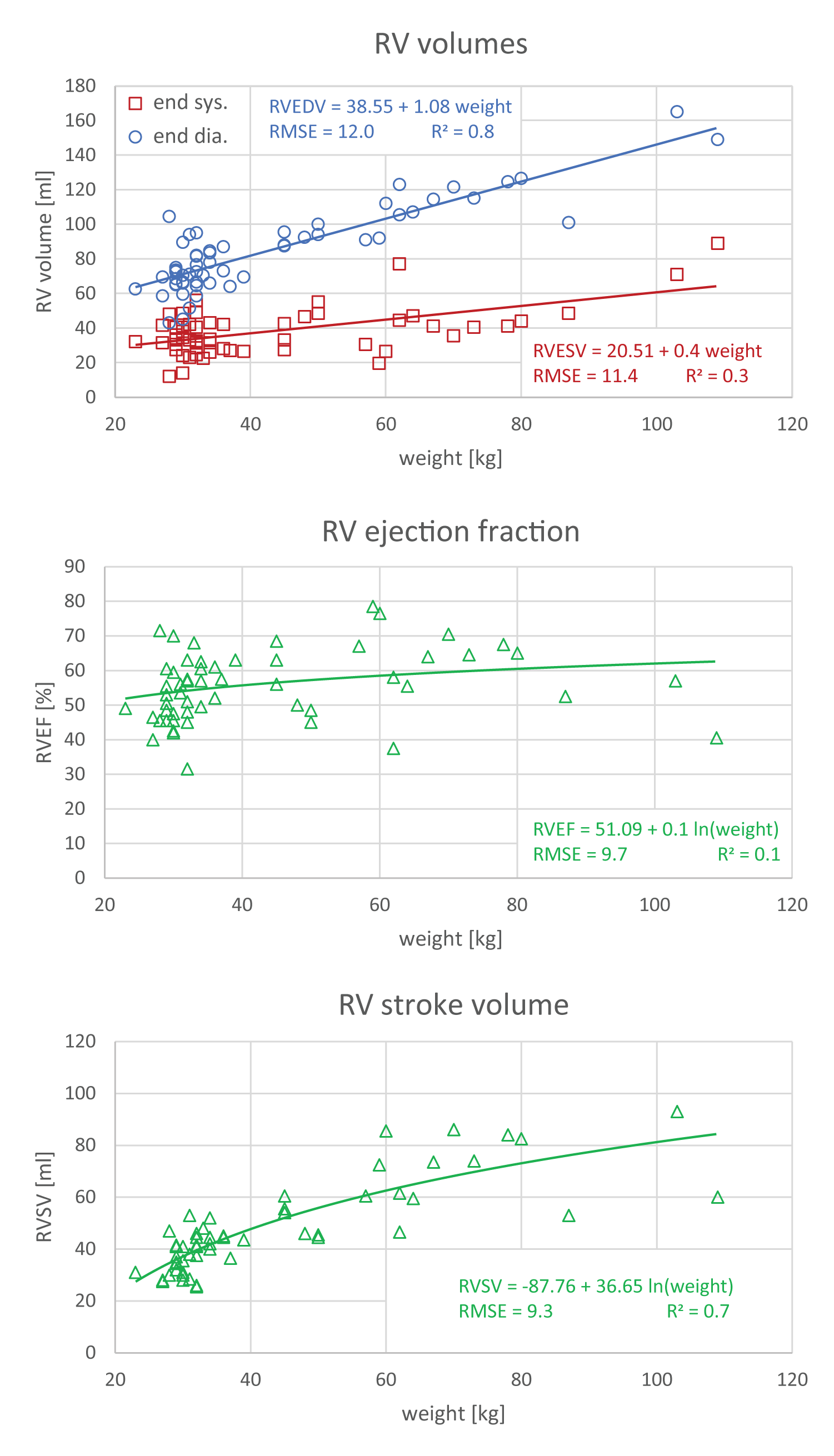
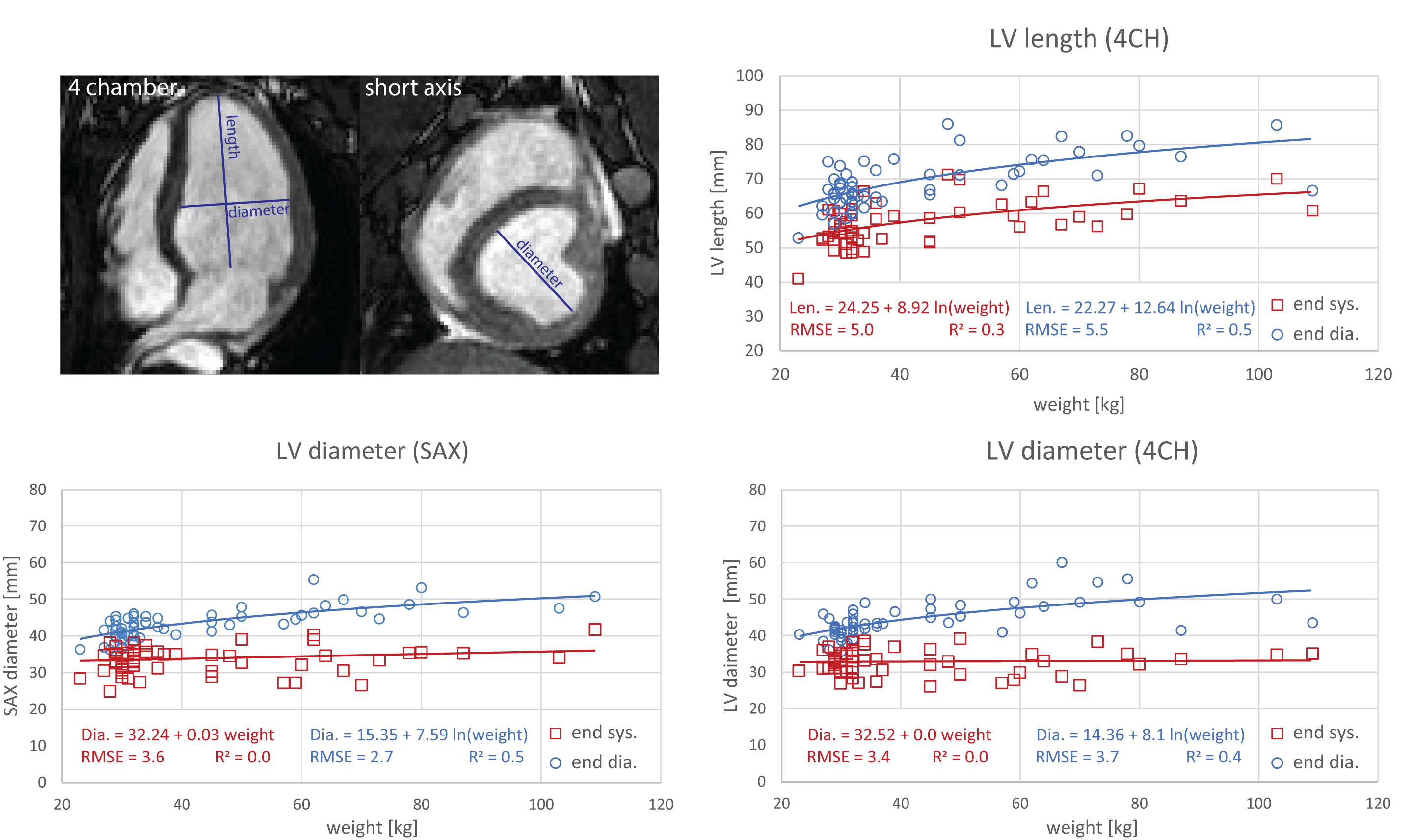
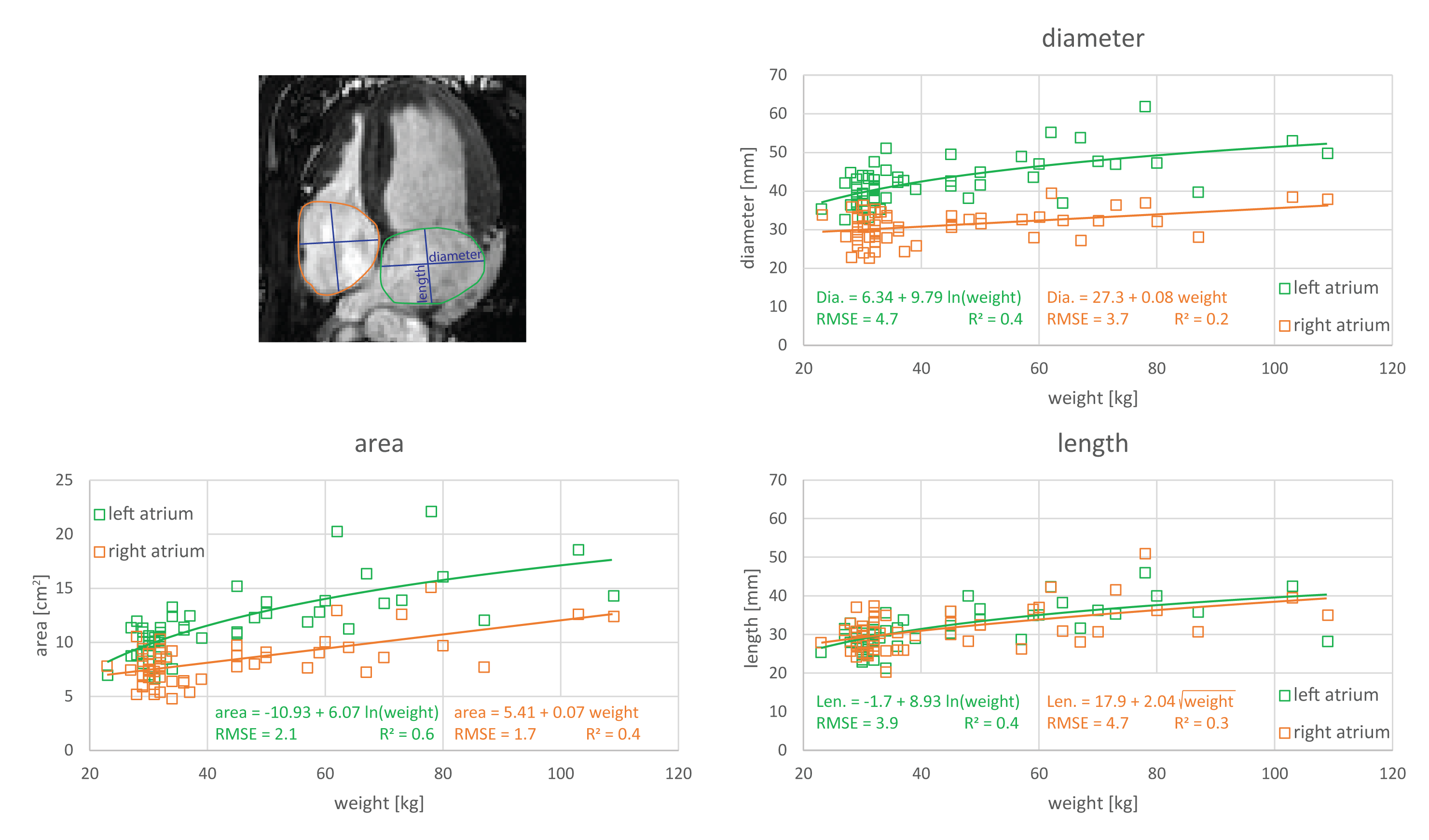
Figure 1 shows LVEDM/LVESM, LVEF, LVEDV/LVESV as well as stroke volume (LVSV) as a function of animal weight. The individual best fitting functions and corresponding RMSE are shown in the individual figure inserts. LVEDM (sqrt, R2=0.8), LVESM (sqrt, R2=0.8), LVEDV (ln, R2=0.8) as well as LVSV (ln, R2=0.7) correlate well with weight. LVESV correlated fairly (ln, R2=0.3) with animal weight and LFEF did not correlate (R2=0.1). No differences between LVEDM and LVESM were found. Figure 2 shows the corresponding RVEDV/RVESV, RVEF, RVSV. RVEDV (linear, R2=0.8) and RVSV (ln, R2=0.7) correlate well with animal weight, while RVESV only fairly correlates (linear, R2=0.3). Similar to LVEF, RVEF does not correlate with animal weight. Figure 3 shows LV length and diameter as function of animal weight. Only a fair correlation was found for end-diastolic/end-systolic length (ln, R2 = 0.5 and ln, R2 = 0.3), and end-diastolic diameter derived from both short-axis and four-chamber view (ln, R2 = 0.5 and ln, R2 = 0.4). Figure 4 shows the end-systolic LA and RA dimensions as well as the area in four-chamber view as a function of animal weight. Left atrial diameter (ln, R2 = 0.4) as well as the LA and RA length (ln, R2 = 0.4 / sqrt, R2 = 0.3) and measured areas (ln, R2 = 0.6/ linear, R2 = 0.4) correlated fairly with body weight. RA diameter correlated poorly (linear, R2 = 0.2) with body weight.Conclusion
In this study, we found animal somatic growth-dependent clinically relevant CMR parameters such as mass, volumes and stroke volumes. Ejection fractions, however, were independent of animal weight. Animals heavier than 60kg presented similar functional, volumetric and dimensional values as presented for humans [4], however pigs tend to outgrow humans at excessive weights (>100kg). RV parameters were slightly below men but correspond well to women. The lightweight animals (<40kg) correspond well to children of age 9-12 years [4]. The findings show that pig models are a good substitute in cardiovascular research, yet it is crucial to choose the right animal size in accordance with the experimental needs and refine the study design early.Acknowledgements
No acknowledgement found.References
[1] K. Haghighi u. a., „Human phospholamban null results in lethal dilated cardiomyopathy revealing a critical difference between mouse and human“, J. Clin. Invest., Bd. 111, Nr. 6, S. 869–876, März 2003, doi: 10.1172/JCI17892.
[2] I. Ginis u. a., „Differences between human and mouse embryonic stem cells“, Dev. Biol., Bd. 269, Nr. 2, S. 360–380, Mai 2004, doi: 10.1016/j.ydbio.2003.12.034.
[3] J. Schulz-Menger u. a., „Standardized image interpretation and post-processing in cardiovascular magnetic resonance - 2020 update: Society for Cardiovascular Magnetic Resonance (SCMR): Board of Trustees Task Force on Standardized Post-Processing“, J. Cardiovasc. Magn. Reson., Bd. 22, Nr. 1, März 2020, doi: 10.1186/s12968-020-00610-6.
[4] N. Kawel-Boehm u. a., „Reference ranges (“normal values”) for cardiovascular magnetic resonance (CMR) in adults and children: 2020 update“, J. Cardiovasc. Magn. Reson., Bd. 22, Nr. 1, Dez. 2020, doi: 10.1186/s12968-020-00683-3.
Figures