3948
Rapid, accurate and precise T1 mapping in rat heart at 9.4 T MRI1Institute of Experimental Medical Research, Oslo, Norway, 2KG Jebsen Center for Cardiac Research, Oslo, Norway
Synopsis
Keywords: Small Animals, Cardiovascular, T1 mapping
Motivation: Preclinical cardiac T1 mapping in high field encountered a great challenge, such as rapid heart rate, B0 and B1 inhomogeneity, which affects both accuracy and precision in T1.
Goal(s): We aim to develop an accurate, fast and reproducible T1 mapping method at 9.4 T MRI.
Approach: We adapted inversion efficiency (IE) to the segmented Look-locker sequence and validated it in a phantom, as well as in aorta banding rats.
Results: Segmented Look-Locker T1 mapping with IE is a fast and accurate T1 mapping method, which corrects the imperfection of inversion pulse in high field.
Impact: We have developed a rapid, highly precise T1 mapping technique for rat hearts using 9.4 T MRI, enabling longitudinal scans and comparisons between different animals.
Background:
Rodent model are indispensable for cardiac basic research. It contributes to increase our knowledge and providing new approaches to investigate the mechanism of cardiac pathologies. Cardiovascular magnetic resonance (CMR) is the modality of choice to study the mechanism of the heart and vasculature, offering detailed images of both structure and function. Myocardial T1 mapping shows to be promising for characterization of myocardium in a wide variety of cardiomyopathies, for example the extent of fibrosis. However, preclinical T1 mapping in high field encountered a great challenge, such as rapid heart rate, B0 and B1 inhomogeneity, which affects both accuracy and precision in T1. In this study, we aim to develop and evaluate a fast multi-slice modified Look-locker for accurate T1 mapping in rodents at 9.4 T scanner. In the current T1 mapping sequence, we adapted modified Look-Locker (MOLLI) and inversion efficiency (IE) from Rodgers et al(1), and validated in NiCl2 doped agar-gel phantom, also healthy and aorta banding rats.Methods:
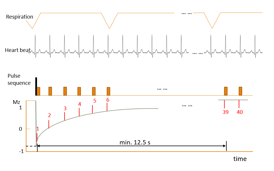
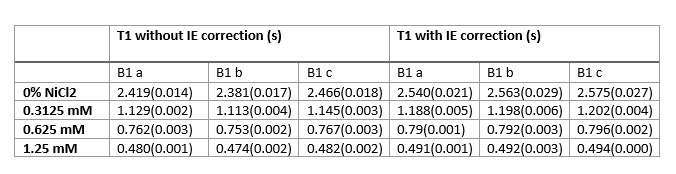
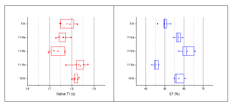
All experiments are conducted at 21 cm bore size 9.4 T MRI magnet (Agilent Technologies, Palo Alto, CA, USA) interfaced to Avance Neo console (Bruker Biospin, Ettlingen, Germany). Scanning software Paravision 360 v3.3. The phantom consisted of 4 tubes containing 2% agarose-gel at different concentration of NiCl2, 0 mM, 0.3125 mM, 0.625 mM, 1.25 mM. 5 rats at different age (5,11,19 month old) went through 5 scanning sessions during 1.5 weeks to validate the precision of T1 mapping and CINE. A rat model of three different degrees of atrial constriction, along with sham-operated controls (n=34 total). MRI was conducted 12 weeks after the operation.T1 mapping protocol is respiratory-triggered, cardiac gated, Look-locker sequence with a spoiled, multi-slice, segmented gradient-echo readout. A delay was added at the end of the look-locker acquisition train to ensure the acquisition of two fully-recovered images for IE correction (Fig.1 ). The three slices were placed on basal, mid-ventricle and apical myocardium. Key acquisition parameters: slice thickness = 1.5 mm, matrix = 128 x 64 (zero filled to 128 x 128), FOV = 45 mm x 45 mm, true TR > 12.5 s, TE = 2.41 ms, flip angle = 8°, segment size = 4, echo images = 40; acquisition time = ~3.5 minutes.
First T1LL,T1*, and A are determined from 1-38 images (except motion corrupted images) as typical look-locker data fitting (2). With the knowledge of the equilibrium magnetization M0 (Image 39) , accurate T1 can be derived as T1 = T1*(M0/A). Image 40 is not used in this abstract, but it can be employed for an even more accurate approach of 4-parameter model fitting for T1 (1).
Results:
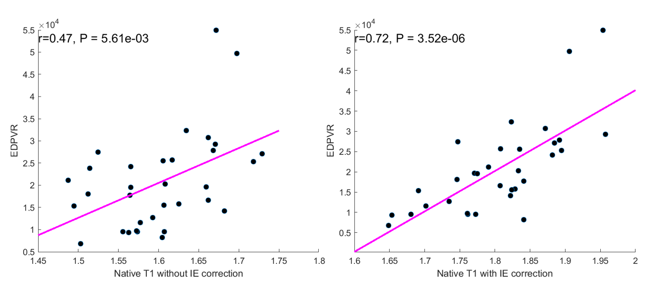
In phantom experiments, T1 times from both methods with IE and without IE compare to gold-standard T1 RARE measurement. While Look-locker corrected T1 values underestimate T1 at different extent at various B1 field strength, T1 with IE correction yields high precision (0.49% ±0.30% ) and accuracy (0.80%±1.02%) compared to T1 RARE measurement (Fig. 2) . In 5 healthy rats at different age, the precision of T1 time with IE correction is better than the eject fraction from cine acquisition (1.70%±0.55%; 4.56%±1.08% ) (Fig. 3). In sham and aorta banding operated rats, native T1 from method with IE correlates well to the end diastolic pressure volume relationship (EDPVR) and better than the method without IE (R=0.72; R=0.48) (Fig. 4).Acknowledgements
No acknowledgement found.References
1. Rodgers CT, Piechnik SK, Delabarre LJ, et al. Inversion recovery at 7 T in the human myocardium: Measurement ofT1, inversion efficiency and B1+. Magn Reson Med 2013;70(4):1038-1046.
2.Deichmann R, Haase A. Quantification of T1 values by SNAPSHOT-FLASH NMR imaging. Journal of Magnetic Resonance (1969) 1992;96(3):608-612.
Figures