3945
Stiffness-Transformable Nanoplatforms Loaded with Irisin: Tumor Microenvironment Responsiveness for MRI and Ferroptosis Induction1Radiology, Zhongda Hospital, Medical School, Southeast University, Nanjing, China
Synopsis
Keywords: Probes & Targets, Tumor, Integrated diagnosis and treatment
Motivation: MnO2 combined with MONs offers potential for accurate tumor diagnosis and treatment. Our MMONs, with adjustable hardness, address issues with hard and soft nanoparticles, showing tumor responsivity and enabling MRI. Irisin loading boosts ferroptosis in drug-resistant tumors.
Goal(s): The synthesis of a stiffness-transformable nanoplatform loaded with Irisin achieves magnetic resonance imaging and induces ferroptosis in the tumor microenvironment.
Approach: MMONs and MMONs-Irisin were synthesized, characterized, and their magnetic resonance imaging and cytotoxic effects were validated at both cellular and animal levels.
Results: MMONs-Irisin is deformable, exhibits excellent magnetic resonance imaging effects, and promotes ferroptosis in tumors.
Impact: MMONs are GSH-responsive, with higher T1 and T2 relaxation rates than Magnevist at equal concentrations. They adjust stiffness for enhanced tumor uptake, loaded with Irisin, combat chemotherapy resistance through ferroptosis. MMONs-Irisin holds potential for safe, integrated cancer diagnosis and treatment.
Manganese (Mn), as one of the essential trace elements in the human body, plays a significant role in various physiological processes1. MnO2 can exhibit GSH-responsive behavior in the tumor microenvironment, releasing Mn2+. Mn2+ not only serves as an excellent MRI contrast agent in the biomedical field but can also induce ferroptosis in tumor cells, overcoming chemotherapy resistance2. Solid mesoporous organosilica nanoparticles (MONs) possess uniform and controllable size, pore volume, large surface area, easily functionalizable surfaces, and good biocompatibility. They can be modified or loaded with various drugs and imaging agents to construct novel nanotherapeutic platforms3. Combining MnO2 with MONs holds great potential for precise tumor diagnosis and therapy. However, the hardness of nanoparticles significantly affects their biological performance. Softer nanoparticles typically exhibit higher macrophage internalization and liver distribution, whereas harder nanomaterials are taken up less by tumors4. Therefore, we synthesized stiffness-transformable manganese oxide hybridized mesoporous organosilica nanoparticles (MMONs), which can achieve magnetic resonance imaging while overcoming the limitations of both soft and hard nanoparticles. MMONs are loaded with Irisin, a peptide involved in lipid metabolism regulation5, to synergistically promote ferroptosis in chemoresistant tumors.
METHODS
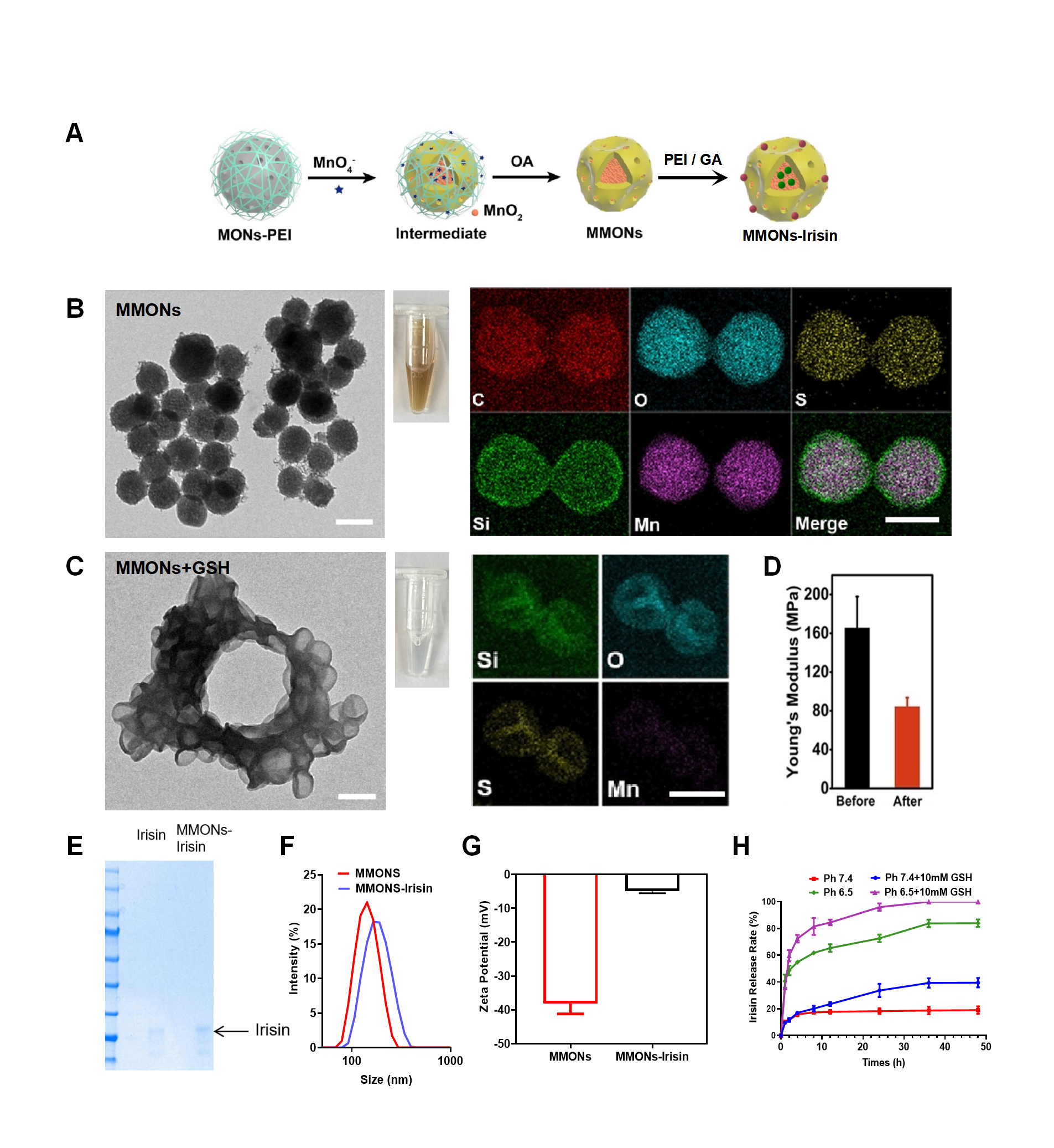
First, MONs were synthesized using CTAB, NH3·H2O, TEOS, and TETS. Then, as shown in Figure 1A, MMONs were synthesized using a PEI solution, KMnO4 solution, and oleic acid. Subsequently, Irisin was tethered to the surface of MMONs through schiff base bonding to create MMONs-Irisin. The morphology, elemental composition, particle size, Zeta potential, and hardness of MMONs and MMONs-Irisin were characterized. Their response to 10 mM GSH and drug encapsulation and release rates under slightly acidic conditions were observed. The T1WI and T2WI effects of MMONs-Irisin were investigated using in vitro models and pancreatic cancer mice. At the cellular and animal levels, the cytotoxicity of MMONs-Irisin against gemcitabine-resistant pancreatic cancer cells was evaluated, and its efficacy in promoting ferroptosis and safety were observed through Western blot experiments.
RESULTS
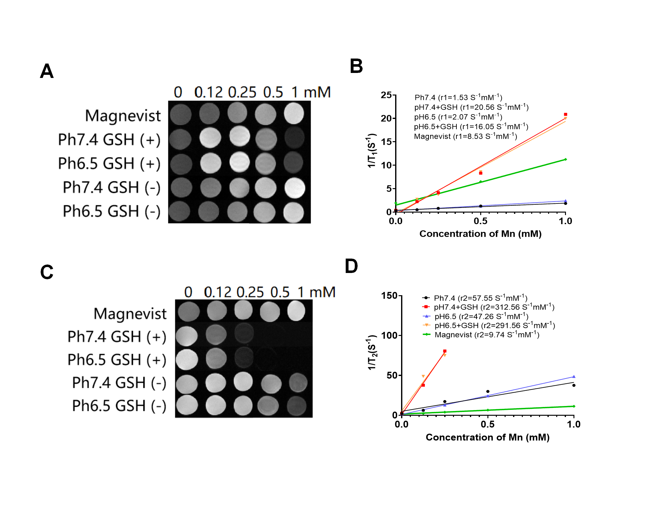
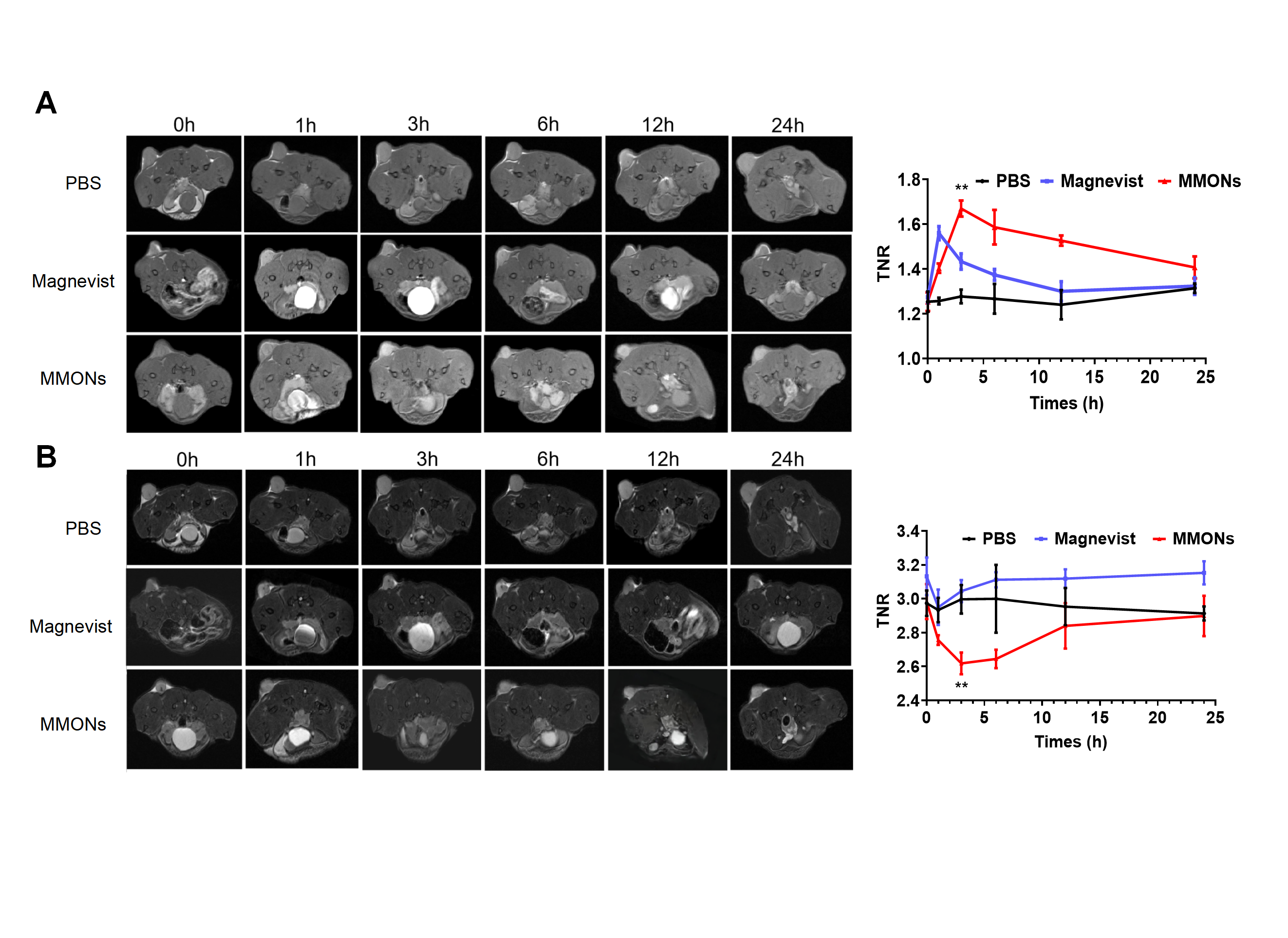
The successful synthesis of spherical MMONs nanoparticles, as depicted in Figures 1B and 1C, led to the release of Mn2+ in a GSH environment. The nanoparticles transformed from spherical to concave spherical, resulting in a significant decrease in hardness and a decrease in Young's modulus from approximately 160 MPa to about 80 MPa (Figure 1D). These nanoparticles had a diameter of approximately 100 nm, carried a negative charge, and exhibited a encapsulation rate of about 98.9% for Irisin. Under mildly acidic conditions at pH 6.5, Irisin was gradually released, with nearly complete release within 24 hours (Figure 1E-F). As shown in the in vitro gradient model in Figures 2A and 2C, MMONs demonstrated excellent T1WI and T2WI imaging effects, especially in a GSH environment. A concentration of only about 28 µg/mL of MMONs (with a Mn concentration of 0.25 mM) saturated the T1 signal. At the same concentration, both the T1 (r1=20.56 S-1mM-1) and T2 (r2=312.56 S-1mM-1) relaxation rates were significantly higher than those of Magnevist. In Figure 3, mice with pancreatic cancer were injected with PBS, Magnevist, and MMONs. The imaging effect was best observed at 3-6 hours after MMONs injection in T1WI and T2WI images. Compared to the PBS group and the Magnevist group, the signal-to-noise ratio changes in tumor and normal tissues were more significant in the MMONs group. As shown in Figures 4A-B, a gemcitabine-resistant pancreatic cancer cell line (GEM-Panc02) was successfully established. MMONs-Irisin exhibited an approximately 70% killing rate against it, a significant increase compared to the gemcitabine-only group (P<0.001). Figures 4C-E illustrate the treatment of gemcitabine-resistant pancreatic cancer tumor-bearing mice with MMONs-Irisin, resulting in a substantial reduction in tumor size. At the animal level, Western blot experiments demonstrated a significant decrease in GPX4 protein expression with MMONs-Irisin treatment, promoting ferroptosis in gemcitabine-resistant cells, as shown in Figure 4F. Within a concentration of 0.04 mg/mL, no significant toxicity was observed in mice (Figure 4G).
DISCUSSION AND CONCLUSION
MMONs exhibit a large surface area, uniform pore size, high drug loading, excellent biocompatibility, and adaptable structure. Their salient feature is GSH responsiveness, resulting in significantly higher T1 and T2 relaxation rates compared to Magnevist at equivalent concentrations. Furthermore, they can modulate their stiffness to enhance tumor uptake. By incorporating Irisin, they promote ferroptosis to overcome chemotherapy resistance. MMONs-Irisin holds promise as a secure, integrated nanoplatform for cancer diagnosis and treatment.
Acknowledgements
This study has received funding by National Nature Science Foundation of China (81871412 and 82272064), Jiangsu Provincial Science and TechniqueProgram (BK20221461), Postgraduate Research & Practice Innovation Program of Jiangsu Province (KYCX21_0159, KYCX22_0297, and KYCX23_0323).References
1.Xu W, Qing X, Liu S, et al. Hollow Mesoporous Manganese Oxides: Application in Cancer Diagnosis and Therapy. Small. 2022;18(15):e2106511.2.Sahoo K, Sharma A. Understanding the mechanistic roles of environmental heavy metal stressors in regulating ferroptosis: adding new paradigms to the links with diseases. Apoptosis. 2023;28(3-4):277-292.
3.Janjua TI, Cao Y, Kleitz F, et al. Silica nanoparticles: A review of their safety and current strategies to overcome biological barriers. Adv Drug Deliv Rev. 2023;14:115115.
4.Peng X, Chen K, Liu W, et al. Soft Mesoporous Organosilica Nanoplatforms Improve Blood Circulation, Tumor Accumulation/Penetration, and Photodynamic Efficacy. Nanomicro Lett. 2020;12(1):137.
5.Bao JF, She QY, Hu PP, et al. Irisin, a fascinating field in our times. Trends Endocrinol Metab. 2022;33(9):601-613.
Figures
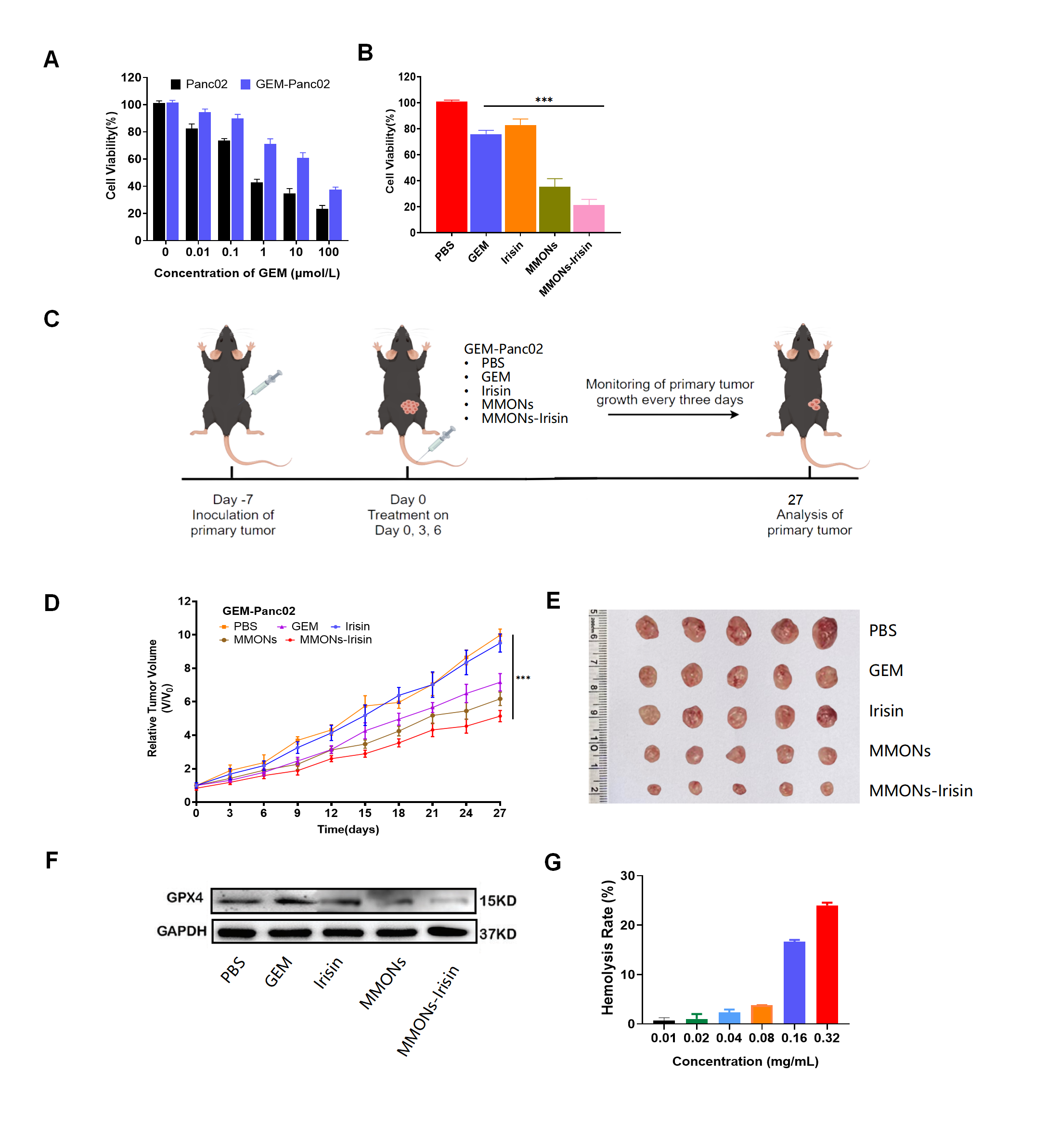
A) Successful gemcitabine-resistant Panc02 mouse pancreatic cancer cell induction; B) Cytotoxicity of different drugs on GEM-Panc02 cells; C) Treatment plan for gemcitabine-resistant pancreatic cancer mice; D) Tumor volume changes in mice; E) Post-treatment tumor images; F) GPX4 ferroptosis marker expression post-treatment in different groups; G) Influence of MMONs-Irisin at varying concentrations on mouse hemolysis rate. ***P < 0.001.