3936
Single-shot 2D spiral imaging of the human brain at 10.5 Tesla using 128 receive channels: initial experience1Radiology, University of Minnesota, Minneapolis, MN, United States, 2Deceased, Minneapolis, MN, United States
Synopsis
Keywords: High-Field MRI, Brain, spiral imaging, field monitoring
Motivation: There has been an increasing interest to pursue spiral imaging at ultrahigh field owing to its improved sampling efficiency.
Goal(s): Our goal was to demonstrate the feasibility of spiral imaging in humans at 10.5 Tesla.
Approach: Highly-accelerated single-shot 2D spiral GRE images were collected using 128 receive channels and a sequence developed in an open source environment. Dynamic field changes associated with the spiral readout gradients were measured in a separate session using 16 NMR probes.
Results: Quality T2*-weighted single-shot spiral imaging of the human brain was achieved by simultaneous corrections of static off-resonances and dynamic field changes through image reconstruction.
Impact: The demonstrated feasibility of spiral imaging in humans at 10.5 Tesla may shed light on how best to implement spiral imaging at ultrahigh field, paving the way for many applications that would benefit from a spiral readout.
Introduction
Ultra-high field (UHF) magnetic resonance imaging is increasingly utilized in research and clinical settings due to the potential gains in signal-to-noise and contrast-to-noise ratios. However, capturing these advantages faces major acquisition challenges due to the shorter T2* and greater sensitivity to static and dynamic field inhomogeneities, the latter largely induced by physiological processes. As such, the choice of sampling strategy is key to realizing the promises of UHF1. Spiral trajectories have been proposed to mitigate these challenges via fast traversal of k-space and favorable point-spread function. Incorporation of field monitoring into UHF MRI applications has been demonstrated to improve the image reconstruction through direct measurement and correction of spatiotemporal phase accrual associated with rapidly changing readout gradients2,3.Here we report our initial experience in pursuing single-shot 2D spiral T2*-weighted (T2*w) imaging of the human brain at 10.5 Tesla (10.5T) using a field camera and Pulseq4. Our image results using a custom 128-channel receive RF array5 show that it is feasible to achieve high-resolution, highly-accelerated, single-shot 2D spiral T2*w brain imaging at 10.5T.
Methods
Data were collected on a Siemens 10.5T Dotplus MR scanner (Siemens, Erlangen, Germany) capable of 16-channel parallel transmission and up to 128-channel signal reception and equipped with a body gradient allowing for 200 T/m/s maximum slew rate and 80 mT/m maximum strength.Human data were obtained using a custom 16-channel transmit and 128-channel receiver RF array (operated in its CP mode for excitation).
A single-shot 2D spiral gradient echo (GRE) sequence with a time-optimal readout waveform6 was implemented using the pulseq framework4. A healthy adult who signed a consent form approved by the local Institutional Review Board was scanned.
T2*w brain images were acquired from a single transverse slice located at the isocenter. Imaging parameters were: FOV=21x21 cm2, in-plane resolution=1 mm, slice thickness=2 mm, k-space under-sampling rate=5 (giving rise to a readout as short as 23 ms), TE=16 ms (chosen for T2* weighting), and TR=1 s. Second-order B0 shimming was performed to reduce susceptibility-induced off-resonance effects inside the target slice before image acquisition.
Dynamic field evolution was measured in a separate session using a Clip-on Camera7 (Skope MRT, Zurich, Switzerland). The Clip-on camera probes (16 19F NMR field probes)8 were optimally distributed inside a scaffold (Fig. 1) which in turn was placed inside the RF coil. Field measurements up to 3rd order were recorded.
Image reconstruction was accomplished using skope-i (Skope MRT, Zurich, Switzerland). Image reconstruction was based on an expanded signal model9 and integrated measurements of field evolution, static dB0, and coil sensitivities. Static dB0 and multi-coil sensitivity maps (Fig. 2) were obtained from a double-echo GRE image acquisition of the same slice at 2-mm resolution. Images were reconstructed by inverting the expanded signal model via an iterative conjugate-gradient SENSE algorithm10-12.
Results
The measured k-space trajectory deviated from the nominal (programmed) trajectory, with the deviation accumulating along the traversal and reaching its maximum toward the end of the trajectory (Fig. 3).Dynamic field fluctuations were also observed for zero-th and second order terms of spherical harmonic basis set, leading to non-negligible phase accrual along the image readout (Fig. 4).
Image reconstruction with nominal k-space trajectory and without dB0 correction led to substantial artifacts across the target slice (Fig. 5). Incorporating dB0 correction provided improvement in image quality, but still presented some artifacts (e.g., those as indicated by a yellow circle). Those artifacts were mitigated by including dynamic field measurements (up to second order) in the reconstruction.
Discussion
We have demonstrated the feasibility of single-shot 2D spiral T2*w imaging of the human brain at 10.5T using an open-source sequence development environment.Critical to this success was the synergistic combination of various techniques, including a field measurement system for magnetic field monitoring, a 128-channel RF coil enabling high acceleration in data acquisition, and an extended iterative conjugate-gradient SENSE algorithm allowing image reconstruction with simultaneous corrections of static dB0 and dynamic field changes.
This work lays the foundation for improved anatomical and functional imaging at UHF using open-source tools, efficient image readouts, and accurate knowledge of system performance. Future work will include expanding this framework to include diffusion imaging and extending to full brain coverage.
Conclusion
It is feasible to perform high-resolution, highly-accelerated, single-shot 2D spiral T2*-weighted imaging of the human brain at 10.5T using a high-channel-count RF coil in combination with dynamic field monitoring and correction.Acknowledgements
The authors would like to thank Cameron Cushing and Paul Weavers from Skope MRT for their indispensable contributions. This work was supported in part by NIH grants NIBIB P41 EB027061, U01 EB025144 and S10 RR029672.
References
1. Feizollah, S. and C.L. Tardif, High-resolution diffusion-weighted imaging at 7 Tesla: Single-shot readout trajectories and their impact on signal-to-noise ratio, spatial resolution and accuracy. Neuroimage, 2023. 274.
2. Kasper, L., et al., Rapid anatomical brain imaging using spiral acquisition and an expanded signal model. Neuroimage, 2018. 168: p. 88-100.
3. Vannesjo, S.J., et al., Retrospective correction of physiological field fluctuations in high-field brain MRI using concurrent field monitoring. Magnetic Resonance in Medicine, 2015. 73(5): p. 1833-1843.
4. Layton, K.J., et al., Pulseq: A rapid and hardware-independent pulse sequence prototyping framework. Magnetic Resonance in Medicine, 2017. 77(4): p. 1544-1552.
5. Lagore, R., et al. 128-channel brain imaging array with improved acceleration at 10.5 Tesla. in Proceedings of the Annual Meeting of ISMRM. 2023.
6. Lustig, M., S.J. Kim, and J.M. Pauly, A fast method for designing time-optimal gradient waveforms for arbitrary k-space trajectories. IEEE Transactions on Medical Imaging, 2008. 27(6): p. 866-873.
7. Dietrich, B.E., et al., A Field Camera for MR Sequence Monitoring and System Analysis. Magnetic Resonance in Medicine, 2016. 75(4): p. 1831-1840.
8. De Zanche, N., C. Barmet, J.A. Nordmeyer-Massner, and K.P. Pruessmann, NMR probes for measuring magnetic fields and field dynamics in MR systems. Magn Reson Med, 2008. 60(1): p. 176-86.
9. Wilm, B.J., C. Barmet, M. Pavan, and K.P. Pruessmann, Higher Order Reconstruction for MRI in the Presence of Spatiotemporal Field Perturbations. Magnetic Resonance in Medicine, 2011. 65(6): p. 1690-1701.
10. Kasper, L., et al., Matched-filter acquisition for BOLD fMRI. Neuroimage, 2014. 100: p. 145-160.
11. Pruessmann, K.P., M. Weiger, P. Börnert, and P. Boesiger, Advances in sensitivity encoding with arbitrary k-space trajectories. Magnetic Resonance in Medicine, 2001. 46(4): p. 638-651.
12. Pruessmann, K.P., M. Weiger, M.B. Scheidegger, and P. Boesiger, SENSE: Sensitivity encoding for fast MRI. Magnetic Resonance in Medicine, 1999. 42(5): p. 952-962.
Figures
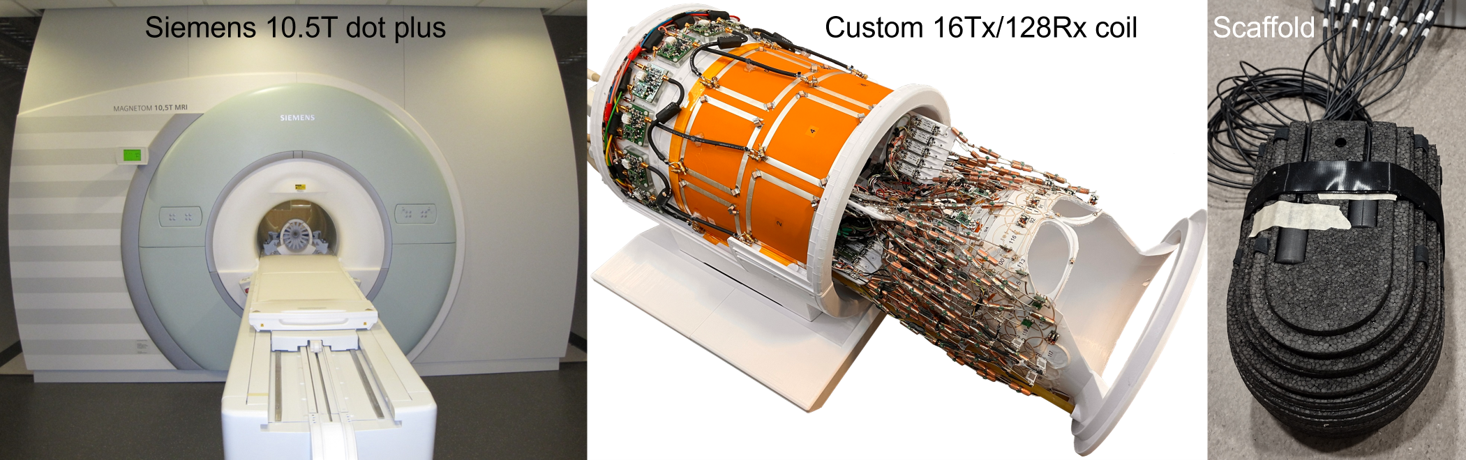
Fig. 1. Experimental setup. Human image data were collected on a 10.5 Tesla (10.5T) Siemens Magnetom Dotplus scanner (left) using a custom 16-channel transmit 128-channel receive (16Tx/128Rx) RF coil (middle). The dynamic field monitoring was conducted in a separate session using a scaffold (right) in which 16 field probes were placed across 3D space to characterize the dynamic field changes associated with the spiral image readout.
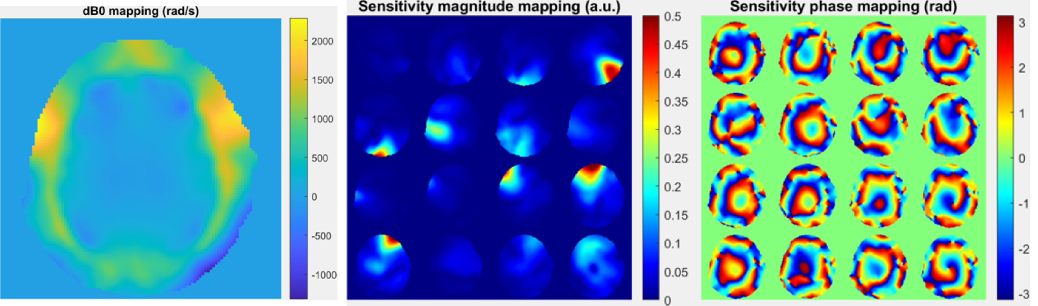
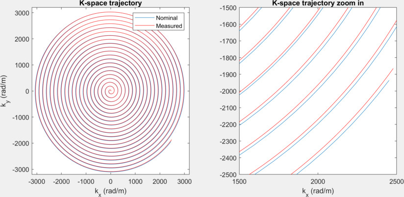
Fig. 3. Measured vs nominal k-space trajectory for the spiral image readout. The 2D spiral trajectory (left) for a 21x21 cm2 FOV, a 1-mm resolution and a 5-fold under-sampling rate was accomplished by designing time-optimal gradient waveforms. The measured k-space trajectory appeared to deviate from the nominal one, especially when approaching the outermost of the k-space (right).
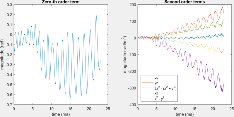
Fig. 4. Measured dynamic field changes for zero-th (left) and second (right) order terms associated with the spiral image readout. Even with a readout as short as 23 ms, non-negligible phase accrual due to higher order dynamic field changes was observed.
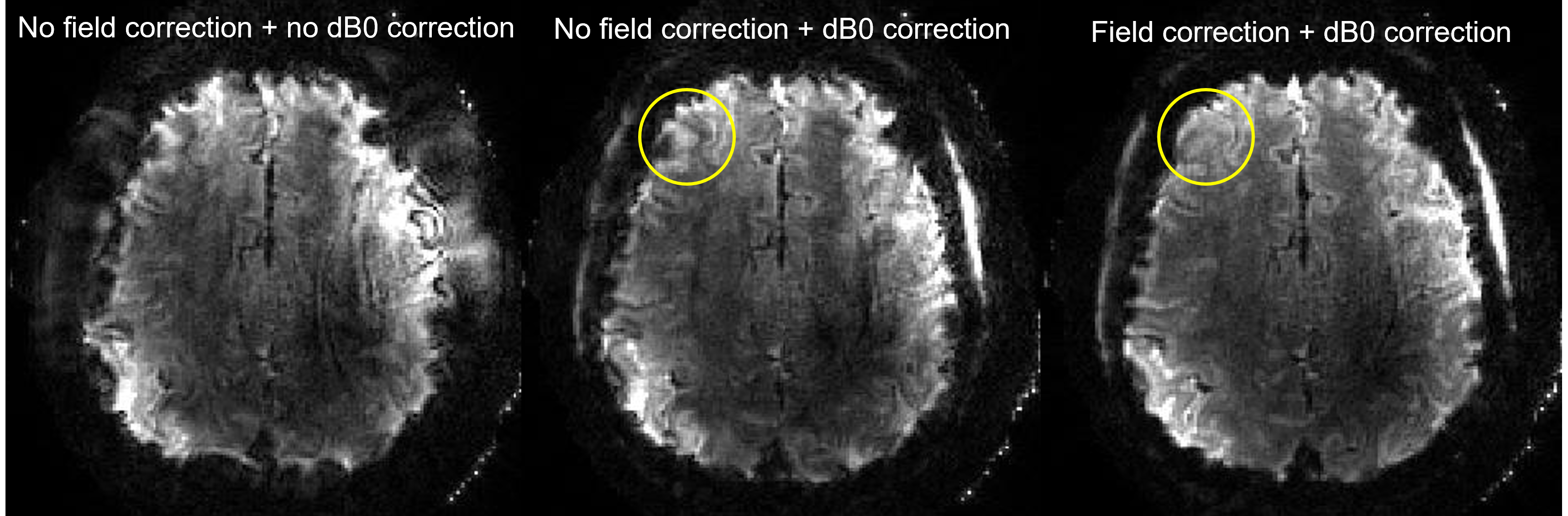
Fig. 5. Single-shot 2D spiral T2*w brain imaging at 10.5T. Data were obtained with TE/TR = 16/1000 ms and reconstructed using conjugate-gradient SENSE. The reconstruction with nominal k-space trajectory and without static off-resonance (dB0) correction (left) presented strong artifacts. Incorporating dB0 correction (middle) largely improved the image quality but still exhibited some image artifacts (e.g., as indicated by the yellow circle). The artifacts were effectively removed by also addressing dynamic field changes through image reconstruction (right).