3933
A dynamic shim approach to correct eddy-currents and concomitant-field using multi-coil AC/DC shim array1Department of Radiology, Stanford University, Stanford, CA, United States, 2Department of Electrical Engineering, Stanford University, Stanford, CA, United States, 3Athinoula A. Martinos Center for Biomedical Imaging, Massachusetts General Hospital and Harvard Medical School, Charlestown, MA, United States, 4Stanford Center for Cognitive and Neurobiological Imaging, Stanford University, Stanford, CA, United States
Synopsis
Keywords: System Imperfections, Diffusion/other diffusion imaging techniques
Motivation: To correct undesirable eddy-current and concomitant-fields effect in MRI acquisitions.
Goal(s): To achieve successful eddy-current and concomitant-fields mitigations, paving the way for high-quality 3D-invivo diffusion MRI with high-SNR.
Approach: We developed a dynamic shim approach with a multi-coil AC/DC shim-array to correct eddy-currents induced-phase in diffusion-prep encoding, and concomitant-field in long spiral in double-oblique positions.
Results: Invivo experiments were performed on the AC/DC shim-array to demonstrate cardiac-gated, multi-shot 3D-diffusion-prepared DTI acquisition without need for SNR-zapping stabilizer. High-quality, high-b-value(2000 s/mm2) acquisition was achieved where a synergistic combination of shim-array and pre-pulse gradient mitigation strategies were employed to minimize eddy-current phase in this difficult case.
Impact: We proposed a dynamic shim approach for robust invivo eddy-current correction in 3D-DTI with short preparation-time and high-SNR. Our results demonstrates the AC/DC coil's effectiveness in compensating challenging concomitant-fields during double-oblique slice acquisitions, especially with long-spiral sampling and high-performance gradients.
Introduction
AC/DC shim-array has been proven to enhance B0 homogeneity1–3. In our previous abstract4, we introduced the innovative application of the AC/DC shim-array for correcting eddy currents, where we validated this concept with single-diffusion-direction acquisition at b-value=600s/mm2. For compensating the concomitant-field, our simulations demonstrated a reduction in blurring when utilizing the AC/DC coil to compensate a conventional spiral-trajectory on an axial slice. Building upon our earlier work, we propose a dynamic shim approach to provide robust eddy-currents correction for invivo diffusion-prepared(DP) 3D-DTI with short preparation-time and full signal-level. High-b-value(2000s/mm2) was achieved through further refinement using a synergistic combination of shim-array and pre-pulse gradient mitigation strategies to minimize eddy-current phase in this difficult case. In terms of concomitant-field compensation, our simulations highlight the capability of the AC/DC coil to correct more challenging concomitant-fields generated in a double-oblique slice-position, especially when used in conjunction with a long-spiral-sampling and a high-performance gradient.Methods
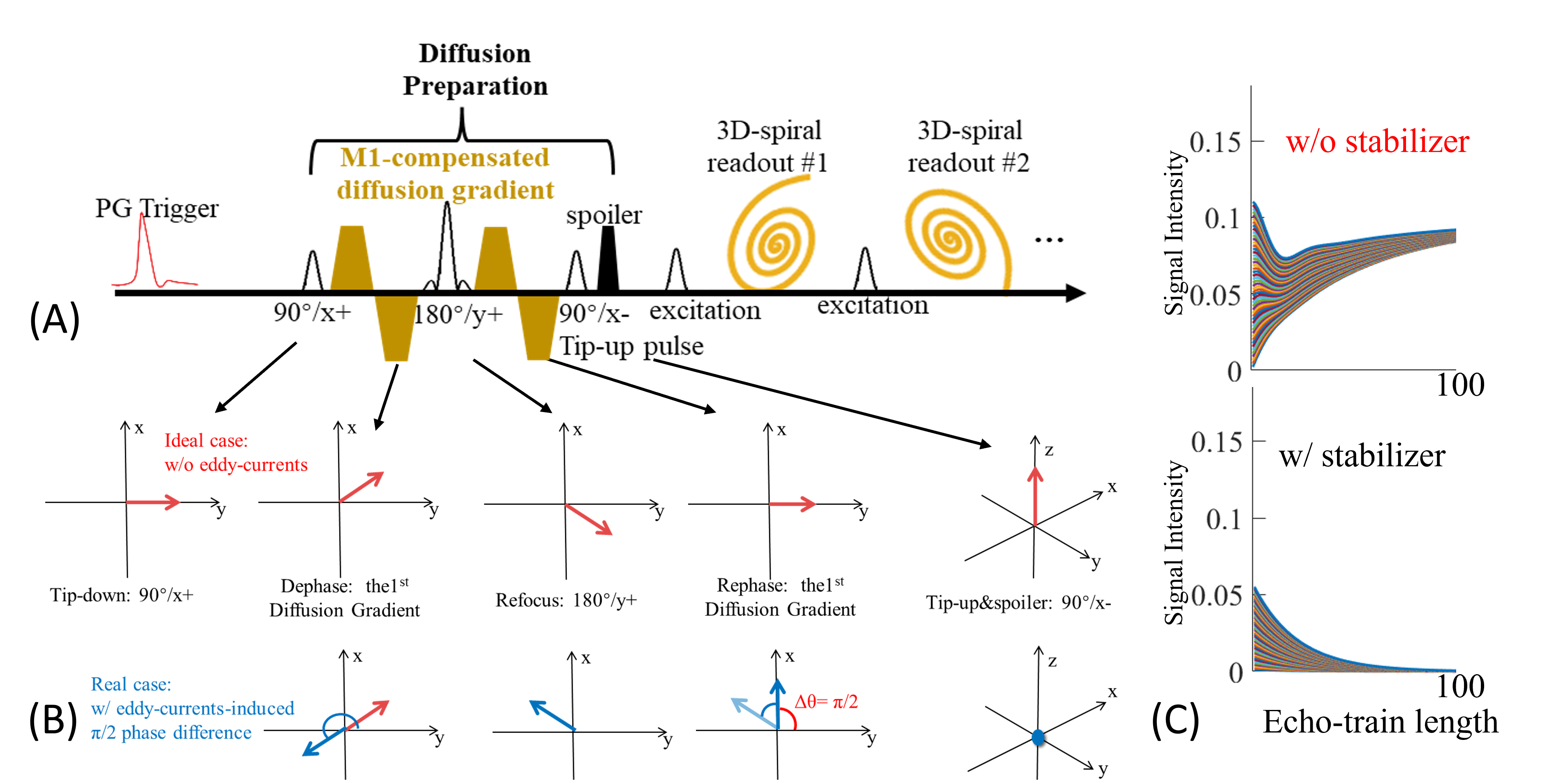
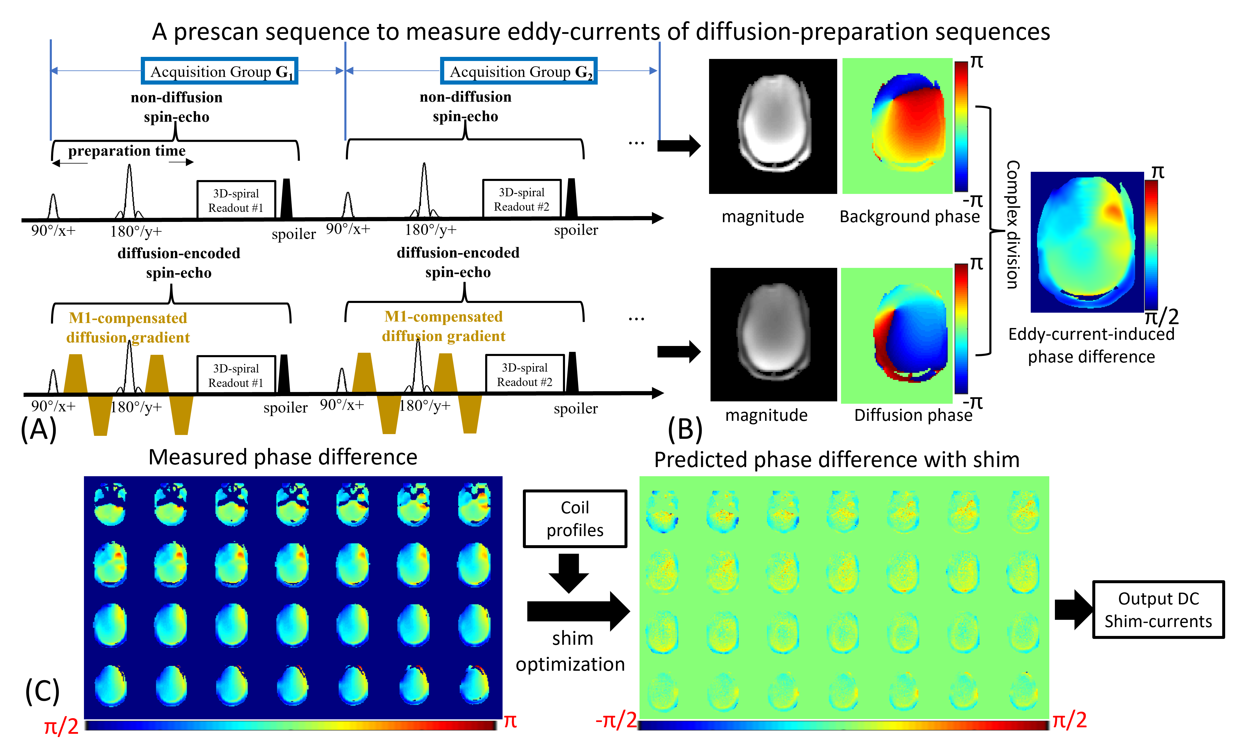
Eddy-currents correction: Figure1(A) depicts a standard DP acquisition. Ideally, all diffusion-encoded signals would transition fully to longitudinal-magnetizations with tip-up pulse, with no eddy-current phase-errors. However, eddy-current-related phase-errors can cause signal-dropout5,6. Figure1(B) demonstrates signal-loss from such phase-errors. Importantly, when the phase-error approaches pi/2, it results in a complete signal loss after tip-up pulse. To mitigate signal loss, two approaches have been proposed: (i) A magnitude stabilizer6 before the tip-up pulse to dephase signal variations, at the cost of reducing the signal by half(Figure1(C)). (ii) Cardiac-gating combined with M1-compensated diffusion-gradients helps minimize phase-disruptions, and a pre-pulse gradient applied before DP is used to counteract eddy-current-related phase-disruptions7. Nonetheless, its efficacy is not robust, particularly at high-b-values. In this study, we combined the AC/DC shim-array with M1-compensated DP and PG to compensate for eddy-current and physiological-induced phase-corruptions:(i) A one-time eddy-current-induced phase characterization: A spin-echo acquisition(Figure2(A)) was implemented as a prescan to measure phase-differences between b=0 and diffusion-weighted acquisitions in a phantom. This reflects an approximation of the eddy-current-induced phase(Figure2(B)).
(ii) Phase variations extracted from (i) were utilized for optimizing shim-currents. This is executed to produce inverse phase maps, thereby compensating for the eddy-current-induced phase-discrepancies(Figure2(C)). With the optimization, the eddy-current-induced phase-differences were minimized to zero.
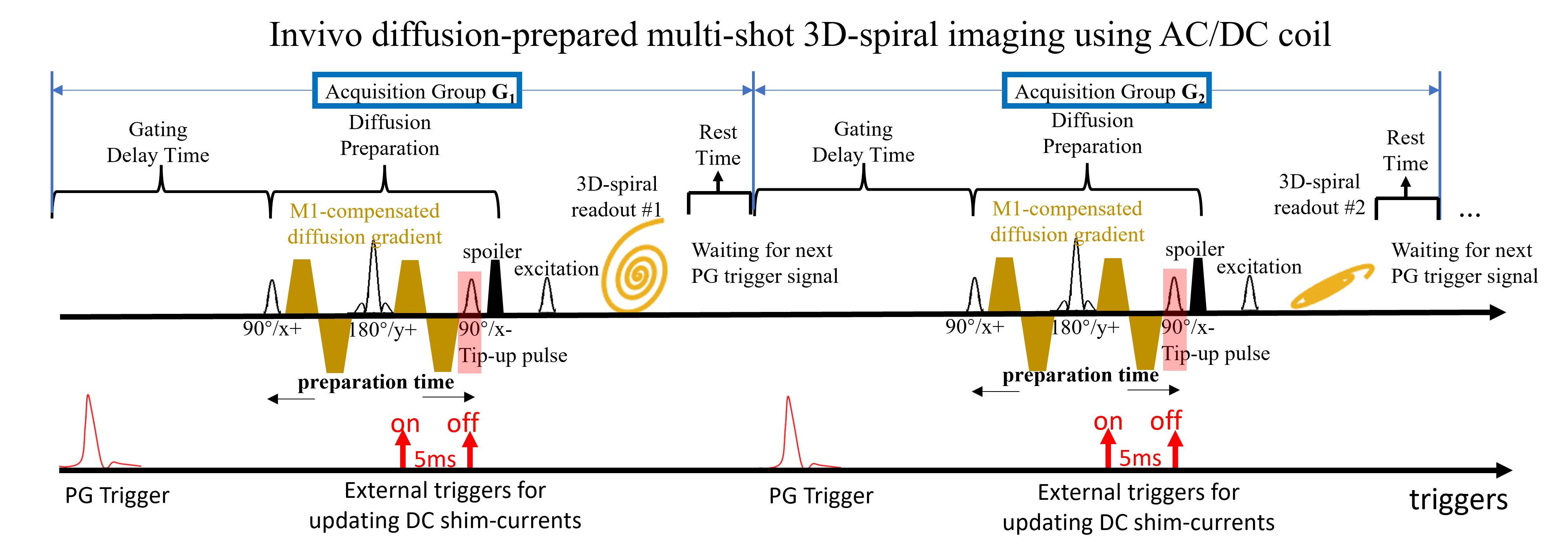
(iii) The calculated shim-currents were applied during the diffusion encodings of the DP sequence, which has the same protocol of the prescan's diffusion encodings in (i). With every TR, the shim-currents were updated via the sequence's external triggers(Figure3). This facilitates the compensation for variable eddy-currents arising from diverse diffusion-directions.
Invivo datasets were acquired: FOV:220mm, 2.0mm-isotropic resolution, 64-shot-3D spiral-projection trajectories were sampled(matrix size: 110×110×110). Six diffusion-directions with b=1000&2000s/mm2 were acquired with TR/DP-time=500/50ms. The shim-duration=5ms.
Concomitant-field correction: The concomitant-fields can cause additional phase-accrual during readouts which resulted in image blurring8,9, particularly in acquisitions on high-performance gradient systems and low-field scanner. The concomitant-field Bc can be expressed as:
$$ B_c≈(\frac{G_z^2}{8B_0})(X^2+Y^2 )+(\frac{G_x^2+G_y^2}{2B_0 }) Z^2-(\frac{G_x G_z}{2B_0 })XZ-(\frac{G_y G_z}{2B_0 })YZ $$
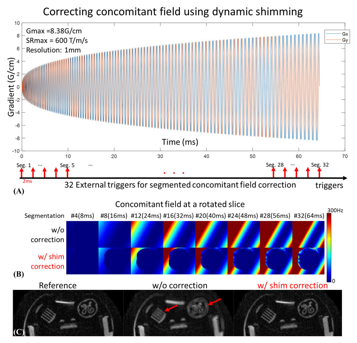
where (Gx,Gy,Gz) are the time-dependent gradient-fields and (X,Y,Z) are spatial coordinates in the magnet. Using this equation, we simulate the concomitant-field of a 64ms single-shot 1mm 2D-spiral trajectory at 3T(Figure 5(A)). Considering the temporal switch limits of the AC/DC shim-array, we divide the 64ms trajectory into 32 segments and calculate the phase-accrual every 2ms. We compensated for the concomitant-field by updating the optimal shim-currents to create opposite phases every 2ms.
Results
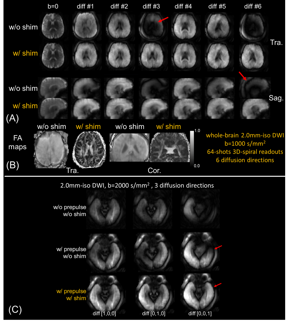
Figure4(A)&(B) show diffusion-weighted images and corresponding FA maps at b=1000s/mm2 with&without shim-correction. Without eddy-currents correction, the eddy-current-induced phase-differences cause signal-loss, as indicated in red arrows, while shim-corrected images show higher signals in these regions without signal dropout and correct FA maps. Moreover, Figure4(C) reveals that combining shim and pre-pulse corrections provides superior signal recovery at b=2000s/mm2 compared to just the pre-pulse correction, highlighting the efficacy of the proposed method at high b-values.Figure5(B) shows the concomitant field maps at Z=+40 mm offset with double-oblique rotation. The phase accumulates across long readout and causes image blurring(Figure5(C)). With masked shim correction, the phase accrual in the region of interest (inside the phantom) were compensated, which resulted in sharper images(Figure 5(C)).
Discussion and conclusion
We developed a dynamic shim approach for eddy-current and concomitant-field corrections. Our results highlight successful eddy-current mitigation within a 3D-DTI acquisition, paving the way for high-quality 3D-invivo diffusion-MRI with high-SNR. As an initial demonstration, we utilized a single-TE spiral-projection readout. However, this approach can be extended to efficient sampling techniques. Additionally, we demonstrated that our approach could resolve the image-blurring from strong concomitant-fields and oblique slice-position, especially in high-performance gradient systems and/or low-field MRI scanners. Future work will explore correction on more complex readout k-space trajectories, where undesirable fields obtained using field probes could provide high-fidelity target for mitigation.Acknowledgements
This study is supported in part by R01MH116173, R01EB019437, U01EB025162, P41EB030006, R01EB033206, U24NS129893References
1. Stockmann JP, Witzel T, Keil B, et al. A 32-channel combined RF and B0shim array for 3T brain imaging. Magn Reson Med. 2016;75(1):441-451. doi:10.1002/mrm.25587
2. Liao C, Bilgic B, Tian Q, et al. Distortion-free, high-isotropic-resolution diffusion MRI with gSlider BUDA-EPI and multicoil dynamic B0 shimming. Magn Reson Med. 2021;86(2):791-803. doi:10.1002/mrm.28748
3. Liao C, Stockmann J, Tian Q, et al. High-fidelity, high-isotropic-resolution diffusion imaging through gSlider acquisition with B1+ and T1 corrections and integrated ΔB0/Rx shim array. Magn Reson Med. 2020;83(1):56-67. doi:10.1002/mrm.27899
4. Liao C, Stockmann J, Cao X, et al. Flexible use of AC/DC coil for eddy-currents and concomitant fields mitigation with applications in diffusion-prepared non-Cartesian sampling. In: ISMRM. ; 2022:1236. https://submissions.mirasmart.com/ISMRM2023/ViewSubmissionTeaser.aspx. Accessed November 7, 2023.
5. Van AT, Cervantes B, Kooijman H, Karampinos DC. Analysis of phase error effects in multishot diffusion-prepared turbo spin echo imaging. Quant Imaging Med Surg. 2017;7(2):238-250. doi:10.21037/qims.2017.04.01
6. Gao Y, Han F, Zhou Z, et al. Multishot diffusion-prepared magnitude-stabilized balanced steady-state free precession sequence for distortion-free diffusion imaging. Magn Reson Med. 2019;81(4):2374-2384. doi:10.1002/mrm.27565
7. Cao X, Liao C, Zhou Z, et al. DTI-MR fingerprinting for rapid high-resolution whole-brain T1, T2, proton density, ADC, and fractional anisotropy mapping. Magn Reson Med. 2023. doi:10.1002/MRM.29916
8. Cheng JY, Santos JM, Pauly JM. Fast concomitant gradient field and field inhomogeneity correction for spiral cardiac imaging. Magn Reson Med. 2011;66(2):390-401. doi:10.1002/mrm.22802
9. Du YP, Zhou XJ, Bernstein MA. Correction of concomitant magnetic field-induced image artifacts in nonaxial echo-planar imaging. Magn Reson Med. 2002;48(3):509-515. doi:10.1002/mrm.10249
Figures