3932
Characterization of Eddy-Currents Associated with Multi-Coil B0 Field Control in an Accessible Head-Only Scanner1Department of Biomedical Engineering, Columbia University in the City of New York, New York, NY, United States, 2Center for Magnetic Resonance Research, University of Minnesota, Minneapolis, MN, United States, 3Department of Radiology and Biomedical Imaging, Magnetic Resonance Research Center, Yale University School of Medicine, New Haven, CT, United States, 4Center for Magnetic Resonance Research, Department of Radiology, University of Minnesota, Minneapolis, MN, United States, 5Department of Radiology, Columbia University Medical Center, New York, NY, United States
Synopsis
Keywords: System Imperfections, Gradients, Eddy-Currents, Multi-Coil
Motivation: The eddy current (EC) behavior of a multi-coil array for image encoding in a novel head-only MR scanner has been estimated theoretically, however, to date, the physical measurements have been outstanding.
Goal(s): Our goal was the spatiotemporal characterization of the ECs of this system.
Approach: We measured the ECs via local field probes by acquiring FIDs at a series of delays after the switching of individual MC channels.
Results: Measurements at over 40 positions in the FOV enabled a comprehensive analysis of the ECs. The longest EC components last for more than a second, however, we show that EC compensation is possible.
Impact: The presented eddy-current characterization is an important stepping stone towards eddy-current compensation for DYNAMITE image encoding in non-traditional MR scanner designs.
Introduction
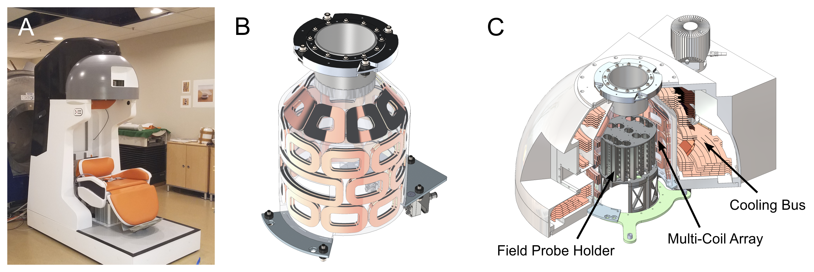
As part of a collaborative effort, we recently implemented a 31-channel multi-coil (MC) array for B0 field control in a novel head-only MR scanner1,2 (Fig. 1A). The dynamic MC technique (DYNAMITE)3 utilizes a number of unspecific coils driven individually to create desired B0 fields. This approach has been shown to enable unrivaled B0 shimming capabilities4–7, and has successfully been used for spatial encoding for MRI8,9. Here, this technology was adopted to enable a range of approaches such as linear and non-linear image encoding together with active shimming of the scanner's inhomogeneous B0 background field.While the MC technique is well established, it has never been adapted to a unique system like the one at hand. A particular challenge was the eddy-current (EC) behavior of the unshielded MC elements in close proximity to the compact magnet. With the magnet bore made from fiberglass composite, the copper bus structure cooling the high-temperature superconducting magnet was identified as the most likeliest place for EC generation (Fig. 1B). While its shape was optimized based on simulations prior to manufacturing, residual ECs are expected to remain10.
Here we present a full experimental characterization of the EC behavior of this system after system integration.
Methods
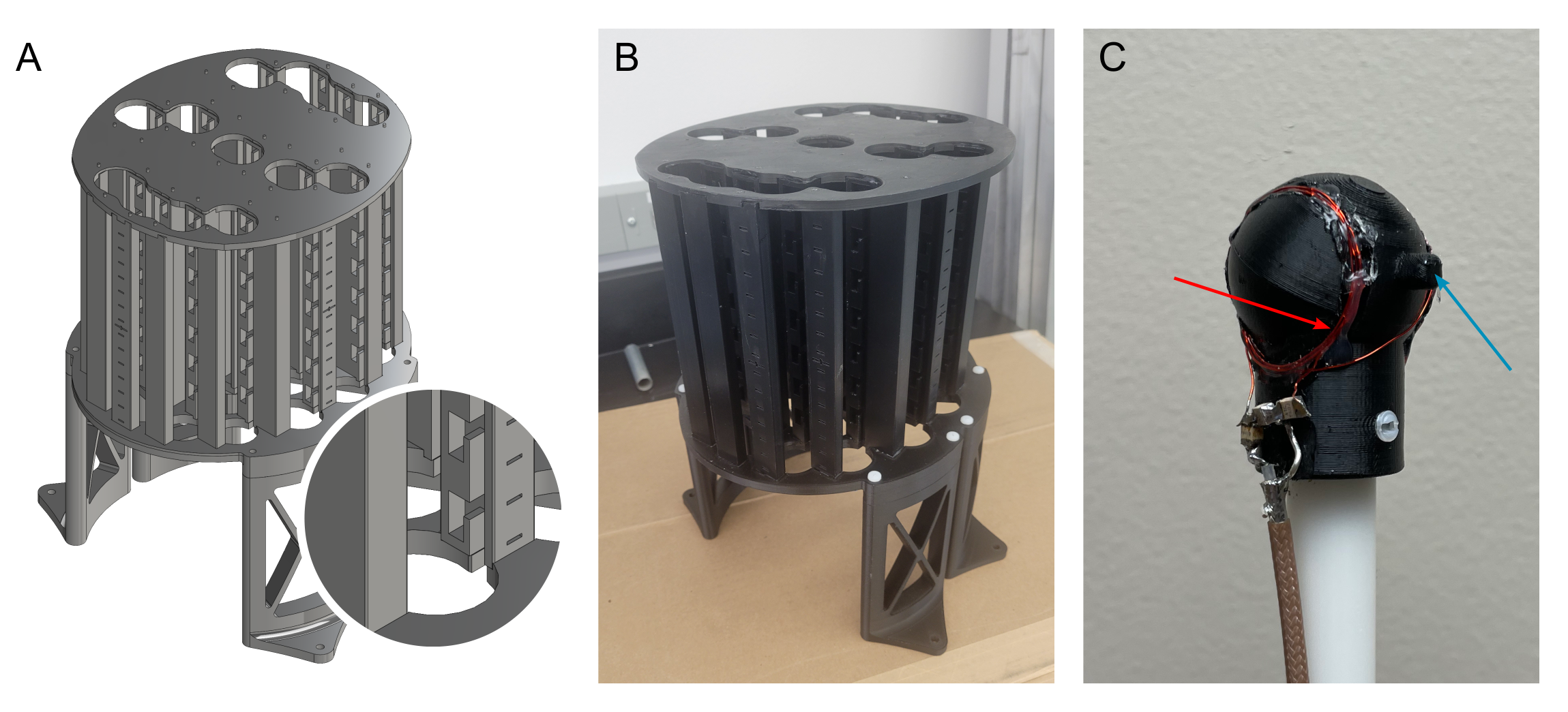
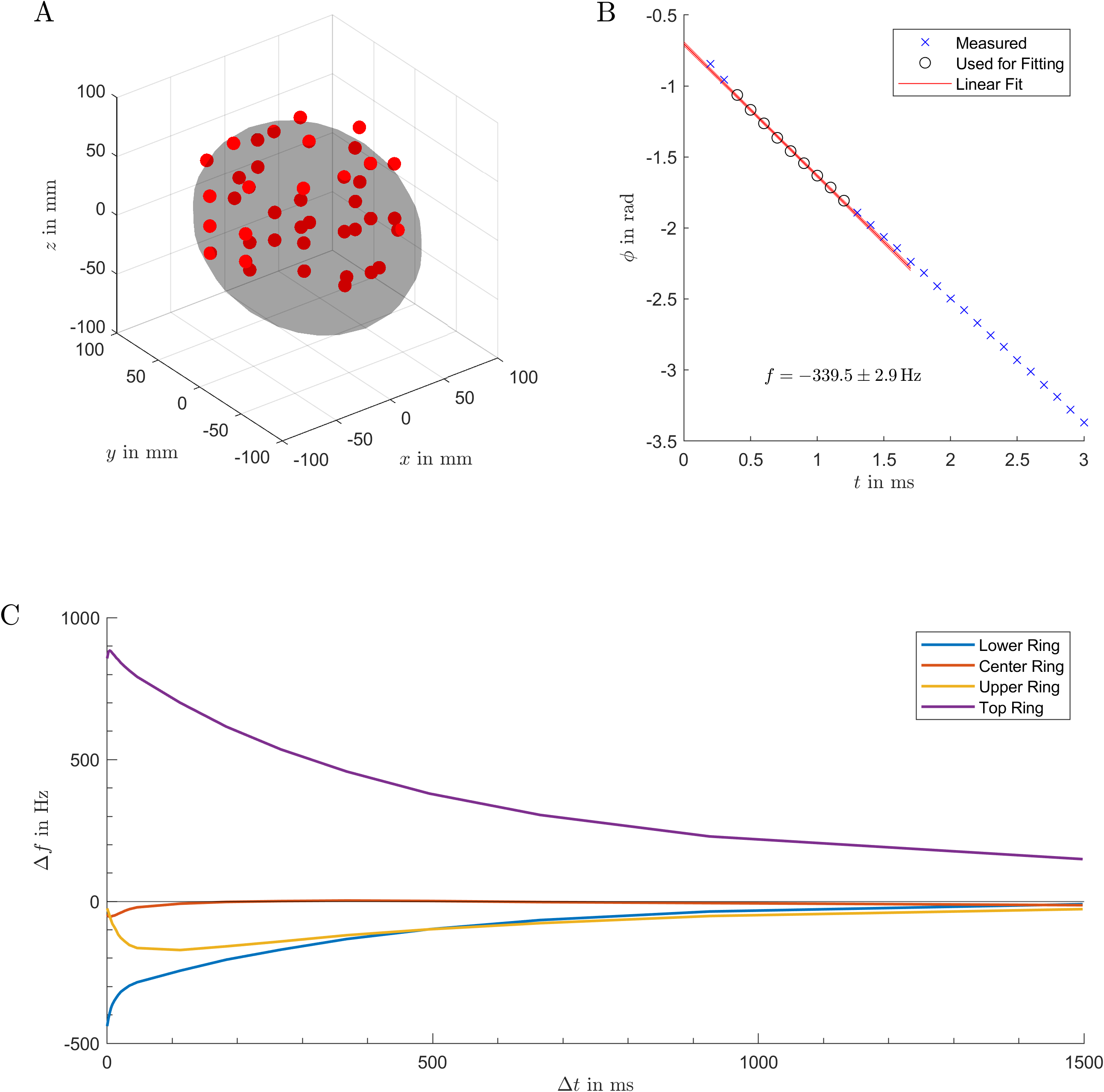
ECs were measured using localized signal probes. We designed a probe holder that fits closely into the magnet bore (Fig. 1C and Fig. 2A/B) and provides 161 unique positions arranged in 23 columns around the transverse plane (Fig. 2A). A water-filled plastic sphere with 1 cm diameter can be positioned in these locations using a fish-ladder approach. A Helmholtz transceive coil was arranged around the probe to maximize SNR (Fig. 2C).The MC array was driven by a custom-designed amplifier system11 and controller12. ECs were characterized by acquiring FIDs at distinct delays after the end of a 500 μs ramp from 2.5 A (50% of the maximum) down to 0 (5 kA/s) and extracting the local frequency from the initial phase of the signal (Fig. 3B). ECs were measured at 41 locations covering a volume of roughly 140x170x100 mm (Fig. 3A) at 20 delays. We measured individual MC elements and the ECs generated by switching linear MC-generated gradients in all three principal directions.
Time-dependent EC fields BEC(r, t) created by the linear gradients were decomposed into previously acquired2 MC basis fields BC(r) with three exponentials:
$$B_{EC}(\vec{r},t)=\sum_{C=1}^{31}\,B_C(\vec{r})\cdot\sum_{i=1}^3A_{i,C}\cdot{}e^{-t/\tau_{i,C}}.$$
The amplitudes Ai,C and time constants τi,C were determined via Matlab’s13 fmincon function while restricting the channel current to 0.5 A (10%).
Results
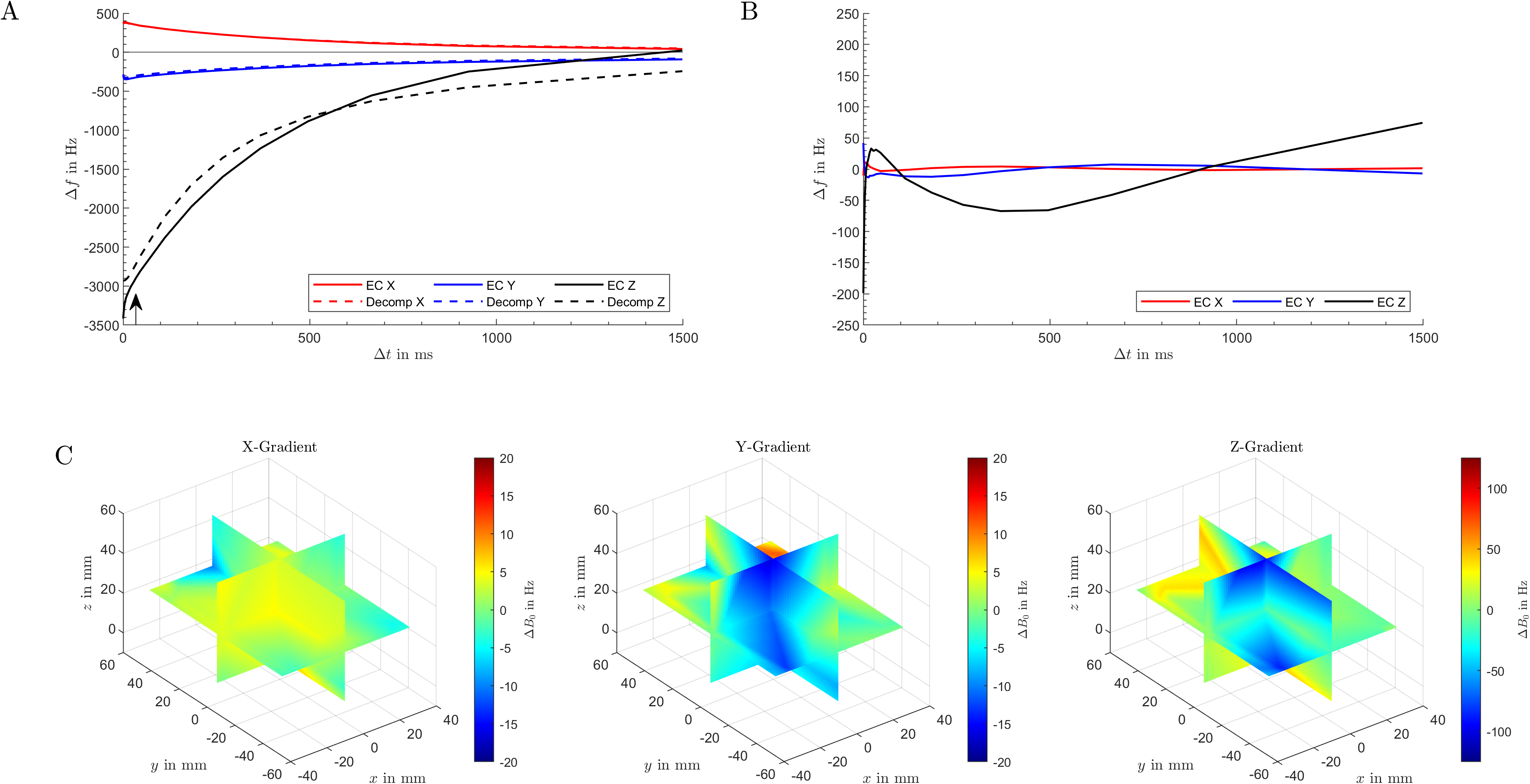
Measured frequencies ranged from -3416 Hz to 1309 Hz with an average uncertainty of 10 Hz (standard deviation extracted from the linear regression, Fig. 3B).All measured conditions exhibited multi-exponential behavior with both short and very long time constants, with the longest EC signals lasting longer than the investigated 1.5 s (Fig. 3C).
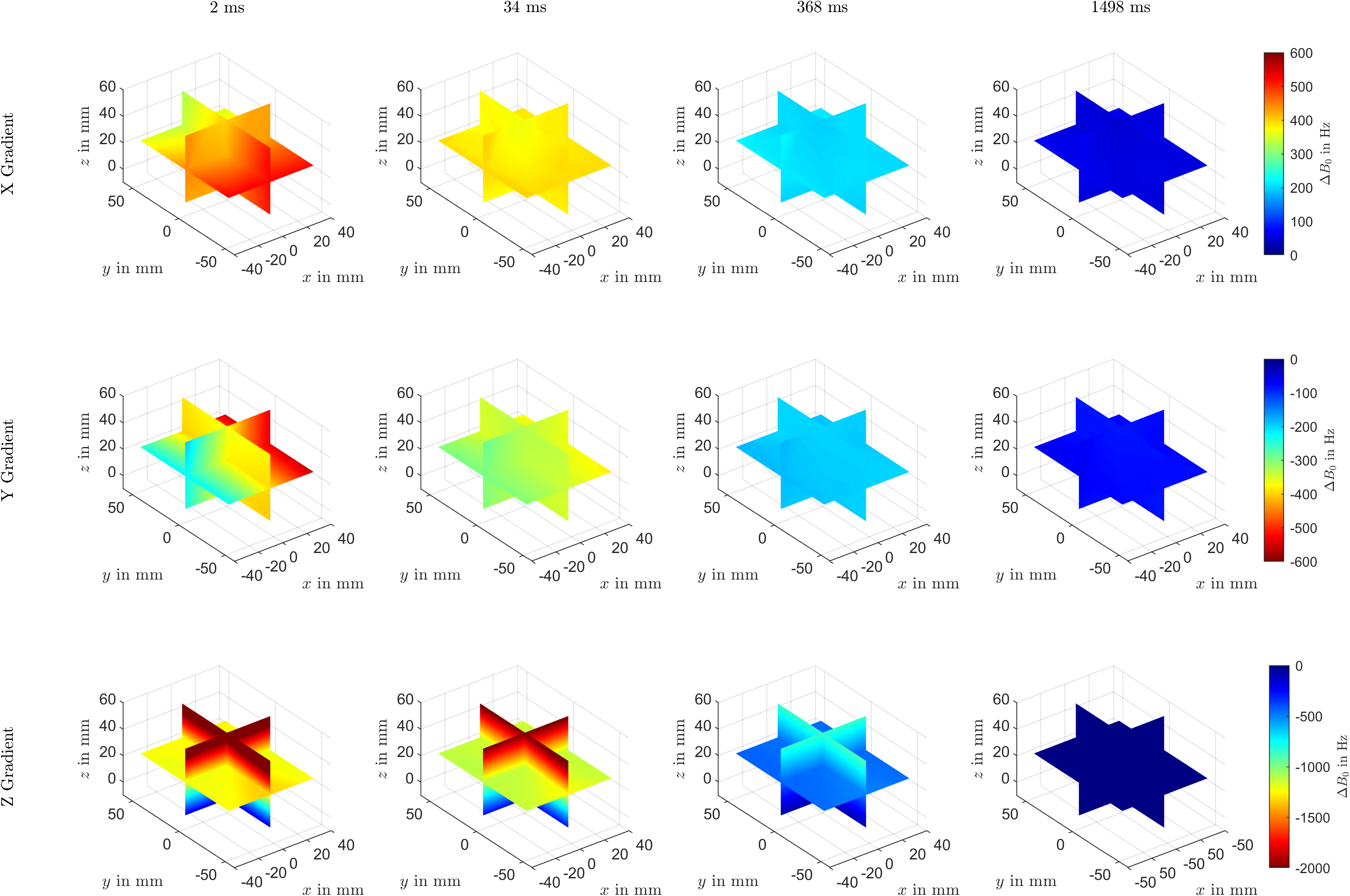
Figure 4 shows the spatial distribution of exemplary EC fields for the switching of MC generated linear gradients. The spatial distribution in earlier time points is governed by linear gradients, while a significant constant offset frequency is dominating the temporal behavior.
A decomposition of the ECs of the linear gradients via MC basis fields (Fig. 5A/B) matched the ECs by 96%, 92%, and 91% for the x-, y-, and z-gradient, respectively (standard deviation of the residual fields). Time constants of the exponentials ranged from 12 ms to 3.3 s with amplitudes up to 0.56 A. An exemplary assessment of ECs throughout an MRI sequence was estimated in Fig. 5C by calculating the resultant EC after a trapezoidal gradient waveform with 5 ms length.
Discussion
We successfully mapped the spatiotemporal EC behavior associated with MC switching in a novel compact head-only scanner. The observed ECs show both short and very long time components with significant amplitudes. The calculated time constants are of similar length than the previously simulated ones10, reinforcing the theory that ECs are primarily generated in the cold bus structure.The effect of the observed ECs on MRI depends on the sequence at hand - long lasting ECs of short gradient waveforms largely cancel out. However, we were also able to show that the EC fields can be generated by the MC array using just 10% of the dynamic range of the system, potentially enabling the implementation of pre-emphasis on the pre-calculated current waveforms. The size and quality of the acquired data set allows for future research into a compensation scheme based on the individual MC coil fields directly.
The characterization of ECs associated with MC hardware presented here is expected to pave the way for further adoption of DYNAMITE image encoding.
Acknowledgements
This research was supported by the National Institute of Biomedical Imaging & Bioengineering of the National Institutes of Health under award numbers U01-EB025153 and R01-EB030560, and was in parts performed at the Columbia MR Research Center.References
1. Garwood M, Mullen M, Kobayashi N, et al. A compact vertical 1.5T human head scanner with shoulders outside the bore and window for studying motor coordination. In: Proc Int Soc Magn Reson Med. 2020;28:1278.
2. Theilenberg S, Shang Y, Ghazouani J, et al. Design and realization of a multi‐coil array for B0 field control in a compact 1.5T head‐only MRI scanner. Magn Reson Med 2023;90:1228–1241.
3. Juchem C, Nixon TW, McIntyre S, Rothman DL, de Graaf RA. Magnetic field modeling with a set of individual localized coils. J Magn Reson 2010;204:281–289.
4. Juchem C, Nixon TW, McIntyre S, Boer VO, Rothman DL, de Graaf RA. Dynamic multi-coil shimming of the human brain at 7 T. J Magn Reson 2011;212:280–288.
5. Stockmann JP, Witzel T, Keil B, et al. A 32-channel combined RF and B0 shim array for 3T brain imaging. Magn Reson Med 2016;75:441–451.
6. Topfer R, Starewicz P, Lo K-M, et al. A 24-channel shim array for the human spinal cord: Design, evaluation, and application. Magn Reson Med 2016;76:1604–1611.
7. Darnell D, Truong T-K, Song AW. Integrated parallel reception, excitation, and shimming (iPRES) with multiple shim loops per radio-frequency coil element for improved B 0 shimming. Magn Reson Med 2017;77:2077–2086.
8. Rudrapatna SU, Fluerenbrock F, Nixon TW, de Graaf RA, Juchem C. Combined imaging and shimming with the dynamic multi‐coil technique. Magn Reson Med 2019;81:1424–1433.
9. Juchem C, Theilenberg S, Kumaragamage C, et al. Dynamic multicoil technique (DYNAMITE) MRI on human brain. Magn Reson Med 2020;84:2953–2963.
10. Theilenberg S, Parkinson BJ, Juchem C. Eddy Current Characterization of a Multi-Coil Array for Imaging in a Head-Only MR Scanner. In: Proc Int Soc Magn Reson Med. 2020;28:4245.
11. Resonance Research, Inc, Bellerica, MA, USA.
12. Nixon TW, McIntyre S, de Graaf RA. The Design and Implementation of a 64 Channel Arbitrary Gradient Waveform Controller. In: Proc Int Soc Magn Reson Med. 2017;25:0969.
13. Mathworks, Natick, MA, USA.
Figures