3928
Translating a Non-invasive Hybrid PET/MR Method of Imaging Cerebral Oxygen Metabolism to Humans1University of Western Ontario, London, ON, Canada, 2Lawson Health Research Institute, London, ON, Canada, 3Centre for Addiction and Mental Health, Toronto, ON, Canada, 4University of Pennsylvania, Philadelphia, PA, United States
Synopsis
Keywords: PET/MR, Arterial spin labelling, Cerebral Oxygen Metabolism
Motivation: To develop a non-invasive method of imaging CMRO2 by hybrid PET/MRI.
Goal(s): Translate a previously validated PET/MR technique capable of imaging CBF, OEF, and CMRO2 to human studies.
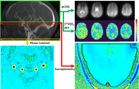
Approach: Technique combines [15O]O2-PET with MRI measurements of whole-brain CMRO2 and arterial spin labeling images of CBF.
Results: Good agreement to literature regional CMRO2 values.
Impact: Hybrid PET/MRI can simplify the PET-only technique of measuring regional CMRO2 by avoiding arterial sampling and only requiring one radiotracer, which reduces scan time and radiation dose, while enabling simultaneous acquisition of perfusion and tissue oxygen extraction.
Introduction
Positron emission tomography (PET) remains the gold standard for imaging the cerebral metabolic rate of oxygen (CMRO2) because of its ability to directly measure the oxygen extraction fraction (OEF).1 However, the procedure is logistically demanding as it requires three radiotracers, invasive as arterial sampling is required for each radiotracer, and long (typically 30-45 min). Hybrid PET/MRI offers the possibility to greatly simplify the procedure by acquiring complementary MRI data.2,3 This hybrid method, termed PMROx (PET/MR imaging of oxidative metabolism), involves measuring whole-brain (WB) CMRO2 by MRI while simultaneously acquiring [15O]O2-PET data. The WB CMRO2 estimate is used a reference to calibrate the [15O]O2-PET data. The last step is to incorporate cerebral blood flow (CBF) images acquired with arterial spin labeling (ASL) to replace [15O]H2O-PET. Using these MRI methods, PMROx is completely non-invasive yet maintains the ability of PET to quantify OEF. The acquisition time is approximately 5 min, which enables CMRO2 to be imaged under different metabolic states, such as baseline and during a functional task, in a single imaging session. The feasibility of PMROx was initially demonstrated on PET-only data4 before being validated in a large animal model.3 The goal of the current work was to present initial data translating PMROx to human participants.Methods
Data were acquired from a healthy subject on a Siemens 3T Biograph mMR scanner. Five minutes of list-mode PET data were acquired after inhalation of approximately 2000 MBq of [15O]O2. [15O]O2 gas, generated from a PETtrace 800 cyclotron (16.5 MeV; GE Healthcare), flowed through a column of activated charcoal and ascarite to remove impurities and delivered to the scanner via a 202 m long stainless steel line. Reference WB CMRO2 was measured by OxFlow, which combines phase-contrast and susceptibility-based MRI to measure WB CBF and OEF, respectively.5 The arterial content of oxygen was estimated by assuming a hemoglobin concentration of 16 g/dL and an arterial oxygen saturation of 98%. The PMROx CMRO2 images were generated from the [15O]O2 and MRI data as shown in Figure 1.4 PET images were reconstructed using an MR-based attenuation correction map, motion-corrected, and smoothed by a 6 mm Gaussian filter. Pseudo-continuous ASL (TR/TE: 4210/37.86 ms, post-labeling delay: 1.7 s, labeling duration: 1.5 s, voxel size: 3.8 mm3) was collected simultaneously with the PET acquisition. A calibration (M0) image was acquired with a TR of 10 s. ASL images were motion-corrected and smoothed by an 8 mm Gaussian filter. All images were pre-processed in SPM12 and calculations were completed with in-house MATLAB (R2023a) scripts.Results
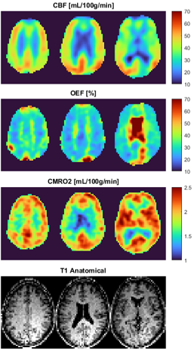
Global CBF and OEF were 32 mL/100g/min and 41%, respectively. Average CMRO2 was 1.9 mL/100g/min. Images of CBF (ASL), OEF, and CMRO2 are displayed in Figure 2. As PMROx directly calculates CMRO2, the OEF images were generated by dividing CMRO2 by CBF.Discussion/Conclusion
This preliminary study demonstrates the feasibility of imaging CBF, OEF and CMRO2 by combining [15O]O2-PET with MRI (ASL and OxFlow). The average CBF and CMRO2 values were in good agreement with literature values.1 Unexpectedly higher OEF values were observed in white matter, likely a result of underestimated white-matter CBF due to transit time errors. This artefact could be corrected using a multi-post-labelling delay ASL technique to measure arterial transit time.6 In summary, this work highlights the ability to generate quantitative images of CMRO2 using hybrid PET/MRI to simplify metabolic measurements by reducing the requisite number of radiotracers and eliminating arterial sampling. Using PET/MRI enables direct comparison with emerging MRI techniques for measuring CMRO2, such as calibrated blood oxygen level dependent imaging.7 Hybrid imaging also avoids the confounding issues of hemodynamic and metabolic fluctuations that can occur when acquiring images sequentially.8Acknowledgements
Special thankyou to Heather Biernaski and Yvonne Huston.References
1. Fan A, et al. Quantification of brain oxygen extraction and metabolism with [15O]-gas PET: A technical review in the era of PET/MRI. Neuroimage. 2020;220(May). doi:10.1016/j.neuroimage.2020.117136
2. Ssali T, Anazodo UC, Thiessen JD, Prato FS, St. Lawrence K. A noninvasive method for quantifying cerebral blood flow by hybrid PET/MRI. Journal of Nuclear Medicine. 2018;59(8):1329-1334. doi:10.2967/jnumed.117.203414
3. Narciso L, Ssali T, Liu L, et al. A Noninvasive Method for Quantifying Cerebral Metabolic Rate of Oxygen by Hybrid PET/MRI: Validation in a Porcine Model. Journal of Nuclear Medicine. 2021;62(12):1789-1796. doi:10.2967/jnumed.120.260521
4. Narciso L, Ssali T, Iida H, St. Lawrence K. A non-invasive reference-based method for imaging the cerebral metabolic rate of oxygen by PET/MR: Theory and error analysis. Phys Med Biol. 2021;66(5):065009. doi:10.1088/1361-6560/abe737
5. Jain V, Langham MC, Wehrli FW. MRI estimation of global brain oxygen consumption rate. J Cereb Blood Flow Metab. 2010;30(9):1598-1607. doi:10.1038/jcbfm.2010.49
6. Ssali T, Anazodo U, Narciso L, Liu L, et al. Sensitivity of Arterial Spin Labeling for Characterization of Longitudinal Perfusion Changes in Frontotemporal Dementia and Related Disorders. Neuroimage Clin. 2022;35:1-2853.
7. Englund E, et al. Calibrated fMRI for dynamic mapping of CMRO2 responses using MR-based measurements of whole-brain venous oxygen saturation. J Cereb Blood Flow Metab. 2020;40(7):1501-1516.
8. Ssali T, Narciso L, Hicks J, et al. Concordance of regional hypoperfusion by pCASL MRI and 15O-water PET in frontotemporal dementia: Is pCASL an efficacious alternative? Neuroimage Clin. 2022;33:102950.
Figures