3926
First experience of pTX RF pulse design at 11.T MRI for whole brain imaging in vivo1BAOBAB, NeuroSpin, University Paris-Saclay, CEA, CNRS, Gif-sur-Yvette, France, 2DACM/IRFU, University Paris-Saclay, CEA, Gif-sur-Yvette, France, 3Siemens Healthineers, Courbevoie, France, 4UNIACT, NeuroSpin, University Paris-Saclay, CEA, CNRS, Gif-sur-Yvette, France
Synopsis
Keywords: RF Pulse Design & Fields, High-Field MRI
Motivation: Following the commissioning of the Iseult CEA 11.7 T whole-body MRI system, first in vivo human brain images have been acquired at 11.7T.
Goal(s): Our aim is to demonstrate the feasibility of whole brain imaging using an 8TX/32RX home-built RF coil and to test subject-tailored pulses as well as calibration-pTX pulses.
Approach: Using 9 in-vivo B1 maps of the brain, we prepared and tested tailored kT-point pulses and universal GRAPE pulses to be used in non-selective 3D sequences.
Results: Our retrospective pulse performance analysis confirms the feasibility of whole brain imaging at 11.7T both using subject tailored and calibration free pTX.
Impact: . At 11.7T, the heterogeneity of the pseudo-CP mode, the transmit efficiency and the SAR level are such that dynamic pTX and extensive RF pulse optimizations are essential for whole brain imaging applications.
Introduction
With the commissioning of the Iseult CEA 11.7 T whole-body MRI system1 and the first ongoing study on healthy volunteers (PREMS), 11.7T MRI of the human brain in vivo has now become a reality. In the framework of this first in vivo protocol, we have acquired quantitative B1 maps in the head of (so far) nine subjects, and this way characterized from the transmit perspective the home-made 8TX/32RX head coil whose design was presented at an earlier ISMRM meeting2. Based on these maps, we tested several pulse design approaches varying Flip Angle (FA) targets, pulse durations, subject-tailored pulses (TP) versus universal pulses (UP), which we could also test in part experimentally. In this work, we present this initial experience of pTX pulse design at 11.7T, and show in particular the importance of dynamic pTX3-5 to deliver whole-brain images at 11.7T (500 MHz).Material and Methods
We performed our acquisitions on the Iseult investigational whole-body 11.7T MRI system equipped with:- A SC72 gradient system (70 mT/m, 200 T/m/s slew rate);
- Second order shimming;
- A 8-channel pTX RF chain with 2 kW per channel (~1.3 kW at the coil plug);
- A 32-channel receive chain;
- A home-build 15(+1)TX/32RX RF coil, with pTx power split on to 180°-phased paired dipoles2.
We applied in nine healthy volunteers a ΔB0 mapping protocol (2.5mm isotropic resolution, TA=90s) and an XFL8 interferometric B1 mapping protocol (5mm isotropic resolution, TA =240s).
For each subject we prepared tailored kT-point9 pulses (TP) to realize:
- 10° and 4° non-selective excitations;
- A 180° non-selective inversion pulse;
- A scalable 0°–105°non-selective refocusing pulse.
Results
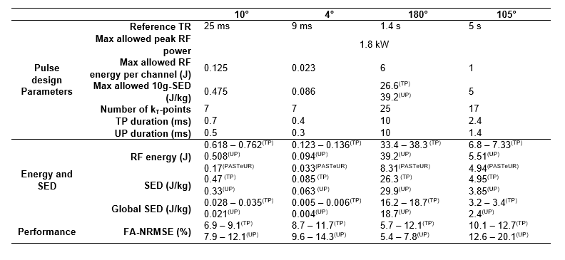
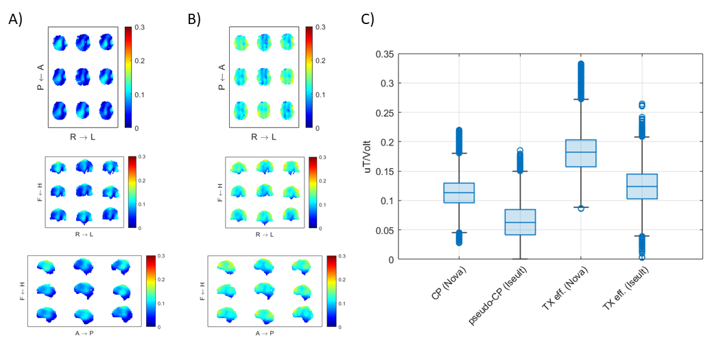
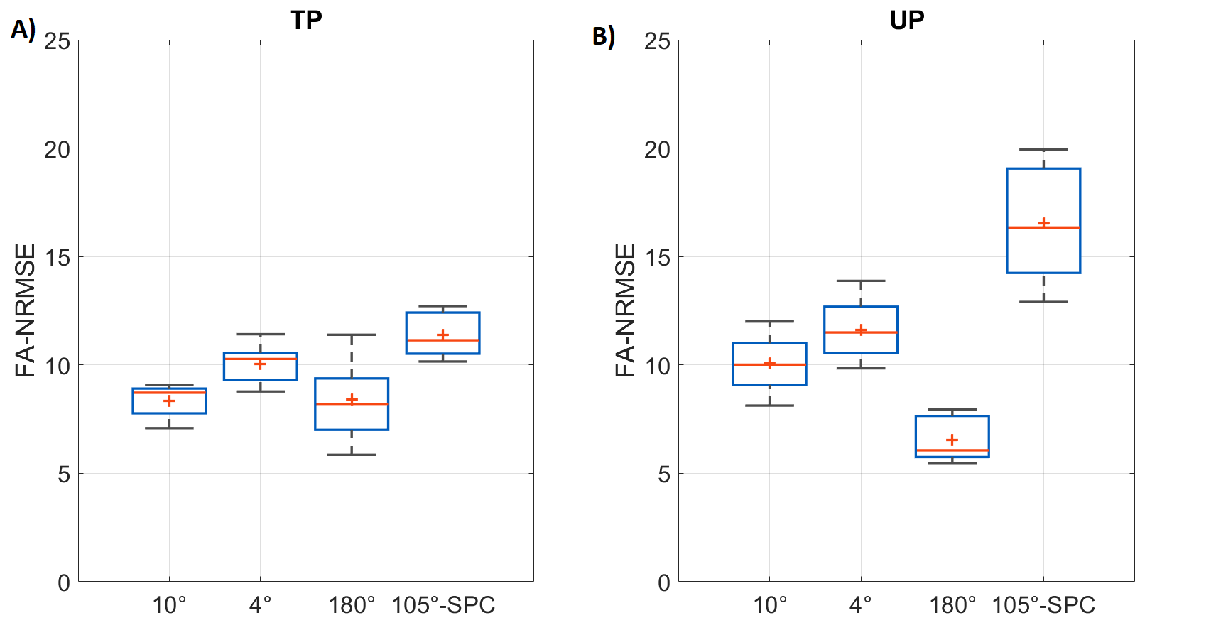
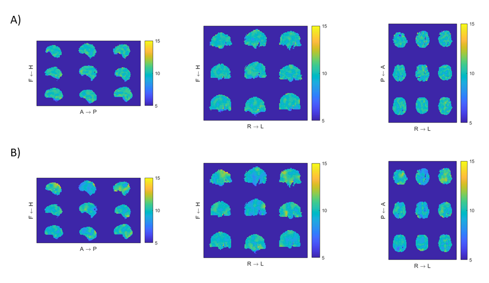
Figure 1 displays the B1 profile of the default phase-shim (or pseudo-CP mode) and the transmit efficiency of the coil array, defined as the sum of magnitudes across channels. Note the increased heterogeneity of the pseudo-CP mode (CV=std/mean=45%) as compared to the typical heterogeneity at 7T (~22%). However the homogeneity of the TX efficiency map (CV=25%), gives evidence that homogeneous excitations are possible using dynamic pTX.The RF pulse energy, specific energy dose (SED) and FA normalized RMS error (FA NRMSE) are shown in Table 1 and in Figure 2. We can observe that the prescribed SED limit (see Table 1) was always reached for the TPs, indicating that local SAR is a limiting factor for pulse performance. The TP (resp. UP) pulses allow achieving a FA-NRMSE value always better than 15 % (resp. 20%). The UPs enabled shorter excitation but did not reach the same level of homogeneity than the TPs.
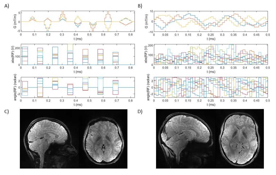
The 10° TP for subject 6 and the 10° UP are displayed in Figure 3, together with exemplary images obtained with these pulses. For the UP, the computation time took several hours on a DELL Precision 7820, making the GRAPE approach inappropriate to compute TPs.
The retrospective FA simulations are shown in Figure 4. After visual inspection of the anatomical images acquired using the TPs and UPs (see ISMRM abstract on the first in vivo brain images acquired at 11.7T), the obtained homogeneity was deemed sufficient to deliver the desired contrast across the whole brain.
Discussion and conclusions
This first pTX pulse design study performed at 11.7T confirms that the degree of heterogeneity of the pseudo-CP mode is significantly increased as compared to 7T. Unsurprisingly, the lower transmit efficiency, higher SAR12, and increased static field heterogeneity at 11.7T challenge our ability to produce uniform spin excitations. This makes dynamic pTX an essential tool to enable whole brain coverage imaging. With the current settings (~1.3 kW peak RF power available at the coil plug and a local SAR limit of 20 W/kg) we could obtain reasonably short RF pulses using GRAPE. For the 180° pulse, the FA-NRMSE was reasonablt good, but the obtained pulses (TP and UP) failed (FA < 90°) in a very localized region in the brain (ventral, left hemisphere). This problem, which remains to be investigated, is believed to mostly result from the RF coil architecture (dual-row, 16 resonators) which performs best on a 16-channel pTX system. But it also puts forward two important and challenging pulse design aspects at 11.7T, namely the quality of the input quantitative B1 maps and the non-convex nature of the pulse optimizations.Acknowledgements
AROMA H2020 FET-Open (885876). ANR-21-ESRE-0006 (“Investissements d'avenir"). Edouard Chazel is thanked for assembling the RF coil.References
- Boulant, N. et al. (2023) ‘Commissioning of the Iseult CEA 11.7 T whole-body MRI: current status, gradient–magnet interaction tests and first imaging experience’, Magnetic Resonance Materials in Physics, Biology and Medicine, 36(2), pp. 175–189. Available at: https://doi.org/10.1007/s10334-023-01063-5.
- Luong, M. et al. (2022) ‘A Compact 16Tx-32Rx Geometrically Decoupled Phased Array for 11.7T MRI’, In proceeeding of the 31st annual conference of ISMRM, UK, London.
- Katscher, U. et al. (2003) ‘Transmit SENSE’, Magn Reson Med, 49(1), pp. 144–150. Available at: https://doi.org/10.1002/mrm.10353.
- Zhu, Y. (2004) ‘Parallel excitation with an array of transmit coils’, Magnetic Resonance in Medicine, 51(4), pp. 775–784. Available at: http://onlinelibrary.wiley.com/doi/10.1002/mrm.20011/full.
- Grissom, W. et al. (2006) ‘Spatial domain method for the design of RF pulses in multicoil parallel excitation’, Magn Reson Med, 56(3), pp. 620–629. Available at: https://doi.org/10.1002/mrm.20978.
- Eichfelder, G. and Gebhardt, M. (2011) ‘Local specific absorption rate control for parallel transmission by virtual observation points’, Magn Reson Med, 66(5), pp. 1468–1476. Available at: https://doi.org/10.1002/mrm.22927.
- Gras V, Boulant N, Luong M, Morel L, Le Touz N, Adam JP, Joly JC. (2023) 'A mathematical analysis of clustering-free local SAR compression algorithms for MRI safety in parallel transmission', IEEE Trans Med Imaging. PP. doi: 10.1109/TMI.2023.3319017.
- Amadon, A. et al. (2012) ‘Validation of a very fast B1-mapping sequence for parallel transmission on a human brain at 7T’, In Proceedings of the 20th Annual Meeting of ISMRM, p. 3358.
- Cloos, M.A. et al. (2012) ‘kT-points: Short three-dimensional tailored RF pulses for flip-angle homogenization over an extended volume’, Magn Reson Med, 67(1), pp. 72–80. Available at: https://doi.org/10.1002/mrm.22978.
- Van Damme, L. et al. (2021) ‘Universal nonselective excitation and refocusing pulses with improved robustness to off‐resonance for Magnetic Resonance Imaging at 7 Tesla with parallel transmission’, Magnetic Resonance in Medicine, 85(2), pp. 678–693. Available at: https://doi.org/10.1002/mrm.28441.
- Massire, A. et al. (2022) ‘PASTEUR: Package of Anatomical Sequences Using Parallel Transmission Universal Pulses Now Available for MAGNETOM Terra’, MAGNETOM Flash (80) 1/2022.
- Hoult, D.I. and Phil, D. (2000) ‘Sensitivity and power deposition in a high-field imaging experiment’, Journal of magnetic resonance imaging: JMRI, 12(1), pp. 46–67.
Figures