3924
Feasibility of local diffusion encoding targeting cortical surface1Martinos Center for Biomedical Imaging, Charlestown, MA, United States, 2Harvard Medical School, Boston, MA, United States
Synopsis
Keywords: Hybrid & Novel Systems Technology, Diffusion/other diffusion imaging techniques, TMS-MRI
Motivation: Mapping of gray matter microstructure by diffusion MRI is limited by gradient strength, which sets the resolution limit toward small cells and axons.
Goal(s): To use a multi-channel 3-axis TMS coil array as a miniature gradient system and generate strong local gradients.
Approach: We ran simulations and ex-vivo experiments using the TMS coil as a strong local gradient for diffusion encoding.
Results: The measured gradient strength matched simulated values. The TMS-gradient system was used to estimate mean diffusivity in ex-vivo tissue. Simulations show it has the potential to generate even stronger gradient fields with higher current and more coil elements.
Impact: Innovations in TMS-MRI gradient design could significantly boost the achievable gradient strength for diffusion MRI in the living human brain, advancing neuroscientific research within a cost-effective design.
Introduction
Traditional diffusion MRI systems rely on whole body or head-only gradient coils. For decades, there has been a drive to achieve higher gradient strengths to probe more intricate brain tissue microstructure. The physical constraints of conventional gradient coil designs often limit this pursuit of higher gradient strength. As an alternative to traditional gradient coils, our recently developed multi-channel 3-axis Transcranial Magnetic Stimulation (TMS) coil array [1] could be used as a miniature gradient system because (i) it can be placed next to the scalp, which can provide a strong local gradient more efficiently. (ii) its low inductance enables a high slew rate, promising the ability to slew up without peripheral nerve stimulation (PNS) and eddy current distortion issues. This preliminary study explores the potential of using multi-channel 3-axis TMS coil elements to produce local diffusion gradients within moderate amounts of current. Our simulations and ex-vivo experiments show that multi-channel 3-axis TMS coil elements can generate independent gradient field bases. Combinations of gradient fields enable probing microstructural information (e.g., mean diffusivity) from ex-vivo tissue samples.Methods
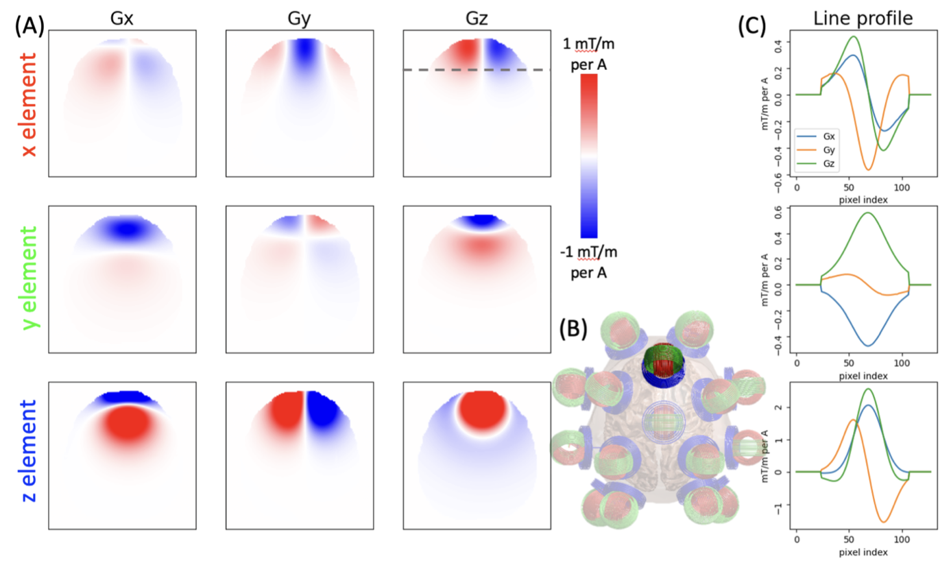
SimulationTo simulate basis gradient maps, We first calculated the magnetic field generated from each TMS coil element applied with 1A; then, we took the derivative of the magnetic field along the z direction to get Gx, Gy, and Gz. Suppose we were to use all 48 channels to achieve the desired gradient; the optimal combination of currents from each channel is determined by minimizing the root-mean-squared-error (RMSE) between target gradient maps and combinations of basis gradient maps. An L1 or L2 regularization was usually applied during the optimization.
Ex-vivo experiment setup
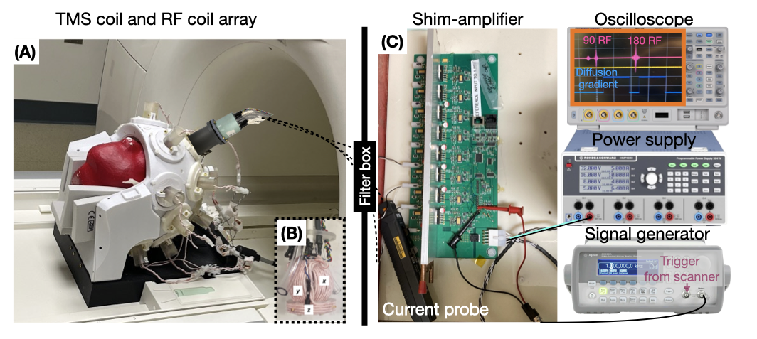
Ex-vivo brain tissue was scanned on a Siemens 3T MRI Skyra with a 28-channel RF coil array designed for concurrent TMS-MRI [2]. In the anterior-superior holder of the RF coil array, we placed a 3-axis TMS coil array. Currents running through coil elements are filtered through the filter box. Outside the room, the signal generator takes external triggers from the EPI pulse sequence and generates a pulse waveform for every trigger. The shim amplifier simultaneously follows the output from the signal generator and amplifies the current amplitude. We used a current probe to measure the current going through TMS coil elements.
Gradient map calibration
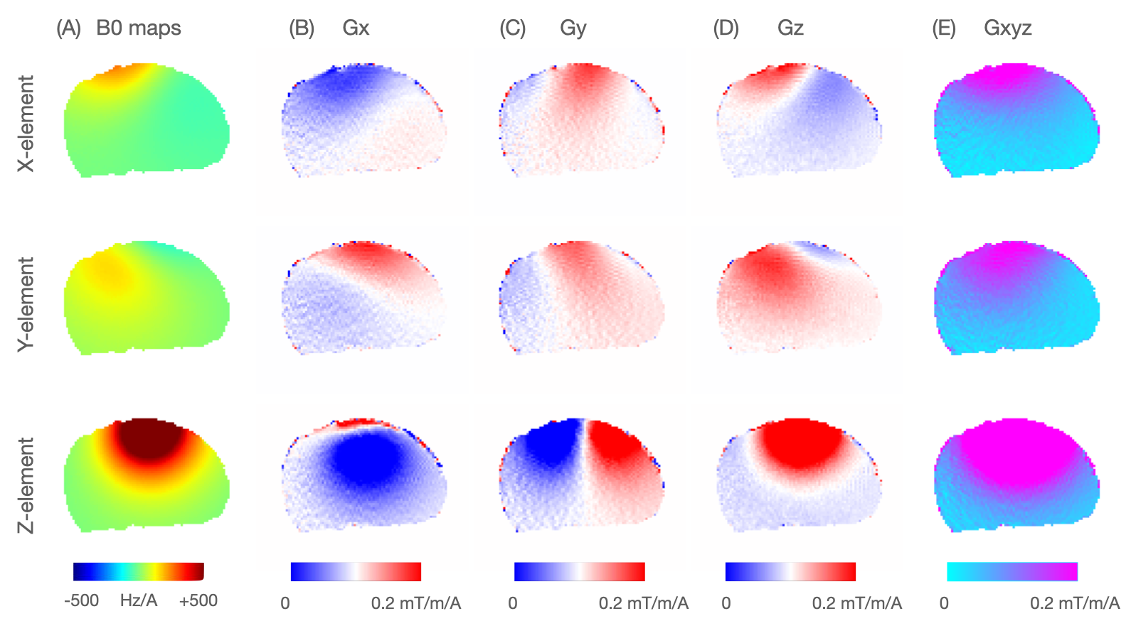
We applied positive and negative 500 mA Direct Current (DC) to each coil element (Ch1, Ch2, Ch3). Under the above six scenarios, B0 field maps were acquired using vendor-provided two echo gradient echo B0 mapping with delta TE of 2.46 ms. B0 maps (Hz/A) were generated by subtracting between the B0 maps from positive 500 mA and B0 maps from negative 500 mA. We then take the derivative on the measured B0 maps. Combined gradient strength was calculated by the sum of squares of Gx, Gy, and Gz (mT/m/A).
Diffusion encoding and model fitting
We applied the current of 9A to Ch1, Ch2, Ch3, Ch1+ Ch2, Ch1+Ch3, and Ch2+Ch3 as six diffusion directions. We used two pulse widths, 16 ms and 20 ms, and a Diffusion time of 24 ms to generate two shells. Because the gradient is not linear, we run voxel by voxel DTI fitting. Mean diffusivity (MD) maps were generated from an ex-vivo tissue sample.
Results
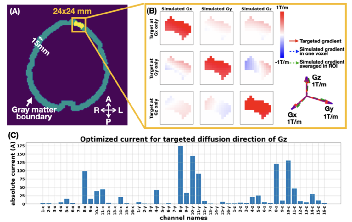
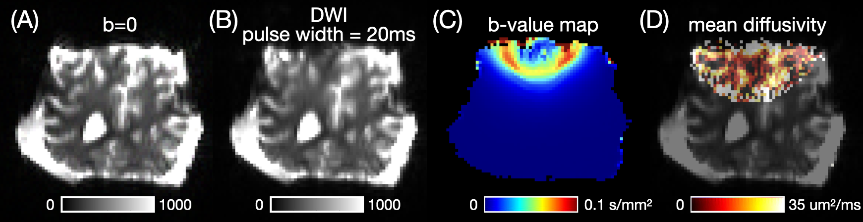
Fig.1 shows the simulated gradient maps (Gx, Gy, Gz) from three channels in one set of 3-axis TSM coil array. The gradient strength ranges around ±1 mT/m/A. Simulations in Fig.2 show that with 175 A of current limit per channel, we can achieve 1000 mT/m of linear gradient in an ROI. Fig.3 displays the setup of ex-vivo experiments, where the 3-axis TMS coil array is used as a diffusion encoding coil. Fig.4 displayed the B0 maps acquired on the scanner and corresponding gradient field maps. Note that the acquired B0 and gradient maps match the simulated gradient maps in Fig.1. Fig.5 showed a b=0 image and a diffusion-weighted image (b ~ 40 s/mm2), and the calculated mean diffusivity maps, where contrast at the pial surface is clearly shown.Discussion and Conclusion
Preliminary experiments demonstrated the feasibility of the concept. Because of the limits from the shim-amplifier and three coil elements available for diffusion encoding, the b-value we could get is quite low. Our simulation shows that with a current limit of 175A and 48 channel TMS coil elements, we could achieve a local diffusion gradient 2x the best current system (Gmax=500mT/m on Connectome 2.0). We believe this novel system will bring an even stronger gradients for diffusion-encoding while maintaining cost-effectiveness; it is expected to push the resolution limit of diffusion MRI and eventually advance neuroscientific studies.Acknowledgements
This work is supported by NIH grants: UG3EB034875, R01MH128421, P41EB030006References
[1] Navarro de Lara, LI et al. "A 3-axis coil design for multichannel TMS arrays." NeuroImage 224 (2021): 117355.APA
[2] Navarro de Lara, LI et al. "A Novel Whole-Head RF Coil Design Tailored for Concurrent Multichannel Brain Stimulation and Imaging at 3T." Brain Stimulation, 2023.
Figures