3921
Practical Utilization of Nonlinear Spatial Encoding: Fast Field Mapping and FRONSAC-wave1Department of Biomedical Engineering, Yale University, New Haven, CT, United States, 2Department of Radiology and Biomedical Imaging, Yale University, New Haven, CT, United States
Synopsis
Keywords: Signal Modeling, System Imperfections: Measurement & Correction, Nonlinear Encoding
Motivation: Nonlinear gradient imaging is impeded by time-consuming field mapping and the encoding ability is yet to be unleashed.
Goal(s): To accelerate nonlinear field mapping and make use of nonlinear encoding.
Approach: We report a fast and robust estimation method for nonlinear field mapping and studied the utility of simultaneously applying nonlinear and linear gradients, referred to as FRONSAC-wave, where the high-frequency nonlinear field waveform is complementary to a lower frequency linear field waveform.
Results: The fast field mapping decreases the field mapping time from 10 hours to <0.5 hour. FRONSAC-wave demonstrates better imaging ability compared to wave at various acquisition settings.
Impact: This study demonstrates the advantage of combined sinusoidal waveforms on linear and nonlinear gradients and proves the feasibility of fast mapping for nonlinear fields. It opens prospects for utilization of nonlinear field encoding in clinical scenarios.
Introduction
Nonlinear gradient channels were leveraged to complement the spatial sensitivity of receiving coils1–3. FRONSAC imaging4 took the approach of interpreting nonlinear gradients as a dynamic modulation in the sampling of k-space. Many emerging opportunities for nonlinear spatial encoding similar to FRONSAC, such as using shimming coil arrays5–8, which are suitable for high slew rates and modest amplitudes.The mechanism of FRONSAC has been described as voxel spreading effect for each voxel9, manifested as Point Spread Function (PSF). This effect of linear-gradient channels was extensively studied in wave imaging10. No previous studies, however, have tested whether FRONSAC nonlinear gradient encoding enhances the parallel imaging performance of wave.
One obstacle to implementing FRONSAC-wave is the time-consuming field mapping. A field camera11,12 does not require a separate scan or reproducibility of imperfections, but this hardware is not available at most sites. Single-slice projection data followed by extrapolation is used for trajectory calibration in wave10,13, but imperfections such as cross-term eddy currents lead to inaccuracies in FRONSAC field mapping.
Here we report a fast and robust estimation method for nonlinear field mapping and studied the utility of simultaneously applying nonlinear and linear gradients, referred to as FRONSAC-wave, where the high-frequency nonlinear field waveform is complementary to a lower frequency linear field waveform.
Theory
FRONSAC Signal ModelAn efficient FRONSAC signal model is to incorporate the concept of PSF, which creates spread-out effects only in the readout dimension. Without loss of generality, the discretized forward model in 2D is described as
$$d_{y,c} = Psf_yS_{y,c}m_y$$
$$Psf_y=iFT[exp^{i2\pi(P_y+k_{linearRO})}]$$
where $$$d_{y,c}$$$ is the $$$y$$$-th line of the spread-out image from the $$$c$$$-th coil; $$$PSF_y$$$ contains the PSF moderated by nonlinear gradient waveform $$$P_y$$$ and linear gradient $$$k_{linearRO}$$$; $$$S_{y,c}$$$ denotes the corresponding sensitivity map; and $$$m_y$$$ is the underlying image.
Nonlinear Field Map Estimation with Sparsity in Frequency
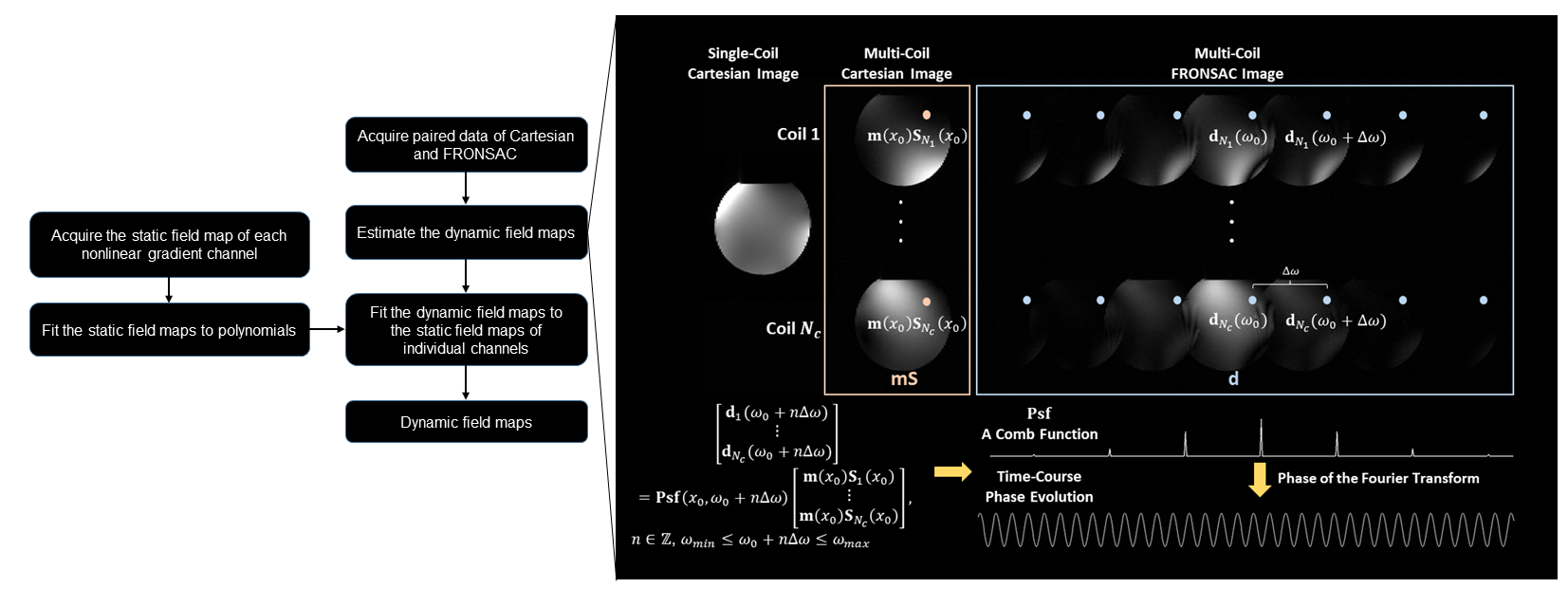
Because the prescribed waveform is a high frequency sinusoid, the PSF is generally sparse in the frequency domain. Estimating the phase and amplitude modulation on this comb function is expected to recover the most significant components. In practice, we extracted the corresponding voxel sets to solve for sparse PSF, shown in Fig. (1). It can also be well solved in the overlapping region by coil sensitivity encoding. This is then transformed into the time domain to yield the phase modulation.
FRONSAC-wave
The performance of wave is mainly determined by the wave amplitude14, which is restricted by the allowable slew rate and lower amplitudes to avoid artifacts caused by eddy-currents, off-resonance spin, or relaxation15.
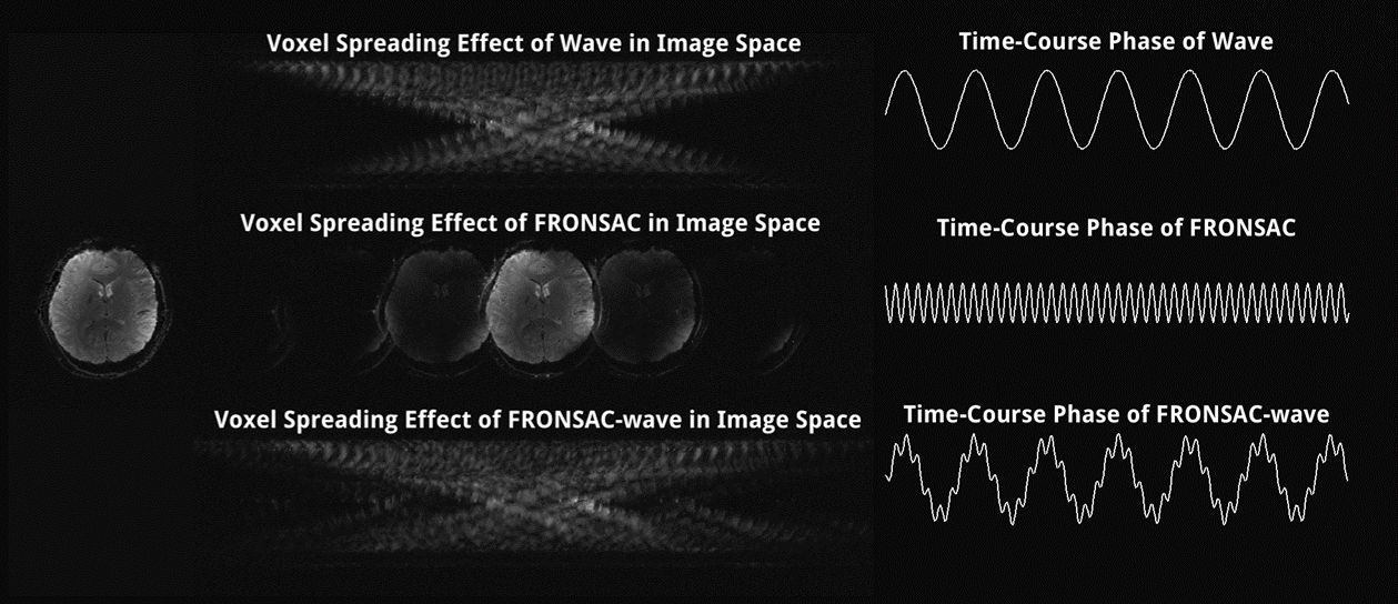
The spatially varying field of FRONSAC, however, creates varying orientations and extent of the local k-space sampling patch. The high-frequency nonlinear field modulation can offer broader and more flexible sampling without causing artifacts or exceeding the slew rate. The phase modulation is shown in Fig. 2.
Methods
The brain data of two healthy volunteers were acquired on a 3T Prisma scanner with a 32-channel RF coil array. The 2D protocol has the following parameters: resolution 1.4x1.4 mm2, FOV 250x250 mm2, TR/TE = 50/35ms, bandwidth = 70Hz/pixel, RO = RL, PE = AP, 6 oversampling rate. The 3D protocol differs on resolution: 1.4x1.4x1.5 mm3, and TR/TE = 24/12ms. We tested two sets of wave parameters, both with 15 cycles and amplitudes of 3mT/m and 5mT/m, respectively. 5mT/m was suggested to be optimal14, and 3mT/m mimicked a slew-constrained acquisition.The nonlinear field of FRONSAC was generated by an insert head gradient with three individual channels of harmonic fields: $$$x^3-3xy^2$$$ , $$$3x^2y-y^3$$$ and $$$x^2+y^2$$$. There are 96 cycles/readout in 2D and 112 cycles/readout in 3D.
Results and Discussion
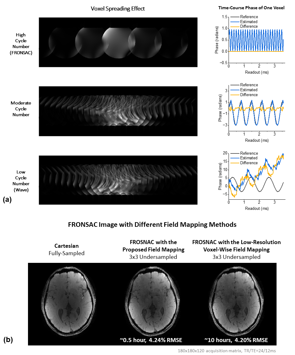
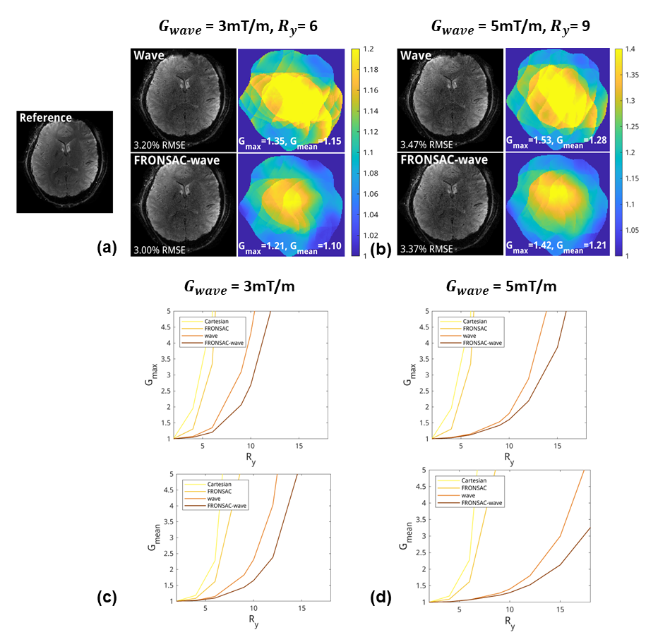
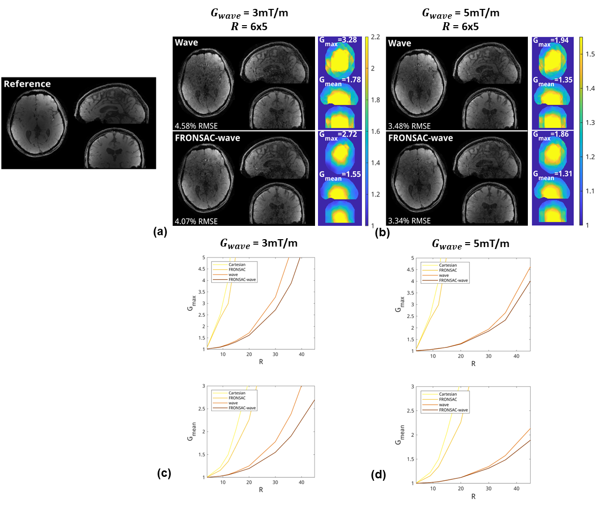
Fig. 3(a) is the condition for the proposed fast field mapping. Higher cycle leads to more sparse ghosts, facilitating the estimation of phase modulation. 3(b) shows much less time required for 3D FRONSAC field mapping with the proposed method (10 hours vs 0.5 hour).Fig. 4(a-b) show two scenarios of 2D imaging, where FRONSAC-wave has lower RMSE and lower g-factor maps compared to wave. (c-d) demonstrate FRONSAC-wave outperform wave, especially at high acceleration factors.
Fig. 5(a-b) show the version of 3D imaging with CAIPI undersampling. FRONSAC-wave also has better performance at high acceleration factors.
Conclusion
We proposed a fast field mapping method that exploits the sparsity of PSF in frequency and used it to study the additional encoding power made possible by combining FRONSAC and wave encoding.Acknowledgements
No acknowledgement found.References
1. Stockmann JP, Ciris PA, Galiana G, Tam L, Constable RT. O-Space Imaging: Highly Efficient Parallel Imaging Using Second-Order Nonlinear Fields as Encoding Gradients with No Phase Encoding. Magn Reson Med. 2010;64(2):447-456. doi:10.1002/mrm.22425
2. Lin FH. Multidimensionally encoded magnetic resonance imaging. Magn Reson Med. 2013;70(1):86-96. doi:10.1002/mrm.24443
3. Gallichan D, Cocosco CA, Dewdney A, et al. Simultaneously driven linear and nonlinear spatial encoding fields in MRI. Magn Reson Med. 2011;65(3):702-714. doi:10.1002/mrm.22672
4. Wang H, Tam LK, Constable RT, Galiana G. Fast rotary nonlinear spatial acquisition (FRONSAC) imaging. Magn Reson Med. 2016;75(3):1154-1165. doi:10.1002/mrm.25703
5. Scheffler K, Loktyushin A, Bause J, Aghaeifar A, Steffen T, Schölkopf B. Spread-spectrum magnetic resonance imaging. Magnetic Resonance in Medicine. 2019;82(3):877-885. doi:10.1002/mrm.27766
6. Xu J, Stockmann JP, Bilgic B, et al. Multi-frequency wave-encoding (mf-wave) on gradients and multi-coil shim-array hardware for highly accelerated acquisition. In: Proceedings of the 28th Annual Meeting of the International Society for Magnetic Resonance in Medicine.
7. Gao Y, Mareyam A, Sun Y, et al. A 16-channel AC/DC array coil for anesthetized monkey whole-brain imaging at 7T. NeuroImage. 2020;207:116396. doi:10.1016/j.neuroimage.2019.116396
8. Juchem C, Theilenberg S, Kumaragamage C, et al. Dynamic multicoil technique (DYNAMITE) MRI on human brain. Magnetic Resonance in Medicine. 2020;84(6):2953-2963. doi:10.1002/mrm.28323
9. Rodriguez Y, Elsaid NMH, Keil B, Galiana G. 3D FRONSAC with PSF reconstruction. J Magn Reson. 2023;355:107544. doi:10.1016/j.jmr.2023.107544
10. Bilgic B, Gagoski BA, Cauley SF, et al. Wave-CAIPI for highly accelerated 3D imaging. Magn Reson Med. 2015;73(6):2152-2162. doi:10.1002/mrm.25347
11. Versteeg E, Klomp DWJ, Siero JCW. Accelerating Brain Imaging Using a Silent Spatial Encoding Axis. Magnetic Resonance in Medicine. 2022;88(4):1785-1793. doi:10.1002/mrm.29350
12. Dispenza NL, Littin S, Zaitsev M, Constable RT, Galiana G. Clinical Potential of a New Approach to MRI Acceleration. Sci Rep. 2019;9(1):1912. doi:10.1038/s41598-018-36802-5
13. Duyn JH, Yang Y, Frank JA, van der Veen JW. Simple Correction Method fork-Space Trajectory Deviations in MRI. Journal of Magnetic Resonance. 1998;132(1):150-153. doi:10.1006/jmre.1998.1396
14. Wang H, Qiu Z, Su S, et al. Parameter optimization framework on wave gradients of Wave-CAIPI imaging. Magnetic Resonance in Medicine. 2020;83(5):1659-1672. doi:10.1002/mrm.28034
15. Polak D, Cauley S, Huang SY, et al. Highly-accelerated volumetric brain examination using optimized wave-CAIPI encoding. Journal of Magnetic Resonance Imaging. 2019;50(3):961-974. doi:10.1002/jmri.26678
Figures