3920
Slice-by-slice B0 Shimming for high-resolution diffusion MRI with an ultra-high performance head-only gradient1Science and Technology Office, GE HealthCare, Royal Oak, MI, United States, 2Technology and Innovation Center, GE HealthCare, Niskayuna, NY, United States, 3Uniformed Services University of the Health Sciences, Bethesda, MD, United States, 4Walter Reed National Military Medical Center, Bethesda, MD, United States, 5Brigham and Women’s Hospital, Boston, MA, United States, 6Mātai Medical Research Institutes, Gisborne, New Zealand, 7GE HealthCare, Melbourne, Australia, 8Stanford University, Stanford, CA, United States
Synopsis
Keywords: Diffusion Acquisition, Shims, Diffusion
Motivation: High-resolution diffusion MRI in high-gradient head-only systems, which is promising for advancing our understanding of human brain microstructure, is still prone to distortion from B0 field inhomogeneity.
Goal(s): To reduce B0 field inhomogeneity for improved image quality of high-resolution diffusion MRI.
Approach: Dynamic slice-by-slice B0 shimming of 0th and 1st order was implemented in diffusion MRI.
Results: Preliminary results showed reduced image shift and distortion of slices at different locations in axial b=0 s/mm2 echo planar image with 1-mm isotropic resolution with dynamic slice-dependent B0 shimming, demonstrating the potential of high image quality of high-resolution diffusion MRI for brain microstructure imaging.
Impact: Neuroimaging scientists and MR physicists, who use high-resolution diffusion MRI for studying brain circuits, connectivity, and microstructure, can benefit from improved image quality with reduced distortion from dynamic slice-by-slice B0 shimming technique in high-performance gradient MRI systems.
Introduction:
Emerging small-bore high-performance gradient MRI systems can simultaneously reach maximum gradient amplitudes of 300-500 mT/m and slew rates of 750-900 T/m/s1–3. The unprecedently high performance enables advanced diffusion encoding that provides image contrast sensitive and specific to brain tissue microstructure4. Furthermore, these systems also benefit image acquisition, including sub-millimeter diffusion MRI and spiral diffusion MRI, both of which play prominent roles for mapping brain connectivity and guiding interventional procedures5–7. Therefore, combining advanced diffusion MRI with high-resolution and/or spiral acquisition in high-performance gradient MRI systems may largely advance our understanding of human brain.Still, both high-resolution single-shot echo-planar imaging and spiral acquisition are more prone to distortion, image blurring, and signal loss from B0 field inhomogeneity. One way to mitigate the artifact is to use B0 shimming. However, conventional static volumetric shimming reduces B0 inhomogeneity for the whole brain, which is limited in its effectiveness for reducing local B0 inhomogeneity in different 2D image slices. Dynamic slice-by-slice shimming has been applied to improve shimming in diffusion MRI in whole-body MRI systems8–11. To further improve the image quality of diffusion MRI in high-performance gradient MRI, we proposed to apply dynamic slice-by-slice B0 shimming in a high-gradient 3T MRI system and preliminarily assess the image quality of diffusion MRI with 1-mm isotropic resolution.
Methods:
Shimming techniques: We compare three shimming techniques, including: (1) static shimming for the whole brain volume; (2) dynamic slice-by-slice adjustment of center frequency to reduce the mean ΔB0; (3) dynamic slice-by-slice shimming of center frequency and linear gradient to reduce the mean and linear ΔB0. High-order dynamic B0 shimming was not included as the time response of these shimming coils are not fast enough to support dynamic shimming.Image acquisition: Two healthy volunteers (male/female, 48/32 years old) were recruited and scanned under a local IRB-approved protocol. MRI scanning was performed in a 3.0T MAGNUS MRI system1 (GE Healthcare, USA) at maximum performance of 300 mT/m and 750 T/m/s, and a 32-channel phased-array receive RF coils (Nova Medical, USA). B0 field map was obtained and analyzed to generate shimming coefficients8 for each slice (Table 1). Axial, 1-mm isotropic, single-shot diffusion MRI (Table 1) were acquired in one subject with the static shimming and the dynamic shimming with both 0th and 1st order. T2 FSE anatomical images were also acquired. Same acquisitions were also obtained in the mini-ACR phantom.
Results:
Figures 2 and 3 show the representative B0 maps of two slices in the mini-ACR phantom and the human brains, using different shimming techniques. Static shimming (Figures 2-3A) failed to correct for the large B0 field inhomogeneity across slices. With slice-by-slice correction for center frequency (Figures 2-3B), the variation of mean ΔB0 across slices were reduced. However, the linear gradient field in each slice (Figures 2-3D) was not neglectable. Dynamic slice-by-slice shimming of 0th and 1st order reduced residual linear ΔB0 field in each slice (Figures 2-3C). The mean of ΔB0 in each slice was reduced to 0 Hz and the standard deviation was reduced from a maximum of 40.6/53.9/58.4 Hz to 13.3/16.8/21.6 Hz across all slices of the phantom/Subject#1/Subject#2, respectively (Figures 2-3E).Figures 4 and 5 show the representative b=0 s/mm2 images in the diffusion acquisitions using static shimming (Figures 4-5A) and dynamic slice-by-slice B0 shimming (Figures 4-5B), and T2 FSE anatomical images in the phantom and one healthy volunteer. Both image shift and distortion (pointed by the red arrows) were observed in the single-shot echo-planar images of different slices with static shimming. After applying dynamic slice-by-slice shimming of 0th and 1st order, both image shift and distortions were reduced in different slices.
Discussions and Conclusions:
This preliminary study demonstrated that dynamic slice-by-slice B0 shimming with 0th order and 1st order compensation can substantially reduce B0 field inhomogeneity for each slice, resulting in improved image quality of diffusion MRI with 1-mm isotropic resolution in high-performance gradient MRI systems. This shimming technique is accessible for all high-performance gradient systems as it does not require extra hardware, as it does not require extra gradient coil and driver which are needed for high-order B0 shimming. The limitation of this study is that the analysis of image shift and distortion was only performed in b=0 s/mm2 images as the structural feature is more prominent. The evaluation of the impact of dynamic slice-by-slice B0 shimming on diffusion-weighted images will be performed in the future. Upon successfully evaluation, this shimming technique will further improve the image quality and the accuracy of sub-millimeter diffusion-based microstructure imaging in both ex-vivo study and in-vivo study of human brain.Acknowledgements
The authors acknowledge the funding support from CDMRP W81XWH-16-2-0054, Massachusetts Life Science Foundation, and the National Institute of Biomedical Imaging and Bioengineering of the National Institutes of Health (NIH) under award Number U01EB034313. The content is solely the responsibility of the authors and does not necessarily represent the official views of the U.S. Department of Defense, Walter Reed National Military Medical Center, Uniformed Services University, or NIH.References
1. Foo TKF, Tan ET, Vermilyea ME, et al. Highly efficient head-only magnetic field insert gradient coil for achieving simultaneous high gradient amplitude and slew rate at 3.0T (MAGNUS) for brain microstructure imaging. Magnetic Resonance in Medicine. 2020;83(6):2356-2369. doi:10.1002/mrm.28087
2. Feinberg D, Dietz P, Liu C, et al. Design and Development of a Next-Generation 7T human brain scanner with high-performance gradient coil and dense RF arrays. In: ISMRM 2021. Accessed November 3, 2023. https://archive.ismrm.org/2021/0562.html
3. Huang SY, Witzel T, Keil B, et al. Connectome 2.0: Developing the next-generation ultra-high gradient strength human MRI scanner for bridging studies of the micro-, meso- and macro-connectome. Neuroimage. 2021;243:118530. doi:10.1016/j.neuroimage.2021.118530
4. Zhu A, Shih R, Huang RY, et al. Revealing tumor microstructure with oscillating diffusion encoding MRI in pre-surgical and post-treatment glioma patients. Magnetic Resonance in Medicine. 2023;90(5):1789-1801. doi:10.1002/mrm.29758
5. Steele CJ, Anwander A, Bazin PL, et al. Human Cerebellar Sub-millimeter Diffusion Imaging Reveals the Motor and Non-motor Topography of the Dentate Nucleus. Cereb Cortex. 2017;27(9):4537-4548. doi:10.1093/cercor/bhw258
6. Su JH, Choi EY, Tourdias T, et al. Improved Vim targeting for focused ultrasound ablation treatment of essential tremor: A probabilistic and patient‐specific approach. Hum Brain Mapp. 2020;41(17):4769-4788. doi:10.1002/hbm.25157
7. Tax C, Rudrapatna U, Mueller L, Jones D. In: Characterizing Diffusion of Myelin Water in the Living Human Brain Using Ultra-Strong Gradients and Spiral Readout. ; 2019:1115. Accessed November 2, 2023. https://cds.ismrm.org/protected/19MPresentations/abstracts/1115.html
8. Lee SK, Tan ET, Govenkar A, Hancu I. Dynamic slice-dependent shim and center frequency update in 3 T breast diffusion weighted imaging. Magn Reson Med. 2014;71(5):1813-1818. doi:10.1002/mrm.24824
9. Stocker D, Manoliu A, Becker AS, et al. Image Quality and Geometric Distortion of Modern Diffusion-Weighted Imaging Sequences in Magnetic Resonance Imaging of the Prostate. Invest Radiol. 2018;53(4):200-206. doi:10.1097/RLI.0000000000000429
10. McElroy S, Winfield JM, Westerland O, et al. Integrated slice-specific dynamic shimming for whole-body diffusion-weighted MR imaging at 1.5 T. MAGMA. 2021;34(4):513-521. doi:10.1007/s10334-020-00898-6
11. Gatidis S, Graf H, Weiß J, et al. Diffusion-weighted echo planar MR imaging of the neck at 3 T using integrated shimming: comparison of MR sequence techniques for reducing artifacts caused by magnetic-field inhomogeneities. MAGMA. 2017;30(1):57-63. doi:10.1007/s10334-016-0582-z
Figures
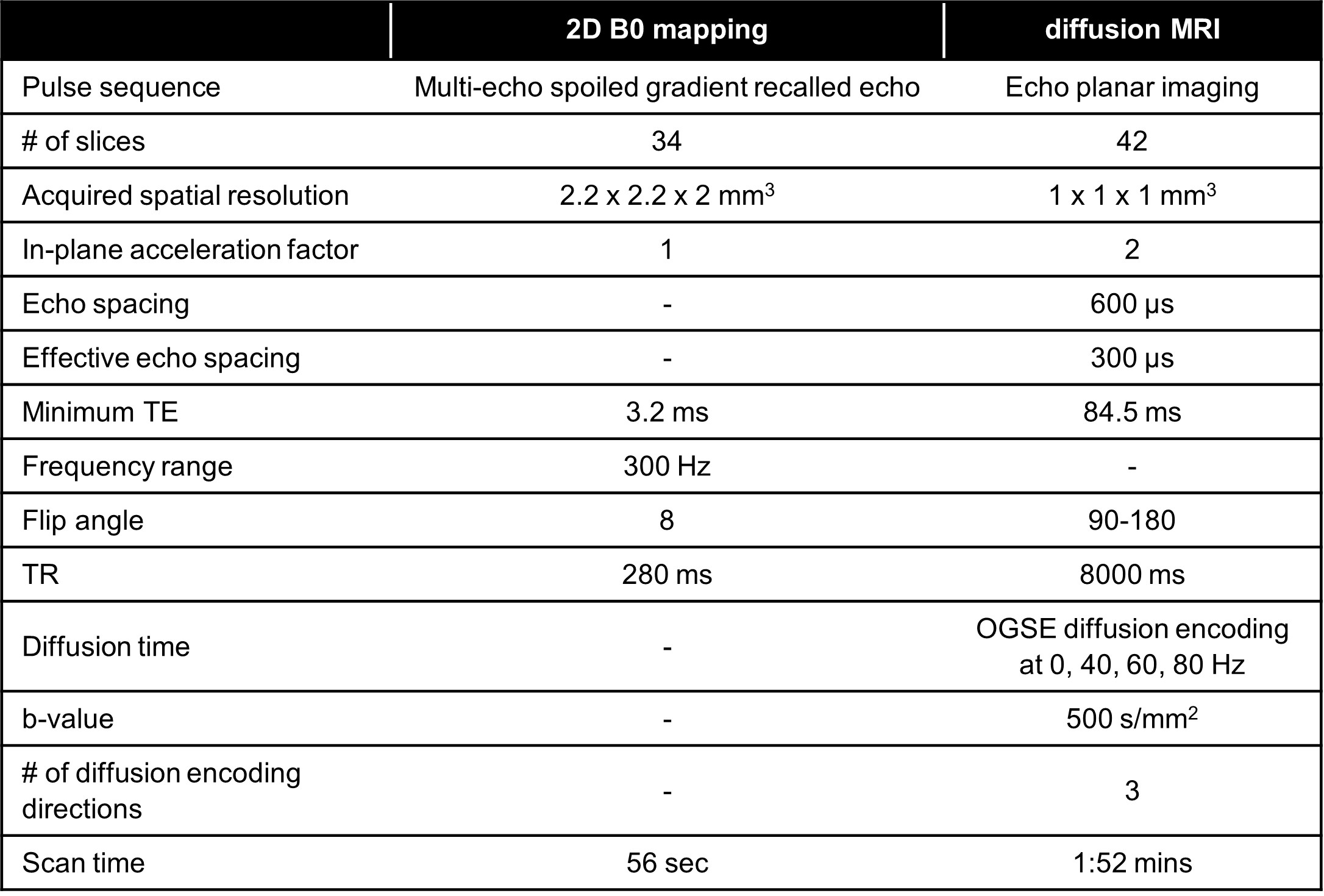
Table 1. Imaging parameters of B0 field mapping and diffusion MRI acquisitions in MAGNUS 3T. OGSE: oscillating gradient spin echo.
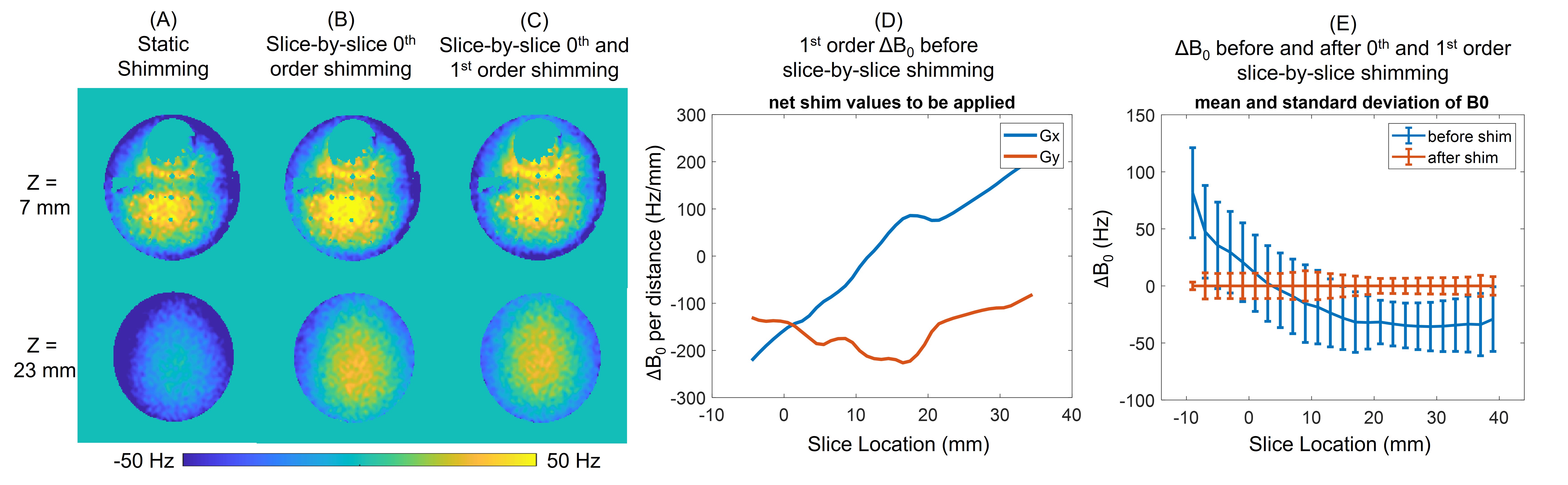
Figure 1. B0 field maps of two representative slices in different slice locations using (A) static shimming for the entire volume; (B) dynamic slice-by-slice 0th order B0 shimming by adjusting center frequency for each slice; (C) dynamic slice-by-slice 0th and 1st order B0 shimming. Slice-dependent in-plane linear gradient field were observed in both X direction and Y direction (D). Mean of ΔB0 was reduced to 0 Hz. The maximum standard deviation across all slices was reduced from 40.6 Hz to 13.3 Hz by applying dynamic shimming (E).
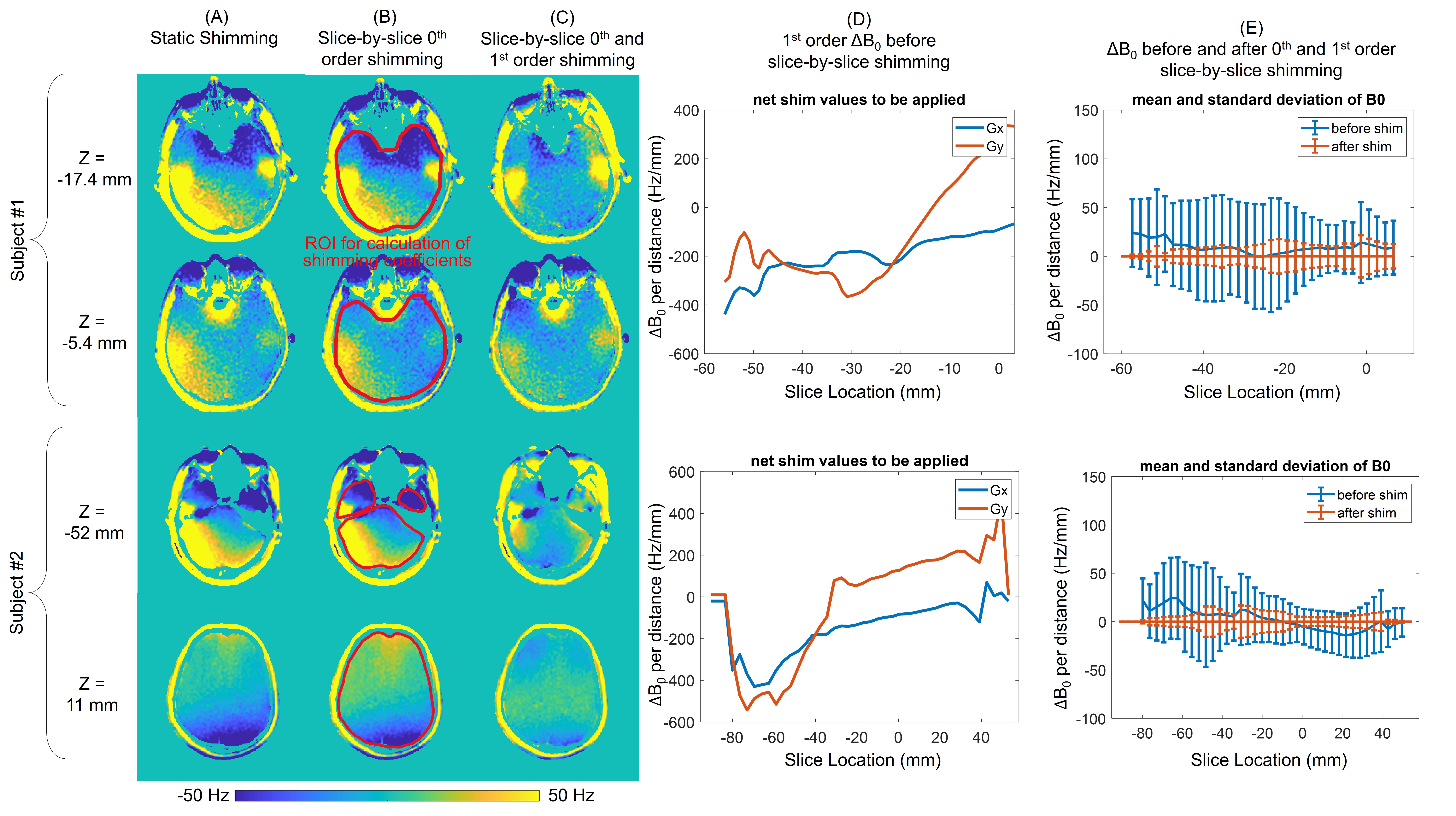
Figure 2. B0 field maps of two representative slices in the two subjects using (A) static shimming for the entire volume; (B) dynamic slice-by-slice 0th order B0 shimming by adjusting center frequency for each slice; (C) dynamic slice-by-slice 0th and 1st order B0 shimming. Slice-dependent in-plane linear gradient field were observed in both X and Y direction (D). Mean of ΔB0 was reduced to 0 Hz. The maximum standard deviations across all slices were reduced from 53.9 Hz (Subject #1) and 58.4 Hz (Subject #2) to 16.8 Hz (Subject #1) and 21.6Hz (Subject #2) by applying dynamic shimming (E).
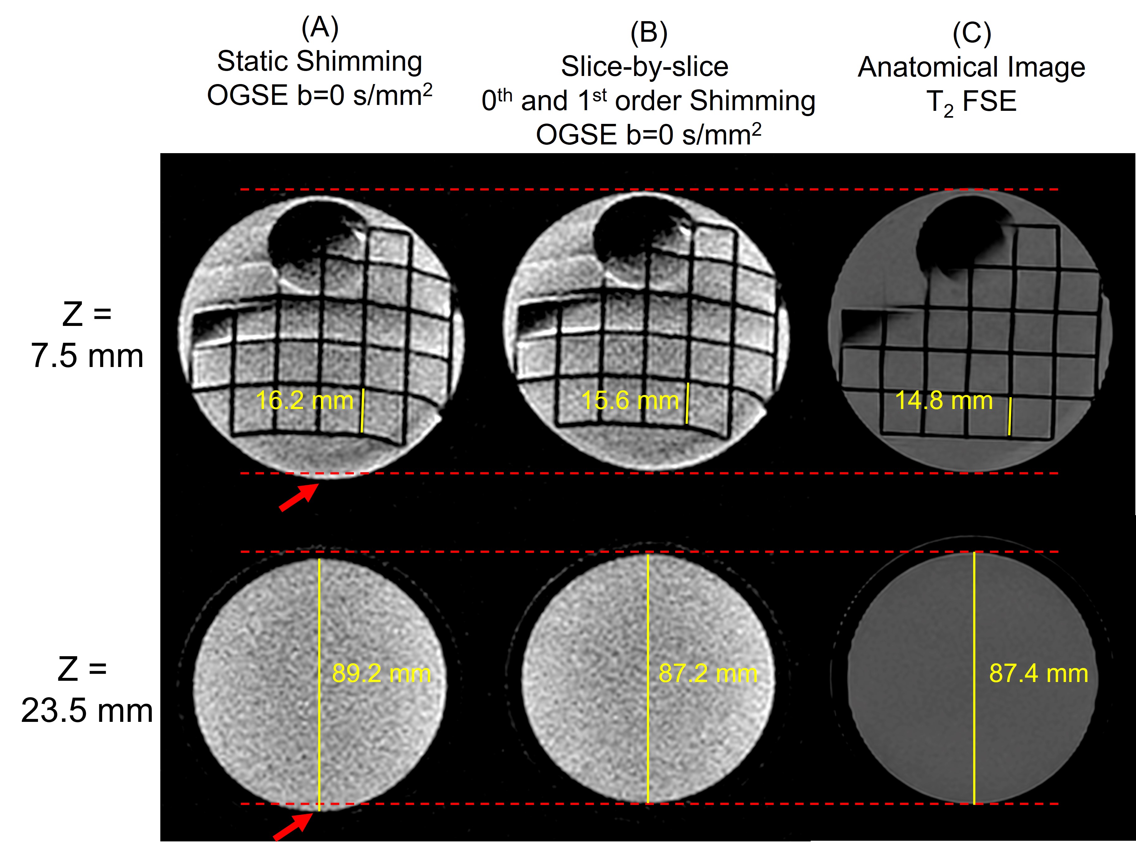
Figure 3. Two representative slices of 1-mm isotropic resolution b=0 s/mm2 images in the diffusion acquisitions of the mini-ACR phantom, using (A) static shimming for the entire volume and (B) dynamic slice-by-slice B0 shimming of 0th order (center frequency) and 1st order gradient. T2 FSE anatomical images (C) are shown as reference. Dynamic slice-by-slice B0 shimming reduces both image shift (pointed by red arrow) and distortion (length measurement as pointed by the yellow lines) in both slices.
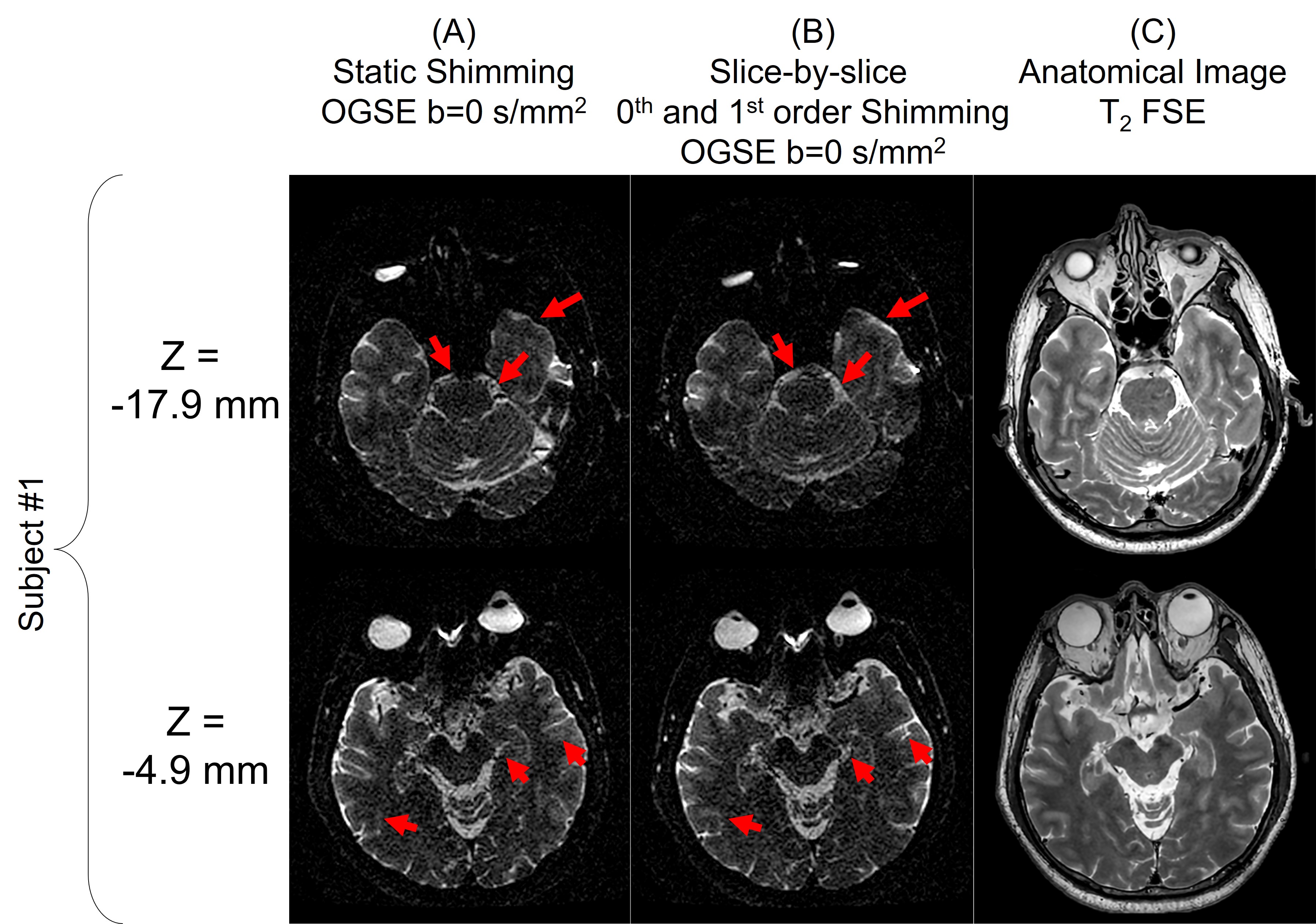
Figure 4. Two representative slices of 1-mm isotropic resolution b=0 s/mm2 images in the diffusion acquisitions of a healthy subject’s brain, using (A) static shimming for the entire volume and (B) dynamic slice-by-slice B0 shimming of 0th order (center frequency) and 1st order gradient. T2 FSE anatomical images (C) are shown as reference. Dynamic slice-by-slice B0 shimming reduces image distortions in both slices (pointed by red arrows).