3918
Dynamic shimming of the spinal cord using a 15-channel AC/DC coil1NeuroPoly Lab, Institute of Biomedical Engineering, Polytechnique Montreal, Montréal, QC, Canada, 2Centre de recherche du CHU Sainte-Justine, Université de Montréal, Montréal, QC, Canada, 3Athinoula A. Martinos Center for Biomedical Imaging, Massachusetts General Hospital, Boston, MA, United States, 4Harvard Medical School, Boston, MA, United States, 5Functional Neuroimaging Unit, CRIUGM, Université de Montréal, Montréal, QC, Canada, 6Mila - Quebec AI Institute, Montréal, QC, Canada
Synopsis
Keywords: Shims, Artifacts, Acquisition Methods, Hybrid & Novel Systems Technology, New Devices, Software Tools, Spinal Cord
Motivation: The static magnetic field around the spinal cord has complex and small-scale non-uniformities, making it difficult to shim with low-order spherical harmonics.
Goal(s): Assess the impact of slice-wise shimming in the cervico-thoracic spinal cord using a 15-channel AC/DC coil.
Approach: Measure the magnetic field and acquire echo-planar images using three different shimming scenarios involving volume-wise 0-2nd order spherical harmonics, volume-wise multi-coil shimming, and slice-wise multi-coil shimming.
Results: Multi-coil shimming decreased magnetic field inhomogeneities by 22% compared to 0-2nd order spherical harmonics when used across the whole imaging volume, and by 36% when used on a slice-wise basis.
Impact: Advanced shimming (using multi-coils) significantly improves the quality of spinal cord MRI images by mitigating susceptibility artifacts. This leap forward facilitates the clinical adoption of challenging imaging techniques like functional MRI in the spinal cord.
Introduction
The spinal cord is one of the most difficult anatomical regions to image. In addition to breathing and cardiac-related motions, this region features numerous small-spatial scale B0 non-uniformities1. Since these inhomogeneities cannot be corrected on a volume basis using the 0-2nd order spherical harmonic (SH) shims available on clinical 3-T scanners, researchers have resorted to state-of-the-art sequences to update the scanner’s gradients on a slice-by-slice basis2–4 and/or using AC/DC coils5. In this study, we characterize various shimming scenarios for spinal cord imaging that combine volume-wise 2nd order shims, a 15-channel AC/DC coil, and slice-wise (dynamic) shimming.Methods
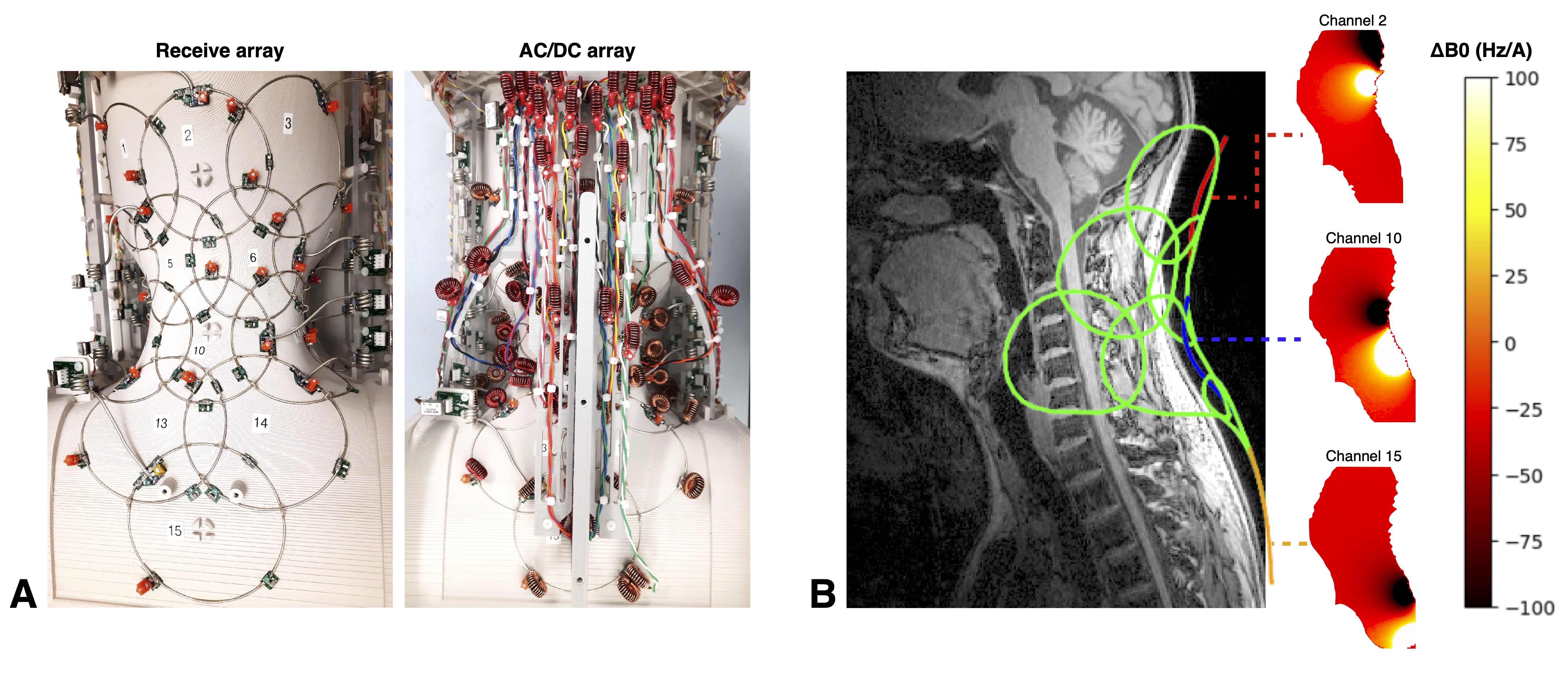
15-channel coilWe built a 15-channel AC/DC 3-T coil that covers the lower brain to the upper thoracic cord (see Figure 1). This coil interfaces with the rev-C version of the shim amplifiers developed by Stockmann et al.6 Currents were capped at ±2.5A per channel, with a combined maximum total current of 25A.
In-vivo procedure
Three male adult subjects, aged 25, 26 and 31, were scanned on a 3-T MRI scanner using the following workflow:
- Connect a laptop to the MRI console for convenient data transfer.
- Acquire a T1-weighted anatomical scan from head to shoulders (MPRAGE, 1mm isotropic, R=3 acceleration, 3:40min)
- Automatically generate a mask of the spinal cord using Spinal Cord Toolbox7, and dilate the mask by 60mm.
- Acquire a “baseline” EPI volume, whose slice prescriptions are used to optimize shimming in the subsequent “real” scans. (i) Manually shim using the scanner’s default shimming protocol over the cervical/upper thoracic spinal cord. (ii) Run a single-shot gradient-echo EPI (axial orientation, TR=1500 ms, TE=45 ms, BW=965 Hz/pixel, FA=90°, resolution=1x1x5mm).
- Acquire a baseline ΔB0 field map. (i) Use the same shim parameters as in (4). (ii) Run a multi-slice dual-echo GRE scan (TR=700 ms, TE=[2.46, 4.92] ms, BW=1500 Hz/pixel, FA=65°, resolution=3.3x3.3x3mm).
- Compute shim coefficients using Shimming Toolbox8 for three different optimizations: volume-wise 0-2nd order SH, volume-wise 0-2nd order SH with multi-coil, and volume-wise 0-2nd order SH with slice-specific multi-coil optimization. The coefficients were fine-tuned to minimize the mean squared error across the entire volume of interest (VOI, cervical/upper thoracic spinal cord).
- Acquire an EPI and a ΔB0 map for every shim scenario. (i) Set the calculated coefficients. (ii) Run an EPI (same acquisition parameters as before). (iii) Run a dual-echo GRE (same acquisition parameters as before).
Results & Discussion
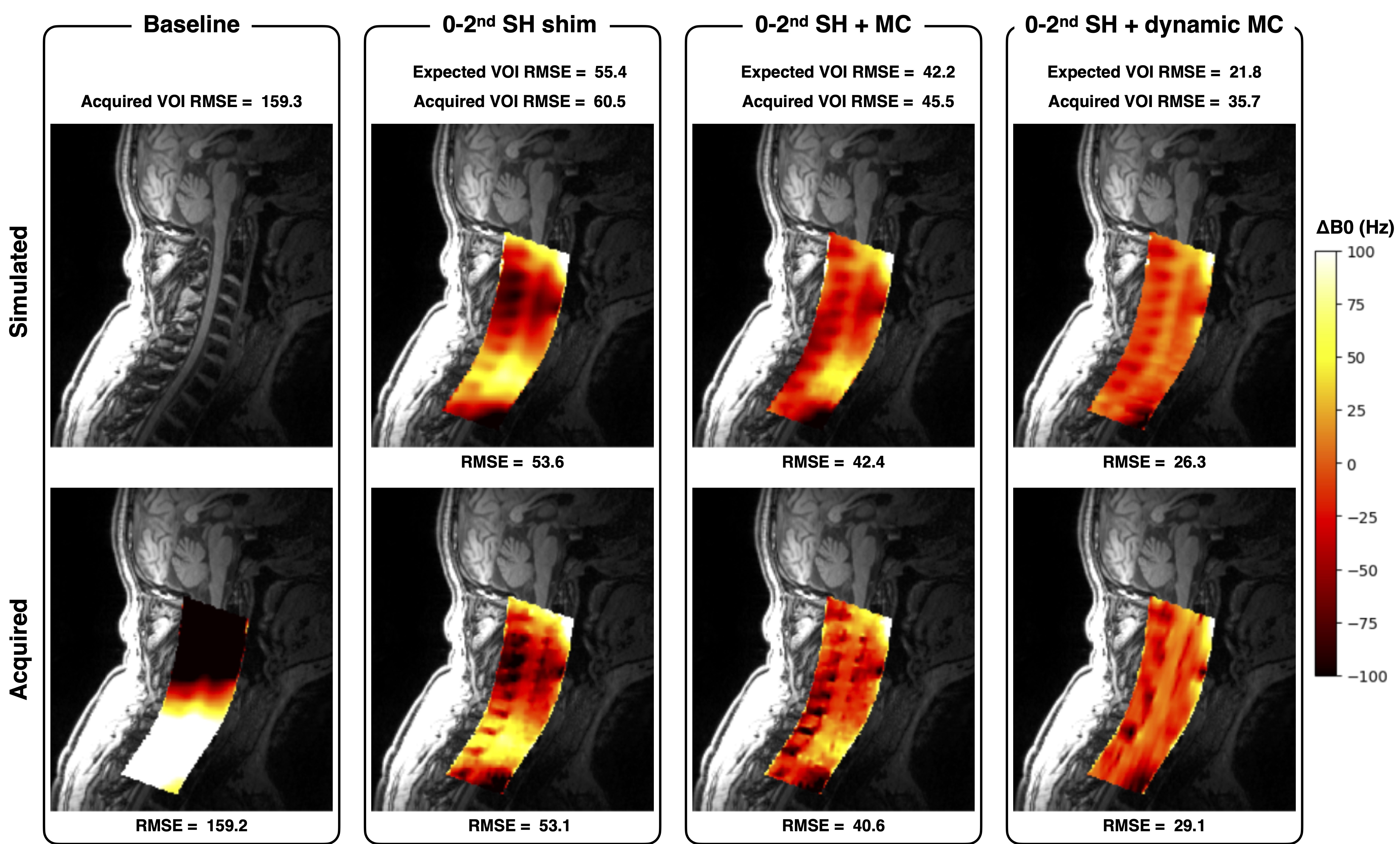
ΔB0 field mapIn Figure 2, the simulated ΔB0 field map generated by Shimming Toolbox (volume-wise shim) closely matches the acquired field map. In dynamic shimming, Shimming Toolbox replicates the field pattern well, except for the highly negative field regions (depicted as black patches) near the vertebrae, leading to an increased Root Mean Square Error (RMSE) within the VOI.
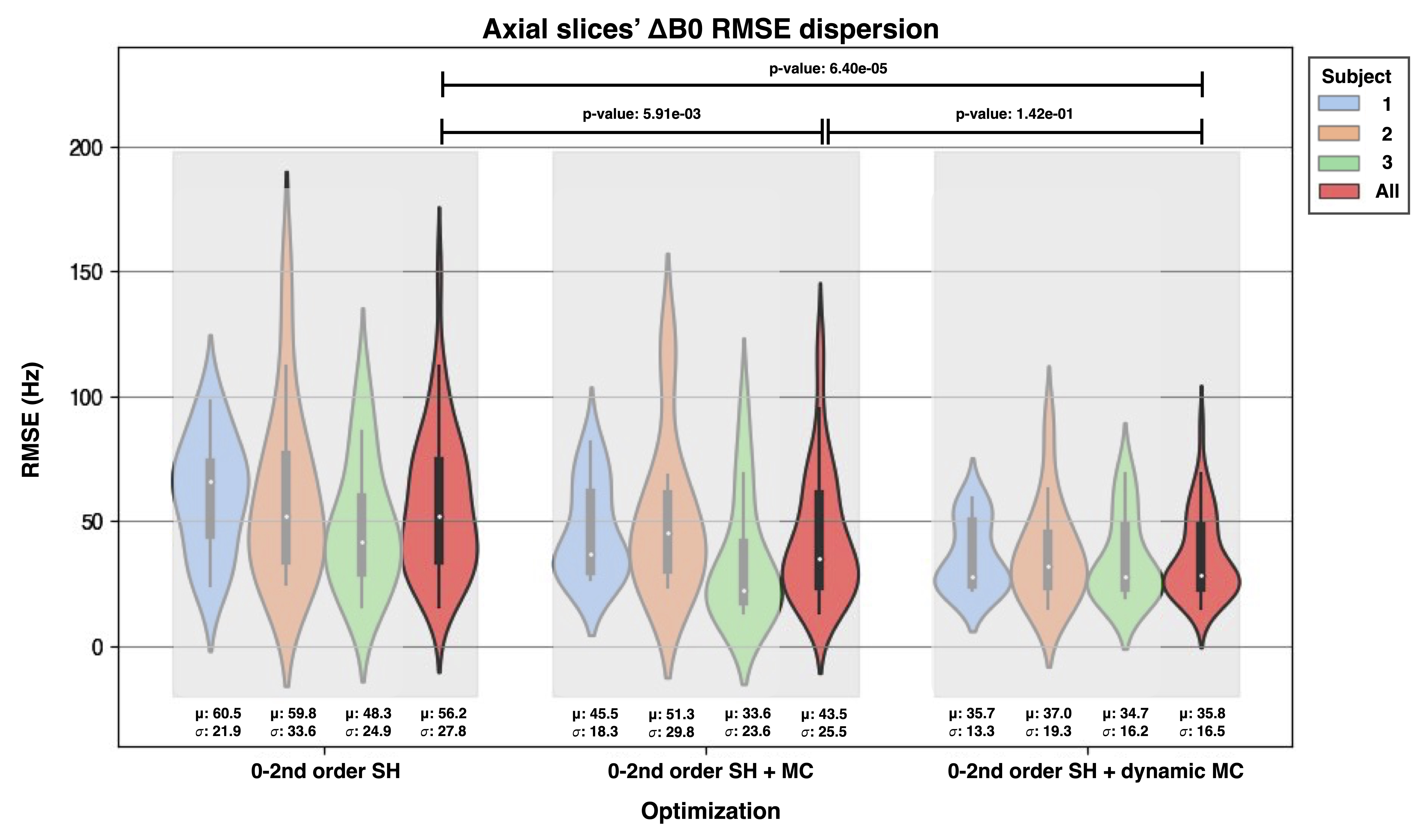
Figure 3 summarizes the acquired ΔB0 field results for all three subjects. The inclusion of multi-coil (both volume-wise and slice-wise) in conjunction with the scanner's 0-2nd order SH demonstrates a significant reduction of the mean ΔB0 offset, as indicated by a unilateral Mann-Whitney test (p < 0.05). However, utilizing multi-coil with slice-wise optimization does not yield a statistically significant improvement (p-value: 0.142) compared to its use in volume-wise optimization.
Overall, when compared to volume-wise 0-2nd order SH optimization, the incorporation of our multi-coil in a volume-wise optimization strategy results in a 22% reduction in VOI RMSE. Optimizing our multi-coil on a slice-by-slice basis provides a further 14% improvement in performance across the three subjects.
EPI
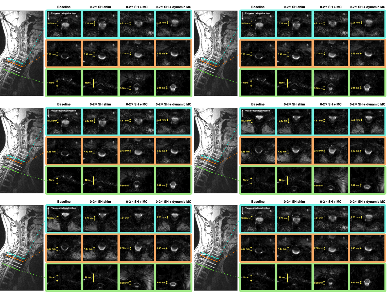
Figure 4 shows an animation illustrating three representative EPI slices acquired from subject 3 in the Anterior-Posterior (AP) and Posterior-Anterior (PA) encoding directions. The application of multi-coil shimming demonstrated its efficacy in recovering spinal cord signals in the lower slices and in reducing the separation between the centers of the spinal cord in AP and PA phase encoding.
Across all slices, the four different optimization approaches depicted in Figure 4 resulted in average spinal cord displacements of 4.4, 5.1, 3.0, and 2.6 mm respectively between AP and PA, representing a reduction of pixel shift by up to 41%. Dynamic shimming proved to be particularly effective in the thoracic region.
Conclusions
Dynamic slice-wise shimming using our AC/DC coil achieves a 36% reduction in B0 inhomogeneity compared to volume-wise 0-2nd order SH shimming. This translates to better quality EPI scans along the cervico-thoracic spinal cord, paving the way to more robust functional imaging.Acknowledgements
We thank Julien Thouveny and Mathieu Boudreau for their insight on the abstract.Funded by the Canada Research Chair in Quantitative Magnetic Resonance Imaging [CRC-2020-00179], the Canadian Institute of Health Research [PJT-190258], the Canada Foundation for Innovation [32454, 34824], the Fonds de Recherche du Québec - Santé [322736, 324636], the Fonds de Recherche du Québec - Nature et technologies [329439], the Natural Sciences and Engineering Research Council of Canada [RGPIN-2019-07244], the Canada First Research Excellence Fund (IVADO and TransMedTech), the Courtois NeuroMod project, the Quebec BioImaging Network [5886, 35450], INSPIRED (Spinal Research, UK; Wings for Life, Austria; Craig H. Neilsen Foundation, USA), Mila - Tech Transfer Funding Program.
References
1. Martin AR, Aleksanderek I, Cohen-Adad J, et al. Translating state-of-the-art spinal cord MRI techniques to clinical use: A systematic review of clinical studies utilizing DTI, MT, MWF, MRS, and fMRI. NeuroImage Clin. 2016;10:192-238. doi:10.1016/j.nicl.2015.11.019
2. Alonso-Ortiz E, Papp D, D’Astous A, Cohen-Adad J. Dynamic shimming in the cervical spinal cord for multi-echo gradient-echo imaging at 3 T. Neuroimage Rep. 2023;3(1):100150. doi:10.1016/j.ynirp.2022.100150
3. Finsterbusch J, Sprenger C, Büchel C. Combined T2*-weighted measurements of the human brain and cervical spinal cord with a dynamic shim update. NeuroImage. 2013;79:153-161. doi:10.1016/j.neuroimage.2013.04.021
4. Islam H, Law CSW, Weber KA, Mackey SC, Glover GH. Dynamic per slice shimming for simultaneous brain and spinal cord fMRI. Magn Reson Med. 2019;81(2):825-838. doi:10.1002/mrm.27388
5. Cuthbertson JD, Truong TK, Stormont R, Robb F, Song AW, Darnell D. An iPRES-W Coil Array for Simultaneous Imaging and Wireless Localized B0 Shimming of the Cervical Spinal Cord. Magn Reson Med. 2022;88(2):1002-1014. doi:10.1002/mrm.29257
6. Stockmann JP, Arango NS, Witzel T, et al. A 31-channel integrated “AC/DC” B0 shim and radiofrequency receive array coil for improved 7T MRI. Magn Reson Med. 2022;87(2):1074-1092. doi:10.1002/mrm.29022
7. De Leener B, Lévy S, Dupont SM, et al. SCT: Spinal Cord Toolbox, an open-source software for processing spinal cord MRI data. NeuroImage. 2017;145(Pt A):24-43. doi:10.1016/j.neuroimage.2016.10.009
8. D’Astous A, Cereza G, Papp D, et al. Shimming toolbox: An open-source software toolbox for B0 and B1 shimming in MRI. Magn Reson Med. 2023;89(4):1401-1417. doi:10.1002/mrm.29528
Figures