3916
Improved characterization of sequence-specific peripheral nerve stimulation (PNS) thresholds for rapid on-scanner monitoring1Martinos Center, Massachusetts General Hospital, Charlestown, MA, United States, 2Harvard Medical School, Boston, MA, United States, 3Harvard Graduate Program in Biophysics, Harvard University, Cambridge, MA, United States, 4Harvard-MIT Division of Health Sciences and Technology, Boston, MA, United States, 5MIT Department of Electrical Engineering and Computer Science, Cambridge, MA, United States
Synopsis
Keywords: Bioeffects & Magnetic Fields, Safety, Peripheral Nerve Stimulation (PNS), gradient coils, fast imaging
Motivation: The on-scanner PNS monitor must estimate each sequence’s stimulation potential. The current SAFE model is overly conservative, unnecessarily restricting gradient performance by up to 1.8X.
Goal(s): Develop a PNS monitor model that rapidly and accurately characterizes a sequence’s PNS thresholds.
Approach: We propose a model (SAFE2) closely inspired by the mechanisms of PNS to capture critical aspects such as E-field cancelations from different gradient axes and extend the training data using detailed PNS modeling reflecting a more diverse set of waveforms.
Results: SAFE2 improves PNS-prediction accuracy by 2X compared to SAFE, boosting usable image encoding performance by up to 30% without hardware changes.
Impact: PNS restricts the usable protocol parameters of EPI, bSSFP, Radial-GRE, etc., yielding suboptimal imaging performance. The current PNS monitoring approach (SAFE) is very conservative, therefore, we propose an improved model yielding up to 30% gradient performance boost without hardware modification.
Introduction
The International Electrotechnical Commission (IEC) sets limits on gradient coil operation in humans to prevent uncomfortable sensations from Peripheral Nerve Stimulations (PNS) [4]. Adhering to the IEC PNS limits requires a PNS monitor model on the scanner that can quickly and reliably estimate the sequence waveform’s PNS thresholds. This is challenging since PNS limits depend on multiple aspects of the imaging sequence, including ramp durations, gradient axes combinations [16,13,1,2,15] and the scan landmark [3, 11,12], and experimental data is only available for a small fraction of this parameter space. The commonly used SAFE model (Safety Assessment by Filtering and Evaluation) [14] was developed more than 20 years ago and was designed purposefully to be conservative due to the lack of detailed modeling tools and the limited experimental data available to train the model. We develop a new model, SAFE2, inspired by the neural dynamics behind PNS to more accurately characterize thresholds boosting usable performance by up to 30%. We propose to train the model using detailed validated PNS predictions [7,8,9,6,10].Methods
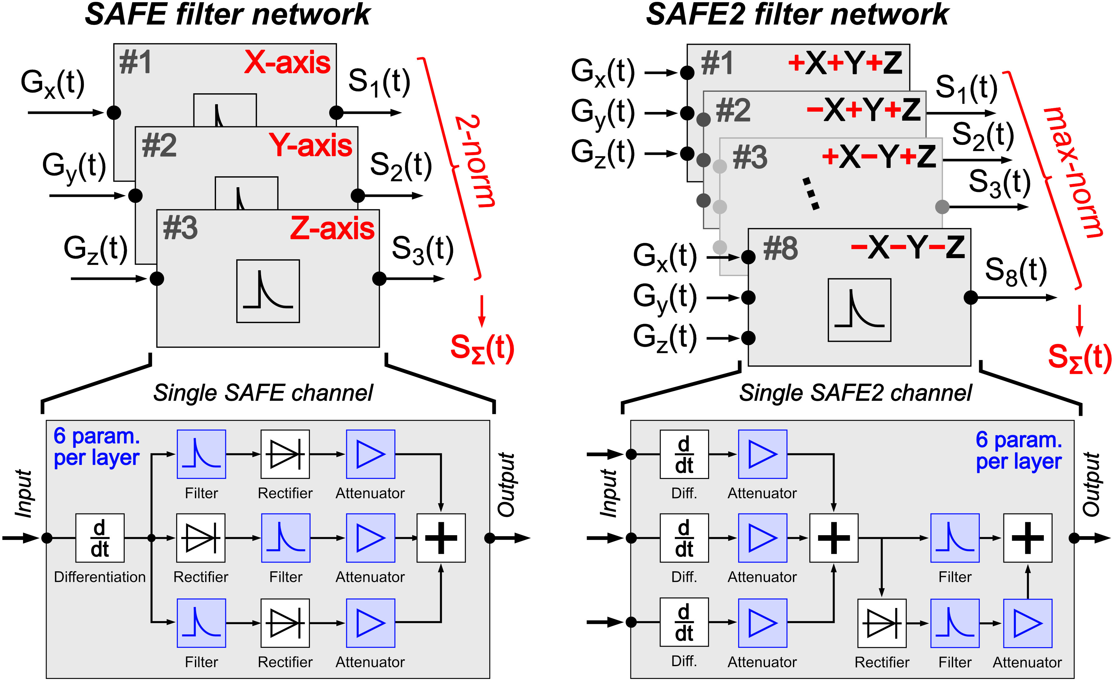
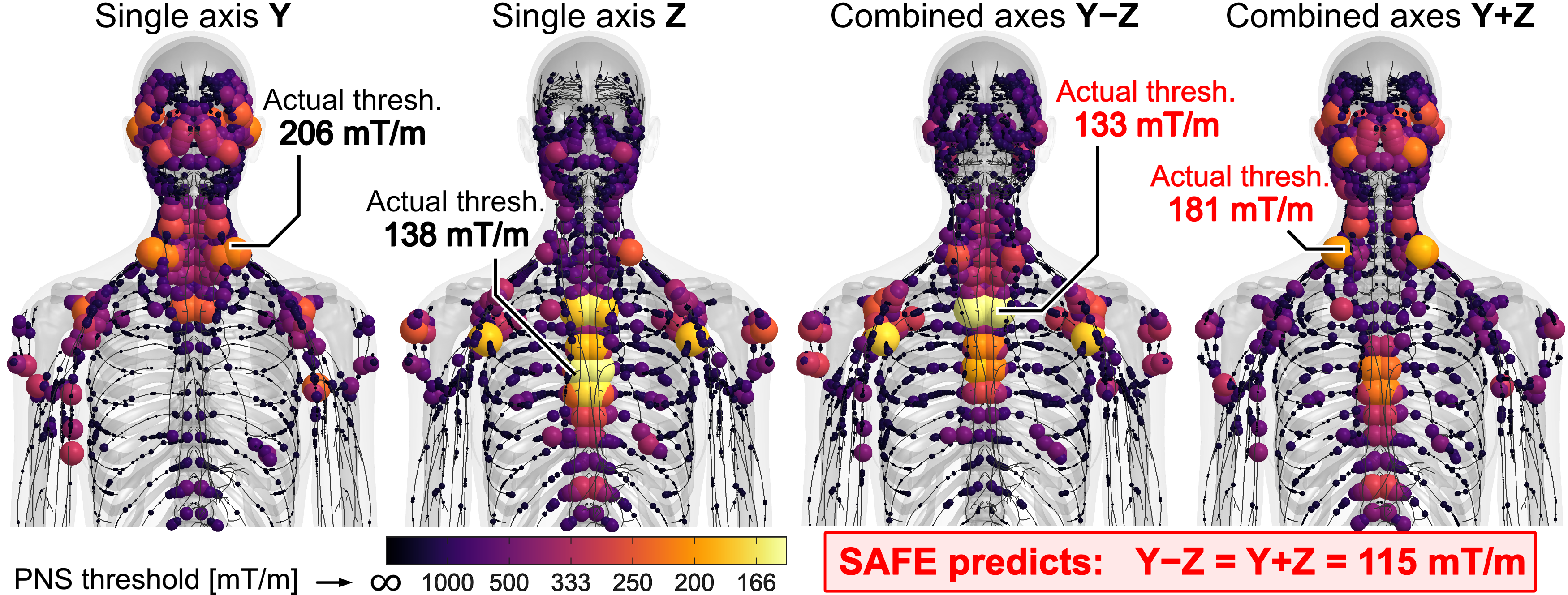
SAFE model: The traditional SAFE model [14] consists of three filter lines (one per gradient coil axis) with a total of 18 parameters (Fig. 1, left). The parameters are calibrated by fitting the model to a set of experimental PNS thresholds spanning different waveforms and axes combinations. Each filter computes a positive unsigned metric $$$S(t)$$$ based on the input gradient waveform $$$G(t)$$$ which is inversely proportional to the PNS threshold, i.e., stimulation occurs for $$$S(t)\geq1$$$. The stimulation metric for combined axes operation ($$$S_\Sigma$$$) is obtained via root sum-of-squares of the single axis metrics. Critically, the use of unsigned metrics does not allow for stimulation contributions (E-fields, nerve membrane charges) of different gradient axes to cancel. Figure 2 shows that, in reality, gradient axes can superimpose destructively, for example, in the Siemens Impulse head gradient [5].SAFE2 model: Our SAFE2 model improves PNS characterization in five important aspects (Fig. 1, right): 1), the PNS propensity is modeled by a signed metric. 2), each filter channel processes all gradient axes simultaneously, allowing PNS contributions to superimpose constructively and destructively. 3), we expand the number of filter channels to 8, with each channel representing a different combination of axes polarities (±X±Y±Z = 23 = 8 combinations). 4), we combine the different filter channels via a maximum-norm (rather than the root sum-of-squares in SAFE) to reflect the fact that stimulations in different body regions are independent of one another. 5) the training experimental data is supplemented with additional threshold data from detailed EM-neurodynamic modeling [8,6,10] thus dramatically expanding the diversity of the training and validation phases.
Generation of training database: We generate a training and validation dataset of predicted PNS thresholds for two gradient systems (Siemens Prisma whole-body gradient and Impulse head-only gradient [5]) using our validated PNS model. The training database includes realistic imaging sequences (EPI, GRE, Diffusion, TSE, Radials, MPRAGE, Spirals) with variations of imaging parameters (TEs, TRs, slice orientations, image resolutions, etc.) per class of sequence, resulting in a total of 1550 waveforms. We use this dataset to calibrate both SAFE and SAFE2 (training) and assess each model’s accuracy (validation).
Results
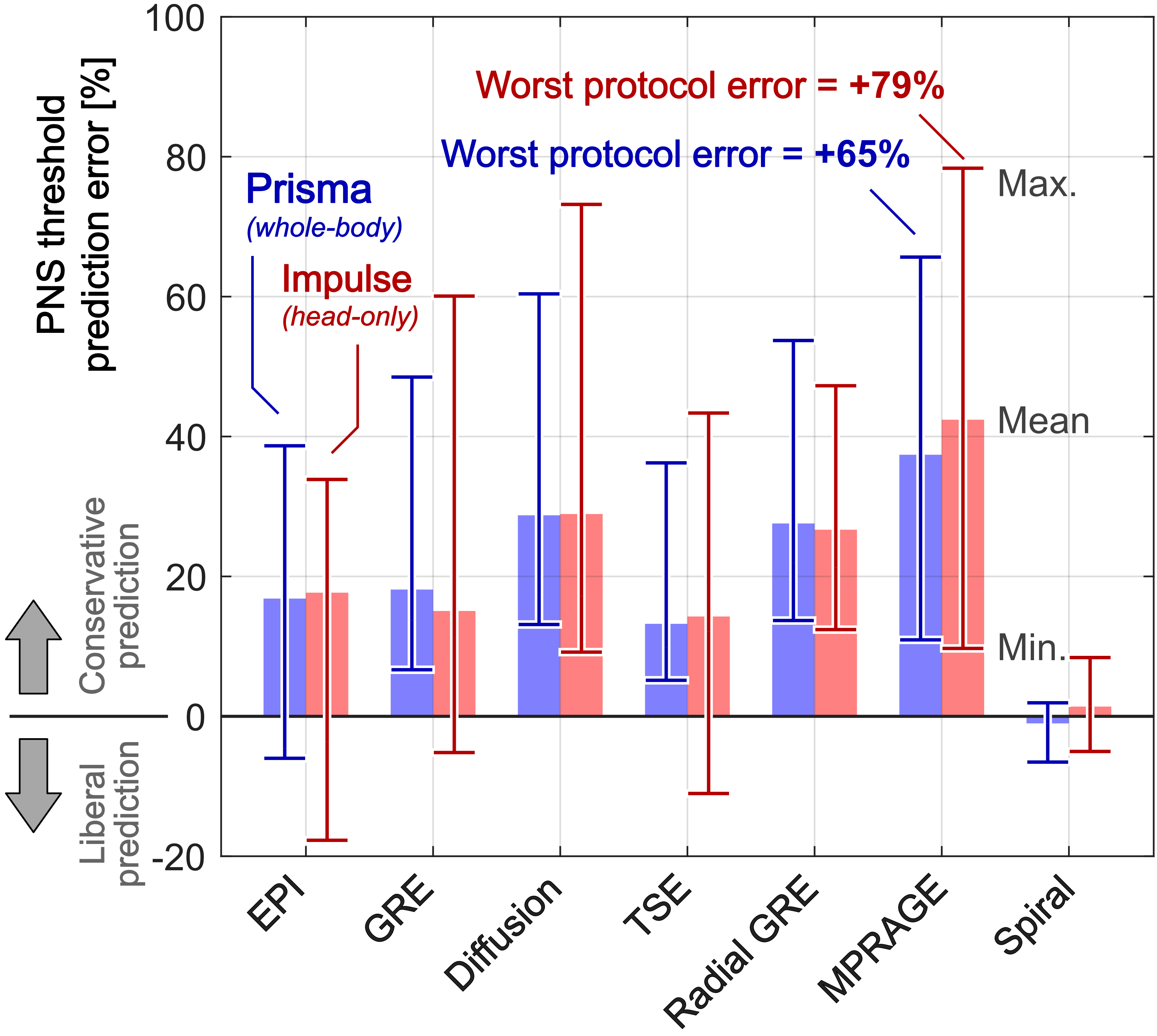
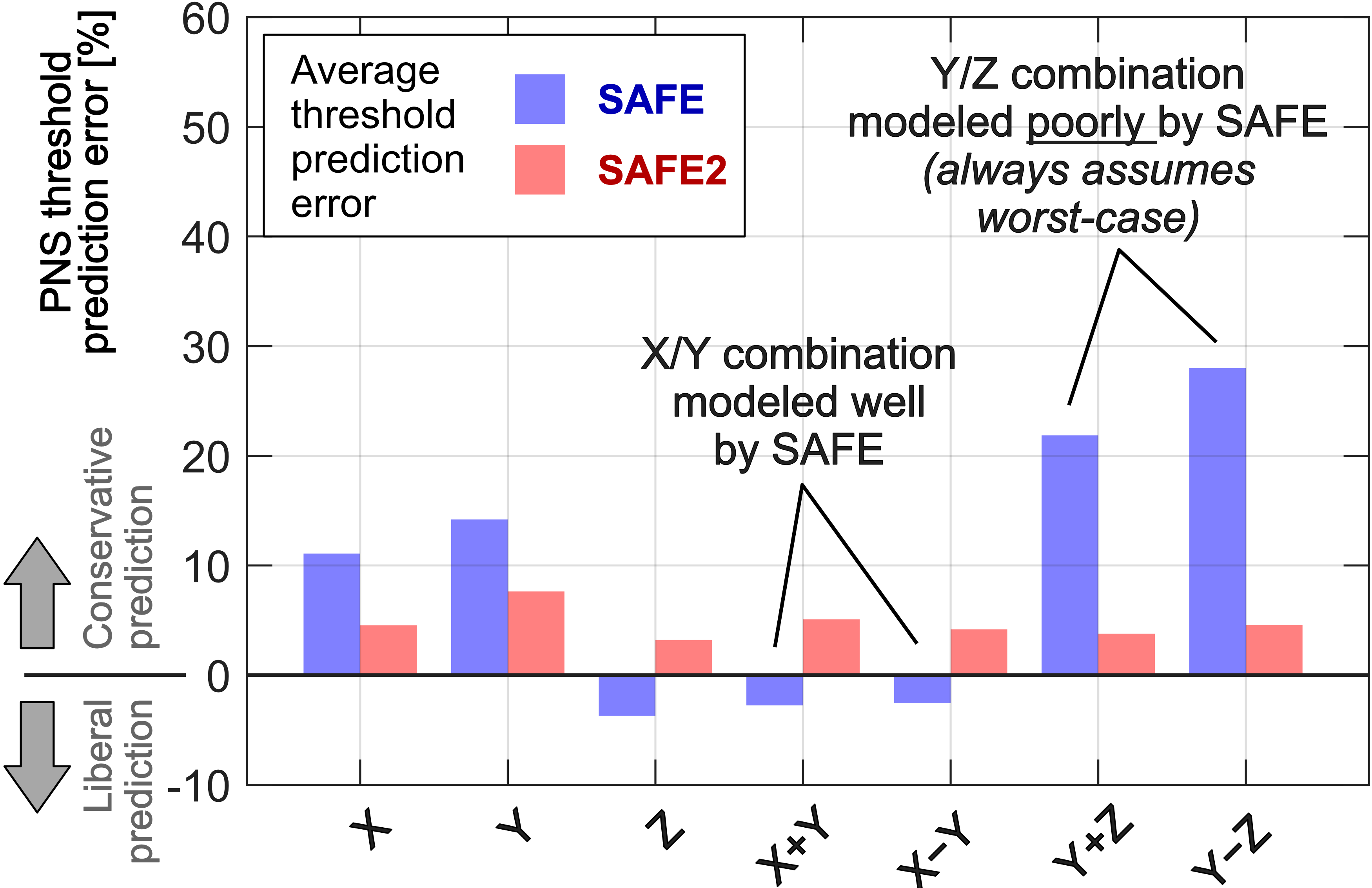
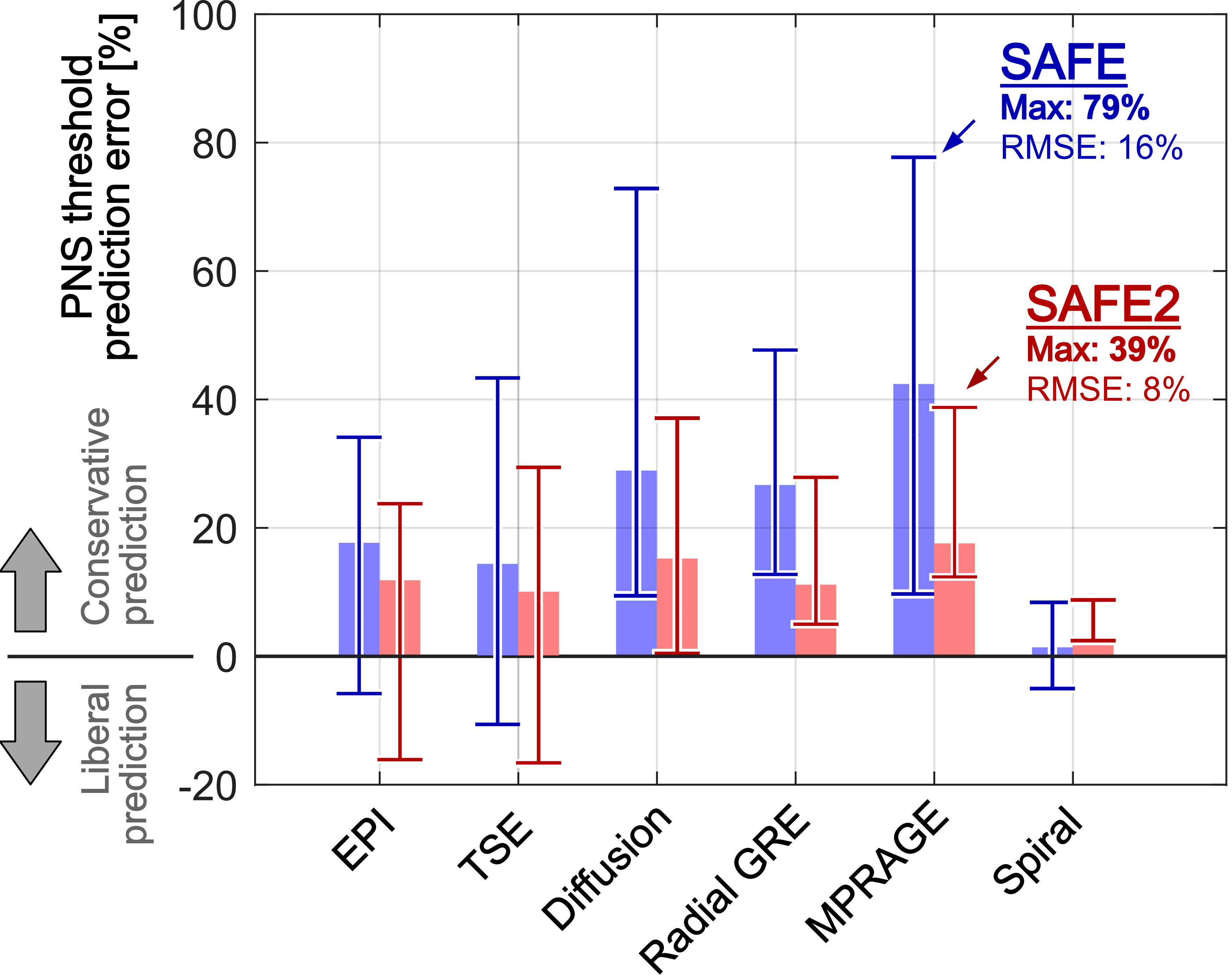
Fig. 3 shows the deviation of the SAFE model’s PNS predictions from the reference thresholds (full EM-neurodynamic model) for the Prisma and Impulse coils for clinically relevant sequences. The SAFE model’s inability to fully capture PNS mechanisms such as cancelations of E-fields and membrane charges from different gradient axes results in prediction errors of up to 65% and 79% for Prisma and Impulse, respectively. Fig. 4 compares the SAFE2 and SAFE predictions for periodic trapezoidal pulse trains played on different axes combinations. The largest SAFE prediction errors occur for the Y±Z combinations, but also for the X and Y cases. SAFE2 achieves an improved and more consistent prediction accuracy across all axes’ combinations. Fig. 5 summarizes the SAFE2 and SAFE prediction accuracy for the Impulse coil. SAFE2 improves the accuracy by 2X in terms of NRMSE (16% to 8%) and maximum prediction error (79% to 39%) across all waveforms.Conclusion
We propose a new PNS monitor, SAFE2, that better captures the detailed mechanisms behind PNS than the conventional SAFE model (including cancelations of E-fields and membrane charges). Training of the more complex SAFE2 model is supported by a comprehensive dataset of predicted PNS thresholds generated using our validated EM-neurodynamic PNS model. SAFE2 achieves 2X more accurate PNS prediction, potentially allowing for a boost in usable gradient performance by 30% without the need for hardware modifications such as redesign of the gradient coil.Acknowledgements
The authors would like to acknowledge the help of past and present members of the gradient coil group at Siemens Healthineers, including Peter Dietz, Axel vom Endt, Gudrun Ruyters, Manuela Roesler, Franz Hebrank, Eva Eberlein. Research reported in this publication was supported by the National Institute of Biomedical Imaging and Bioengineering, and the National Institute for Mental Health of the National Institutes of Health under award numbers U01EB025162, P41EB030006, U01EB026996, R01EB028250, U01EB025121 and R01EB033853. The content is solely the responsibility of the authors and does not necessarily represent the official views of the National Institutes of Health.References
[1] Joachim Abart, Knuth Eberhardt, Hubertus Fischer, Walter Huk, Evelyn Richter, Thomas Storch, Eberhard Zeitler, et al. Peripheral nerve stimulation by time-varying magnetic fields. Journal of computer assisted tomography, 21(4):532–538, 1997.
[2] JD Bourland, JA Nyenhuis, and DJ Schaefer. Physiologic effects of intense mr imaging gradient fields. Neuroimaging clinics of North America, 9(2):363–377, May 1999.
[3] Mark S. Cohen, Robert M. Weisskoff, Richard R. Rzedzian, and Howard L. Kantor. Sensory stimulation by time-varying magnetic fields. Magn. Reson. Med., 14(2):409–414, 1990.
[4] International Electrotechnical Commission et al. Iec 60601-2-33. Medical Electrical Equipment-Part, 2, 2002.
[5] Mathias Davids, Peter Dietz, Gudrun Ruyters, Manuela Roesler, Valerie Klein, Bastien Guérin, David A. Feinberg, and Lawrence L. Wald. Peripheral nerve stimulation informed design of a high-performance asymmetric head gradient coil. Magnetic Resonance in Medicine, 90(2):784–801, 2023.
[6] Mathias Davids, Bastien Guérin, Valerie Klein, Martin Schmelz, Lothar R Schad, and Lawrence L Wald. Optimizing selective stimulation of peripheral nerves with arrays of coils or surface electrodes using a linear peripheral nerve stimulation metric. Journal of neural engineering, 17(1):016–029, 2020.
[7] Mathias Davids, Bastien Guérin, Lothar R. Schad, and Lawrence L. Wald. Predicting magnetostimulation thresholds in the peripheral nervous system using realistic body models. Scientific reports, 7(1):1–14, 2017.
[8] Mathias Davids, Bastien Guérin, Lothar R. Schad, and Lawrence L. Wald. Peripheral Nerve Stimulation Modeling for MRI, pages 87–102. Wiley, 2019.
[9] Mathias Davids, Bastien Guérin, Axel vom Endt, Lothar R. Schad, and Lawrence L. Wald. Prediction of peripheral nerve stimulation thresholds of mri gradient coils using coupled electromagnetic and neurodynamic simulations. Magn. Reson. Med., 81(1):686–701, 2019.
[10] Mathias Davids, Bastien Guérin, and Lawrence L. Wald. A huygens surface approach to rapid characterization of peripheral nerve stimulation (pns). Magnetic Resonance in Medicine, 87(1):377–393, 2021.
[11] James C. Ehrhardt, Chin-S Lin, Vincent A. Magnotta, David J. Fisher, and William T. C. Yuh. Peripheral nerve stimulation in a whole-body echo-planar imaging system. J. Magn. Reson. Imaging, 7(2):405–409, 1997.
[12] Sonja C Faber, Alexander Hoffmann, Christof Ruedig, and Maximilian Reiser. Mri-induced stimulation of peripheral nerves: dependency of stimulation threshold on patient positioning. Magn. Reson. Imaging, 21(7):715 – 724, 2003.
[13] C. L. G. Ham, J. M. L. Engels, G. T. van de Wiel, and A. Machielsen. Peripheral nerve stimulation during mri: Effects of high gradient amplitudes and switching rates. J. Magn. Reson. Imaging, 7(5):933–937, 1997.
[14] Franz X Hebrank and Matthias Gebhardt. Safe model – a new method for predicting peripheral nerve stimulation in mri. In Proceedings of the 8th Annual Meeting of ISMRM, Denver, page 2007, 2000.
[15] JA Nyenhuis, JD Bourland, KS Foster, GP Graber, AV Kildishev, and DJ Schaefer. Magnetic stimulation in humans by mri pulsed gradient fields. In IEEE Engineering in Medicine and Biology 21st Annual Conference and the 1999 Annual Fall Meeting of the Biomedical Engineering Society, volume 2, page 1074. IEEE, 1999.
[16] MICHAEL L. ROHAN and RICHARD R. RZEDZIAN. Stimulation by time-varying magnetic fields. Ann. N.Y. Acad. Sci., 649(1):118–128, 1992.
Figures