3914
Experimental Investigation of Interfering Factors in Cardiac Sensing of Beat Pilot Tone1School of Biomedical Engineering, Shanghai Jiao Tong University, Shanghai, China, 2National Engineering Research Center of Advanced Magnetic Resonance Technologies for Diagnosis and Therapy (NERC-AMRT), Shanghai Jiao Tong University, Shanghai, China
Synopsis
Keywords: Hybrid & Novel Systems Technology, Motion Correction, Cardiovascular
Motivation: Beat Pilot Tone (BPT) has been proposed as a non-contact and accurate cardiac sensing method seamlessly integrated with MR systems. However, BPT is sensitive to interference, thereby limiting its ability to robustly obtain fine-grained cardiac waveform.
Goal(s): Our goal was to demonstrate main factors that could lead to corrupted BPT signal.
Approach: We conducted a series of comparative experiments on the hypothesized influencing factors.
Results: The experiments suggested that BPT transmit power, imaging orientation and other physiological motion like respiration caused varying degradation to the BPT signal.
Impact: The demonstration of interfering factors of BPT cardiac sensing could guide the correct BPT setup and interference suppression method design. This helps BPT to robustly obtain fine-grained cardiac waveform, which could be used for MR motion correction and clinical diagnosis.
Introduction
Cardiac MRI requires synchronization of the MR measurement to cardiac activity which is commonly achieved by the use of electrocardiogram (ECG). However, the setup of ECG is time consuming and the electrode pads may pose a risk of skin burns[1].Beat Pilot Tone (BPT) is a novel and promising alternative for cardiac motion sensing[2,3]. It is non-contact, sensitive and easy to integrate with MR systems. Previous studies have demonstrated that the BPT can detect cardiac motion with improved SNR and more abundant information compared to Pilot Tone (PT)[4]. However, BPT is also sensitive to interference, making it challenging to robustly extract fine-grained cardiac waveform during normal MR scans.
In this work, we hypothesized that BPT transmit power, other physiological motions such as respiration, and imaging sequence parameters related to RF and gradients are the main factors influencing the SNR of BPT acquired cardiac waveform. Therefore, we conducted a series of comparative experiments on a 1.5T commercial scanner (MAGNETOM Aera, Siemens Healthcare, Erlangen, Germany) to assess the impact of these factors.
Methods
Hardware setupFigure 1 shows the hardware setup. The BPT transmitter was placed in the control room. It generated two RF tones by ADF4351 modules, with one tone operating at a frequency of 2.4GHz and the other at 2.463534GHz. The two tones were combined, bandpass filtered, amplified by 30dB and transmitted to a UWB log periodic antenna in the magnet room. We used a 5-meter-long coaxial cable with balun and it causes approximately 15dB attenuation of the signal. The antenna was placed above the chest on top of the subject’s heart to enhance cardiac motion modulation. Then, the modulated two tones were picked up by a 6-channel surface coil, mixed via nonlinear intermodulation in the MRI receiver chain. The subject was simultaneously equipped with ECG suite as the ground truth.
Acquisition experiments
We conducted a series of comparative acquisition experiments on a healthy volunteer using a 2D Golden-angle (GA) GRE (Gradient Echo) sequence. Each experimental group was designed to assess the impact of an assumed factor on the cardiac waveform obtained by BPT. Table 1 shows the specific conditions and sequence parameters of different experimental groups. Transmit power, breath-holding condition, RF, gradients and imaging orientation were changed separately in experimental groups 1 to 4.
Data processing
The BPT signal was extracted using the method described previously[5]. We selected the BPT raw signal received by the surface coil closest to the heart and applied a 5-15 Hz bandpass filter to it.
Results and Discussion
Figure 2a shows the results of experimental group 1. The higher BPT transmit power scan produced sharper cardiac-modulated raw signal. After bandpass filtering, a dBCG-like signal with obvious I and J waves was obtained and the heartbeat interval corresponded well to the ECG ground truth. The raw BPT signal obtained through lower transmit power scan appeared to be noisier, and after bandpass filtering, certain I and J waves became challenging to detect. Figure 2b shows the results of experimental group 2. The raw BPT signal was dominated by respiratory modulation. After bandpass filtering, I and J waves exhibited periodic disturbances synchronized with respiratory cycles.Figure 3 shows the results of experimental group 3. For the 2D GA sequence, gradient switching did not show obvious impact on the performance of BPT sensing. RF interference became apparent in the absence of slice selection gradient because the signal from the entire volume is excited. In the presence of slice selection gradient, RF interference was less noticeable. The impacts of RF interference and gradient switching remains to be verified on other commonly used sequences.
Figure 4 shows the results of experimental group 4. Performing a Fourier transform along the readout direction of the original MR data, the BPT signal appeared as a single peak in the transverse scan, whereas in sagittal and coronal scans, the BPT signal appeared as two peaks. Therefore, the extracted BPT signal is subject to additional modulation apart from motion, preventing the acquisition of fine-grained cardiac waveform. The underlying cause of this phenomenon remains to be investigated
Conclusions
Our experiments suggest that BPT transmit power and respiration motion affect the characteristics of the sensed cardiac waveform. Sufficient BPT transmit power and respiratory suppression algorithm are needed to robustly obtain fine-grained cardiac waveform in normal scans. For the 2D GA sequence, RF interference and gradient switching cause neglectable impacts. However, sagittal and coronal scans result in significant distortion of the BPT signal. Experiments are needed to carry out to find out the reason.Acknowledgements
This work is supported by the National Natural Science Foundation of China National Science Foundation of China (No. 62001290 and 62301309), Shanghai Science and Technology Development Funds (21DZ1100300) and sponsored by the National Science and Technology Innovation 2030 Major Project (2022ZD0208601).
References
[1] Salvaraji, Loganathan, et al. "Electrical safety in a hospital setting: A narrative review." Annals of Medicine and Surgery 78 (2022): 103781.
[2] Anand, Suma, and Michael Lustig. "Beat pilot tone: exploiting preamplifier intermodulation of UHF/SHF RF for improved motion sensitivity over pilot tone navigators." Proc Intl Soc Mag Reson Med. Vol. 29. 2021.
[3] Lamar, Katie, and Michael Lustig. "Respiratory and Cardiac Motion Correction Using the Beat Pilot Tone." (2023).
[4] Anand, Suma, and Michael Lustig. "Beat Pilot Tone: Versatile, Contact-Free Motion Sensing in MRI with Radio Frequency Intermodulation." arXiv preprint arXiv:2306.10236 (2023).
[5] Solomon, Eddy, et al. "Free‐breathing radial imaging using a pilot‐tone radiofrequency transmitter for detection of respiratory motion." Magnetic resonance in medicine 85.5 (2021): 2672-2685.
Figures
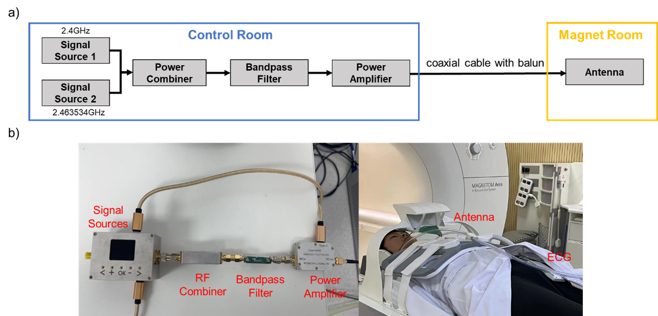
Figure 1: a) A schematic of the hardware setup. The two tones were generated, combined, filtered and amplified in the control room. Then they were transmitted to the antenna in the magnet room through a coaxial cable with balun. b) Photographs of the hardware setup. The UWB log periodic antenna was placed above the chest on top of the subject’s heart to enhance cardiac motion modulation.
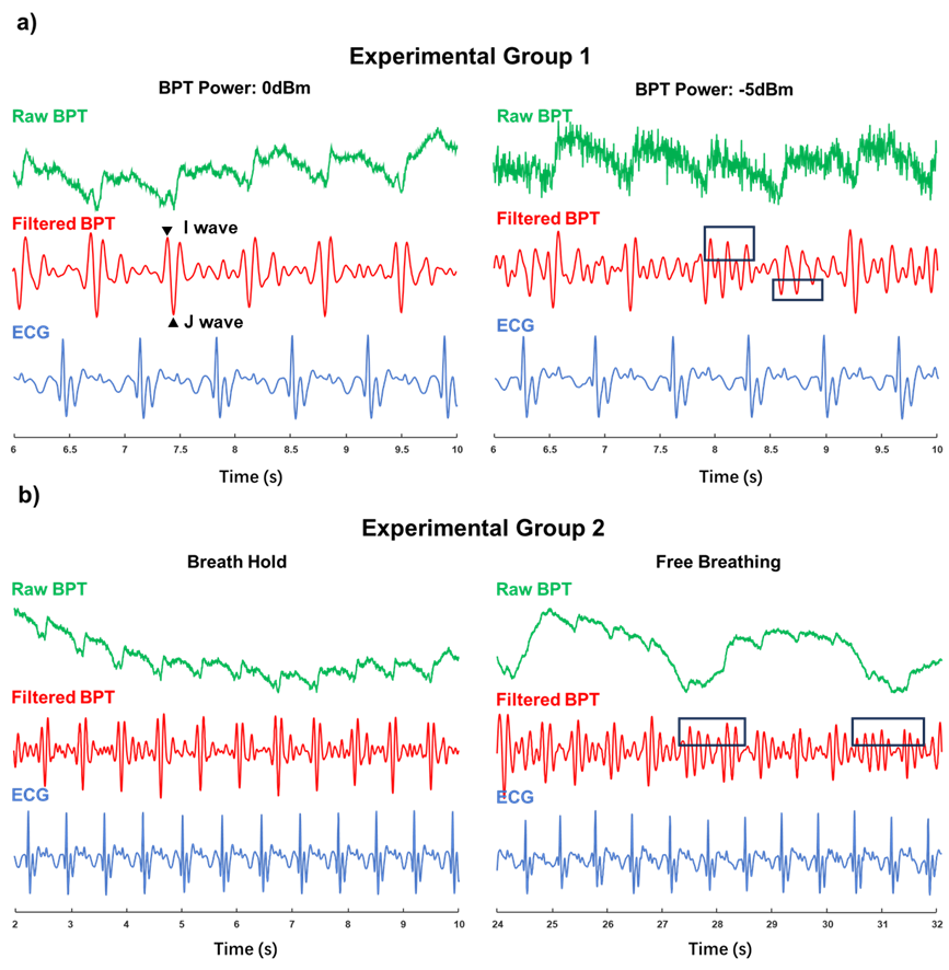
Figure 2: The raw BPT signal, bandpass filtered dBCG-like signal and corresponding ECG ground truth from a) experimental group 1. and b) experimental group 2 . The I and J waves were marked with a black triangle. The reduction in BPT transmit power made I and J waves harder to detect while respiratory motion introduced periodic disturbance (Marked with black boxes).
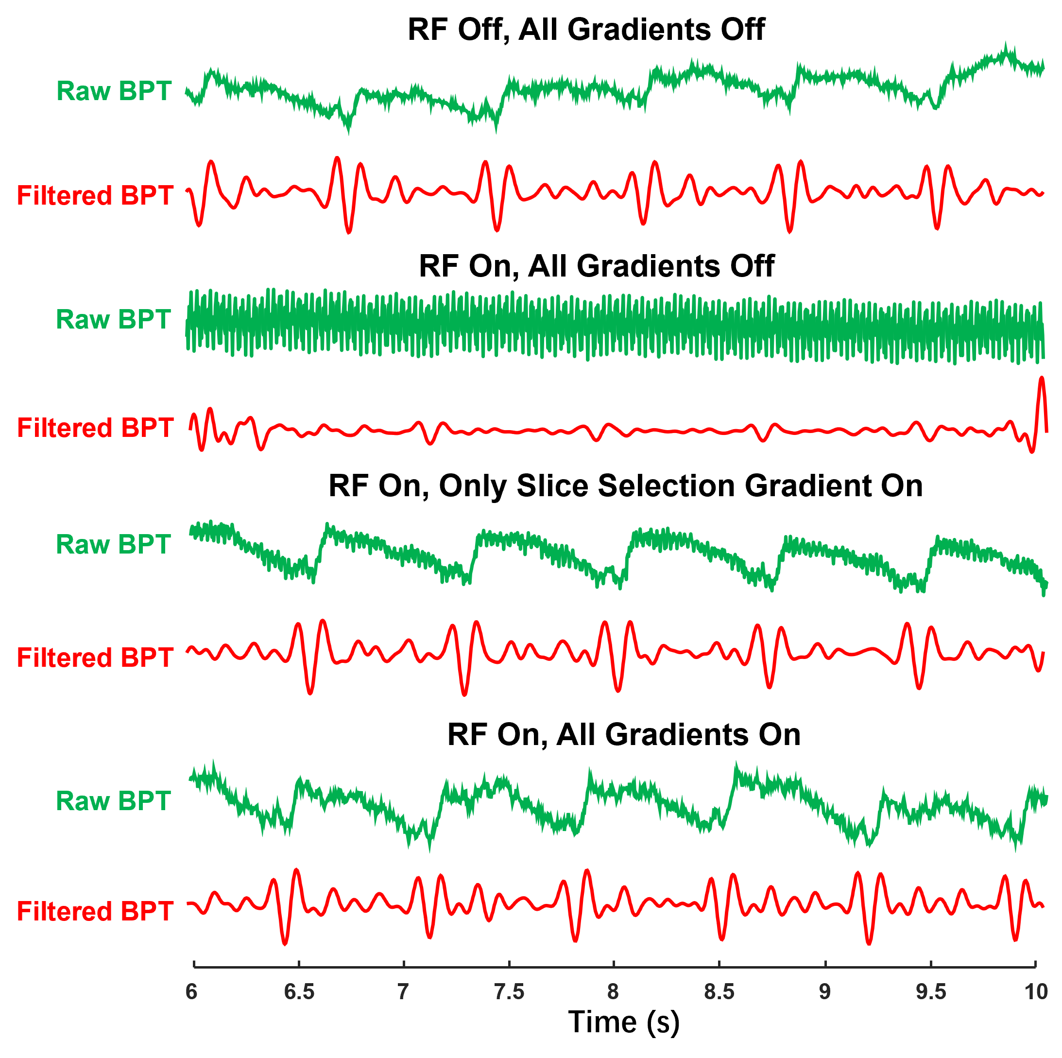
Figure 3: The raw BPT signal and bandpass filtered BPT signal from experimental group 3. RF and gradient switching introduced neglectable interference. In the absence of slice selection gradient, RF excited energy caused distortion to the BPT signal.
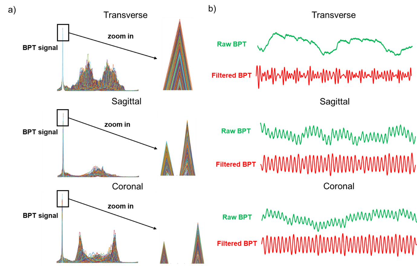
Figure 4: a) BPT signal in frequency domain. Transverse scan showed single BPT peak while sagittal and coronal scans showed double. b) Extracted BPT signal from experimental group 4. In sagittal and coronal scans, the signal is subject to strong distortion.
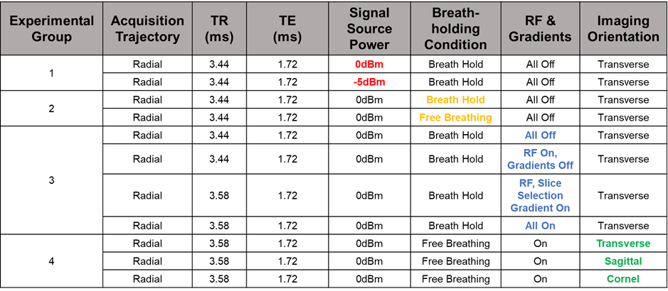
Table 1: Specific conditions and parameters of the experiments performed. Each experimental group only changed the color-labeled condition and other remain consistent.