3909
Potential MRI and PET imaging patterns of decreased Alzheimer's-disease pathological protein clearance in patients with cognitive impairment1Huaxi MR Research Center (HMRRC), Department of Radiology, West China Hospital of Sichuan University, ChengDu, China
Synopsis
Keywords: Alzheimer's Disease, Neurodegeneration, CSF clearance, Choroid plexus
Motivation: Impaired Alzheimer's disease (AD) pathological protein clearance plays a critical role in the progression of cognitive impairment.
Goal(s): To explore whether microscale abnormalities in clearance pathways exhibit macroscopic imaging patterns.
Approach: Data collection was performed, and quantitative features were calculated for intergroup differences and correlation analysis.
Results: The specific imaging patterns represented by choroid plexus and its associated alterations are associated with increased brain AD pathological protein burden and decreased clearance capacity of the glymphatic system.
Impact: This study provides a novel perspective on the decreased clearance capacity of amyloid-beta and tau proteins in cognitively impaired patients' brains. The discovery of distinct imaging patterns aids in accurate diagnosis, treatment, and deepens our understanding of Alzheimer's disease mechanisms.
Background
Deposition of amyloid-beta and tau proteins in the brains of Alzheimer's disease (AD) patients leads to neurodegeneration and progressive cognitive and memory impairment [1]. Evidence suggests that impaired clearance capacity of AD pathological proteins is a key factor in the pathogenesis of AD [2]. Cerebrospinal fluid (CSF) circulation serves as a convergence point for multiple clearance pathways, including interstitial fluid circulation, glymphatic system transport, and clearance across the blood-brain barrier and blood-CSF barrier [3]. The choroid plexus, which forms the blood-CSF barrier, generates CSF. Pathological changes in the choroid plexus have been observed in older individuals and AD patients, affecting CSF production efficiency [4, 5]. Choroid plexus alterations may serve as potential imaging markers of AD-related cognitive impairment [6]. However, the relationship between choroid plexus dysfunction and impaired clearance capacity of AD pathological proteins through multiple clearance pathways is not fully understood. Identifying macroscopic imaging patterns on MRI and PET that correspond to microscale abnormalities within the brain could aid in the diagnosis, differentiation, and treatment of AD patients. Therefore, our aim is to explore distinct patterns of decreased clearance capacity of AD pathological proteins on existing imaging techniques, which may contribute to the precise diagnosis, treatment, and understanding of AD pathogenesis.Methods
We retrospectively collected cognitive assessment and imaging data, including MRI brain structural data, brain PET imaging data of AD pathological proteins, and CSF data, from 122 elderly individuals with or without cognitive impairment. Data was obtained from the Alzheimer's Disease Neuroimaging Initiative database (http://adni.loni.usc.edu). Participants were divided into three groups based on cognitive assessment data. Radiologists manually delineated the regions of interest of the choroid plexus on MRI images. The most discriminative quantitative -features were extracted and selected based on IBSI standards. We compared volumes of the choroid plexus, quantitative features, CSF volumes, and concentrations of amyloid-beta and tau in different brain regions, as well as intergroup differences in CSF amyloid-beta and tau. Correlations between choroid plexus volume, quantitative features, CSF volumes, and amyloid-beta and tau measured through PET and CSF were analyzed.Results
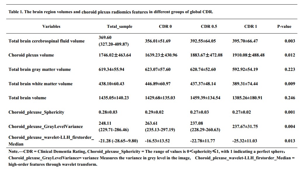
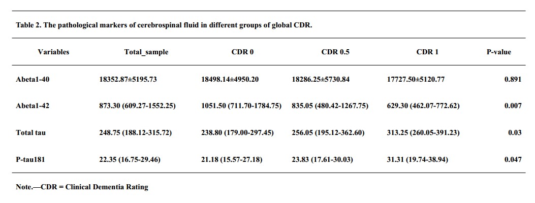
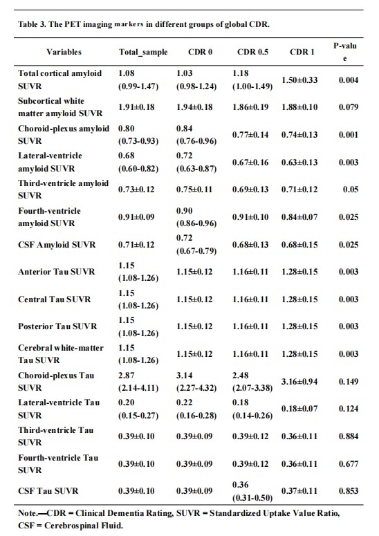
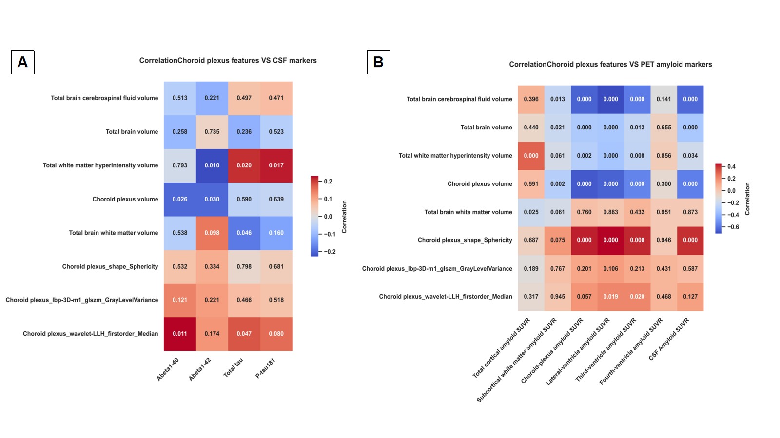
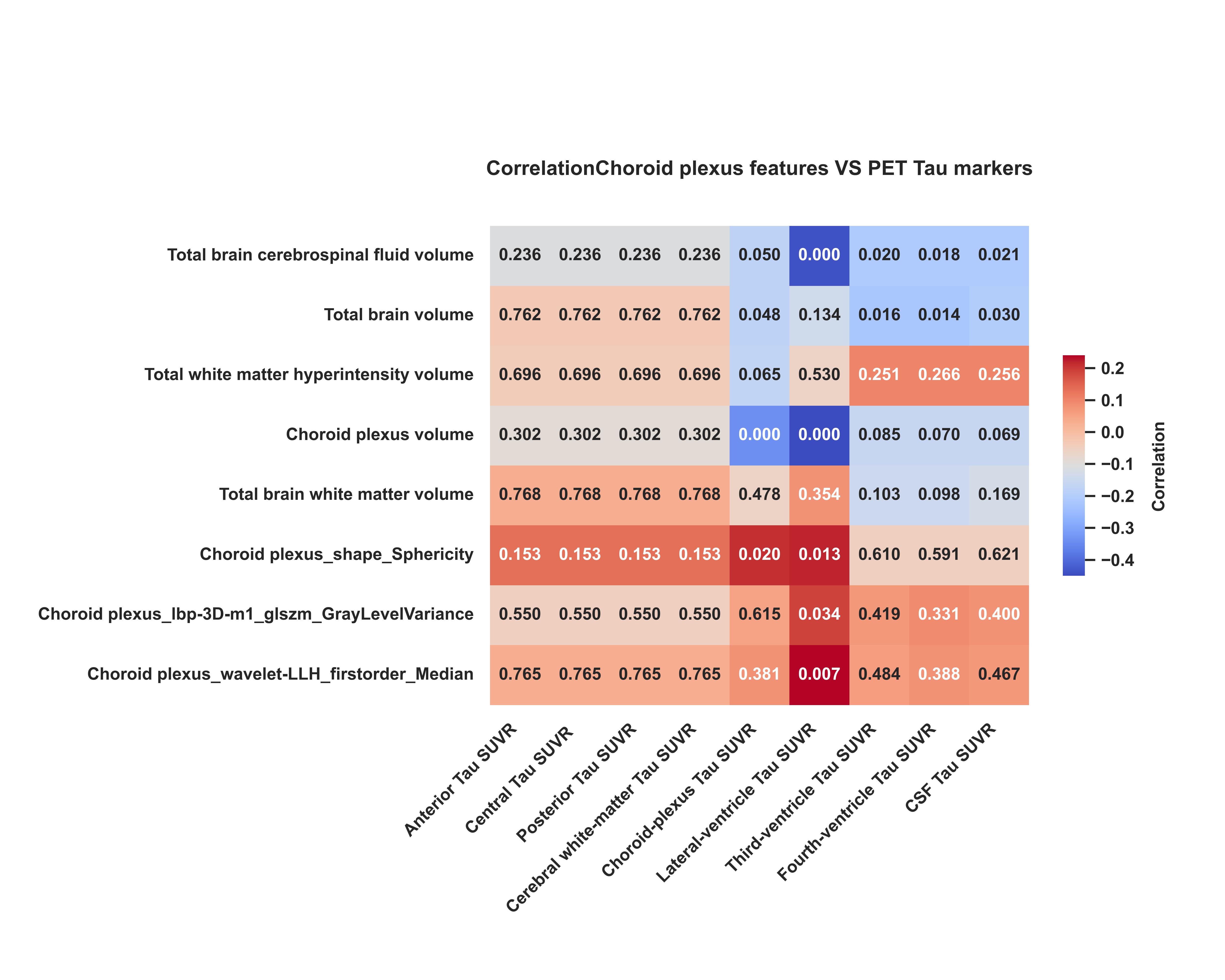
Significant differences were observed in the volumes of the choroid plexus, quantitative features, CSF volume, brain white matter volume, and amyloid-beta and tau levels among the three groups. For the groups with more severe cognitive impairment, the choroid plexus volume was larger, had a less spherical shape, lower internal grayscale variation, larger CSF volume, and smaller brain white matter volume (Table 1-3). Correlation analysis showed that choroid plexus volume was negatively correlated with amyloid-beta and tau levels in the choroid plexus, subcortical white matter, CSF, and ventricular systems. Choroid plexus sphericity showed the opposite trend. Texture and higher-order features of the choroid plexus were positively correlated with amyloid-beta and tau levels in some ventricular systems, as well as with specific amyloid-beta and tau levels in the CSF (Figure 1-2).Discussion
Our study demonstrated significant differences in the volumes of the choroid plexus, quantitative features, and associated secondary brain changes among individuals with different degrees of cognitive impairment. These specific imaging pattern changes accompanying worsening cognitive impairment are related to increased AD pathological protein deposition in the brain and reduced clearance of AD pathological proteins through the glymphatic system into the ventricular system. The results of our study revealed potential macroscopic imaging patterns resulting from abnormal clearance pathways in the brain. The increase in choroid plexus volume observed in our study is consistent with previous research [6]. We speculate that the increase in choroid plexus volume, changes in texture and higher-order features, increased CSF volume, and decreased brain white matter volume may represent a macroscopic imaging pattern of impaired glymphatic clearance of AD pathological proteins, reflecting a decline in brain water circulation capacity. Aquaporin proteins are widely distributed in the blood-brain barrier and the blood-cerebrospinal fluid barrier of the choroid plexus, and in AD patients, there is a decrease in the quantity and relocation of aquaporin proteins [7]. Some studies using MRI water imaging techniques also support the presence of widespread impairments in water exchange capacity in the brains of AD patients [8, 9].Conclusion
The preliminary results of our study support the notion that the distinct imaging pattern represented by the choroid plexus and its associated changes may serve as potential imaging biomarkers for cognitive impairment, reflecting alterations in the clearance capacity of AD pathological proteins in the brain. Further validation of this imaging pattern is needed through the integration of clinical imaging and basic research, as well as multi-center studies.Acknowledgements
None
References
[1] R. van der Kant, L.S.B. Goldstein, R. Ossenkoppele, Amyloid-β-independent regulators of tau pathology in Alzheimer disease, Nature Reviews Neuroscience 21(1) (2019) 21-35.
[2] K.G. Mawuenyega, W. Sigurdson, V. Ovod, L. Munsell, T. Kasten, J.C. Morris, K.E. Yarasheski, R.J. Bateman, Decreased clearance of CNS beta-amyloid in Alzheimer's disease, Science (New York, N.Y.) 330(6012) (2010) 1774.
[3] J.M. Tarasoff-Conway, R.O. Carare, R.S. Osorio, L. Glodzik, T. Butler, E. Fieremans, L. Axel, H. Rusinek, C. Nicholson, B.V. Zlokovic, B. Frangione, K. Blennow, J. Ménard, H. Zetterberg, T. Wisniewski, M.J. de Leon, Clearance systems in the brain-implications for Alzheimer disease, Nature reviews. Neurology 11(8) (2015) 457-70.
[4] J.M. Serot, M.C. Béné, B. Foliguet, G.C. Faure, Morphological alterations of the choroid plexus in late-onset Alzheimer's disease, Acta Neuropathol 99(2) (2000) 105-8.
[5] M.O. Dietrich, C. Spuch, D. Antequera, I. Rodal, J.G. de Yébenes, J.A. Molina, F. Bermejo, E. Carro, Megalin mediates the transport of leptin across the blood-CSF barrier, Neurobiol Aging 29(6) (2008) 902-12.
[6] J.D. Choi, Y. Moon, H.-J. Kim, Y. Yim, S. Lee, W.-J. Moon, Choroid Plexus Volume and Permeability at Brain MRI within the Alzheimer Disease Clinical Spectrum, Radiology 304(3) (2022) 635-645.
[7] D.M. Zeppenfeld, M. Simon, J.D. Haswell, D. D’Abreo, C. Murchison, J.F. Quinn, M.R. Grafe, R.L. Woltjer, J. Kaye, J.J. Iliff, Association of Perivascular Localization of Aquaporin-4 With Cognition and Alzheimer Disease in Aging Brains, JAMA Neurology 74(1) (2017).
[8] Y. Uchida, H. Kan, K. Sakurai, Y. Horimoto, E. Hayashi, A. Iida, N. Okamura, K. Oishi, N. Matsukawa, APOE ɛ4dose associates with increased brain iron and β-amyloid via blood–brain barrier dysfunction, Journal of Neurology, Neurosurgery & Psychiatry 93(7) (2022) 772-778.
[9] C.R. Joseph, C.M. Benhatzel, L.J. Stern, O.M. Hopper, M.D. Lockwood, Pilot study utilizing MRI 3D TGSE PASL (arterial spin labeling) differentiating clearance rates of labeled protons in the CNS of patients with early Alzheimer disease from normal subjects, Magnetic Resonance Materials in Physics, Biology and Medicine 33(4) (2020) 559-568.
Figures