3900
Dynamic Changes in Automatic Generated Quantitative Biomarkers and Correlation with Pathology of Alzheimer’s Disease – A Longitudinal Study1Peking University Shenzhen Hospital, Shenzhen, China, 2The Chinese University of Hong Kong, Hong Kong, China
Synopsis
Keywords: Alzheimer's Disease, Alzheimer's Disease
Motivation: AD-RAI is a novel MRI-based machine-learning derived biomarker and the value of longitudinal AD-RAI remains unclear.
Goal(s): We aimed to assess longitudinal changes of the MRI biomarkers (i.e., AD-RAI, HV, HF, BPV, BPF) in correlation with change in time and conversion status with and without A+T+.
Approach: We selected 168 CU and MCI in ADNI with four-year follow-up with serial MRI scans and corresponding CSF and used linear mixed-effects models to estimate and compare.
Results: AD-RAI of subjects with A+T+ increased significantly faster than non-A+T+ over time and AD-RAI has the potential to track CSF Aβ1–42 as an effective longitudinal surrogate biomarker.
Impact: If the serial AD-RAI change over time is associated with conversion status and AD pathologies. It may be used as a surrogate marker for monitoring disease progression or treatment response in AD.
Introduction
Developing a biomarker of AD that can be used to monitor disease progression or to serve as a surrogate outcome measure in clinical trials is important in the management and research of AD. Changes in such biomarkers over time should correlate with changes in cognitive function and pathological type. Subjects with A+T+ (i.e., Aβ+ and Tau+) tend to progress faster than those without A+T+ in AD continuum. Although serial measurements of biomarkers based on CSF and PET may be used to monitor disease progression.[1] CSF is invasive while PET is associated with radiation exposure, and both are relatively expensive. Previous studies have commonly utilized change in HV as a surrogate marker for monitoring disease progression.[2,3] However, apart from HV, progressive atrophy of other cognitive-relevant brain regions is also present.[4] AD-RAI is an MRI-based machine learning-derived composite index reflecting multi-region atrophy severity of AD. In this study, we aimed to investigate the association between the serial change of AD-RAI and cognitive progression overtime among CU and MCI subjects with and without A+T+ (i.e., Aβ+ and Tau+) and to compare it with other conventional MRI biomarkers (i.e., hippocampal volume (HV), hippocampal fraction (HF), brain parenchymal volume (BPV) and fraction (BPF)), which have been used in the past as surrogate markers for AD.Methods
We selected subjects from the ADNI databases and included those aged 55 to 90 years, diagnosed with CU and MCI at baseline with high-quality volumetric MRI data and CSF at baseline, and at least once matched T1W scans and CSF during follow-up. We defined converters as those who had significant clinical cognitive progression in the 4-year follow-up (i.e., CU to MCI, or MCI to AD dementia), while non-converters were those who remained stable in the 4-year follow-up. We define A+T+ based on the threshold value of Ab1-42 and ptau in CSF [5]. A total of 168 subjects, including 108 non-converters and 60 converters, were recruited with 457 serial MRI scans and corresponding CSF in total. We also considered the influences from age at baseline, gender, education, and APOE4 genotypes. All 3D T1W images were analyzed using the AccuBrain software system. In this study following data were extracted for further analysis: AD-RAI, HV, HF, BPV, and BPF. We used a linear mixed-effects model (LMM) in SPSS 26.0 to explore the serial changes of the imaging biomarkers and interaction effects with and without A+T+ over time, and further explore the association between dynamic CSF Aβ1–42 and dynamic changes in the imaging biomarkers’ interaction effects over time.Results
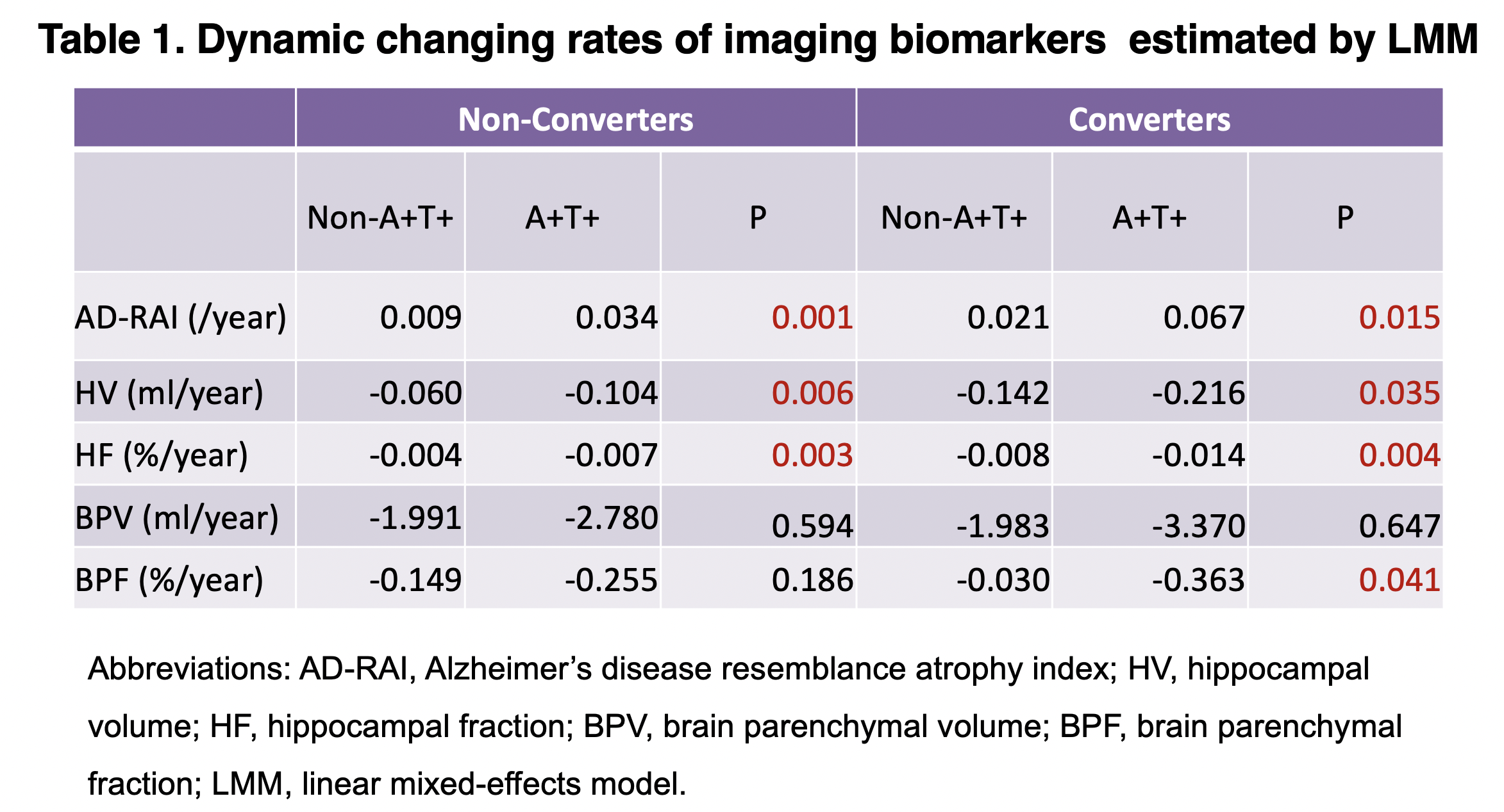
In either converters or non-converters, AD-RAI with A+T+ increased significantly faster than non-A+T+ over time (P < 0.05), while HV and HF with A+T+ decreased significantly faster than non-A+T+ over time (P < 0.05, respectively) irrespective of whether the adjustment was made for the baseline covariates (age at baseline, gender education, and APOE4), which performed better than BPV/BPF (Table 1). Serial AD-RAI was significantly associated with change in CSF Aβ1–42 over time, which outperforms other imaging biomarkers.Discussion
To our knowledge, this is the first study showing that serial changes in machine learning-derived MRI biomarkers had the best association over other conventional imaging biomarkers with underlying AD pathologies. Given that deposition of Aβ is generalized in the brain and Aβ on its own can induce brain atrophy, AD-RAI as a biomarker reflecting multiple AD-relevant regions outperformed hippocampal measures was reasonable. Our finding that serial change in AD-RAI outperformed that of hippocampal and global brain measures in correlating with cognitive progression supports our hypothesis that serial change of a biomarker that can capture the multi-brain regional atrophy characteristic to AD is better than that of a single region in correlating with cognitive progression. Our finding that changing rate of HV and HF was faster in converters than in non-converters was in agreement with previous studies.[2-4,6,7] We also found that the rate of changes of hippocampal measures (HV, HF) had a better correlation with that of whole-brain measures (BPV, BPF) was also consistent with results from previous studies.[2] In the present study, we further found that the rate of change in AD-RAI outperformed both hippocampal and whole-brain measures in correlating with cognitive progression. Noteworthy is that the superiority of the association between change of AD-RAI with cognitive progression over other imaging measures was present even after adjusting to age, gender, education, and APOE4.Conclusion
Serial AD-RAI outperforms other conventional biomarkers in correlating with cognitive progression and AD pathologies. It may be used as a surrogate marker for monitoring disease progression or treatment response in AD.Acknowledgements
The data used in the preparation of this article were obtained from the Alzheimer's Disease Neuroimaging Initiative (ADNI) (adni.loni.usc.edu). ADNI is funded by the National Institute on Aging, the National Institute of Biomedical Imaging and Bioengineering. We are grateful for the research volunteers, their families, and the investigators at the ADNI databases.References
1. Jack CR, Jr., Knopman DS, Jagust WJ, et al. Tracking pathophysiological processes in Alzheimer's disease: an updated hypothetical model of dynamic biomarkers. Lancet Neurol. Feb 2013;12(2):207-16. doi:10.1016/S1474-4422(12)70291-0
2. den Heijer T, van der Lijn F, Koudstaal PJ, et al. A 10-year follow-up of hippocampal volume on magnetic resonance imaging in early dementia and cognitive decline. Brain. Apr 2010;133(Pt 4):1163-72. doi:10.1093/brain/awq048
3. Franko E, Joly O, Alzheimer's Disease Neuroimaging I. Evaluating Alzheimer's disease progression using rate of regional hippocampal atrophy. PLoS One. 2013;8(8):e71354. doi:10.1371/journal.pone.0071354
4. Jack CR, Jr., Shiung MM, Gunter JL, et al. Comparison of different MRI brain atrophy rate measures with clinical disease progression in AD. Neurology. Feb 24 2004;62(4):591-600. doi:10.1212/01.wnl.0000110315.26026.ef
5. Shaw LM, Vanderstichele H, Knapik-Czajka M, et al. Cerebrospinal fluid biomarker signature in Alzheimer's disease neuroimaging initiative subjects. Ann Neurol. 2009;65:403-413. doi:10.1002/ana.21610
6. Nadal L, Coupe P, Helmer C, et al. Differential annualized rates of hippocampal subfields atrophy in aging and future Alzheimer's clinical syndrome. Neurobiol Aging. Jun 2020;90:75-83. doi:10.1016/j.neurobiolaging.2020.01.011
7. Rana AK, Sandu AL, Robertson KL, et al. A comparison of measurement methods of hippocampal atrophy rate for predicting Alzheimer's dementia in the Aberdeen Birth Cohort of 1936. Alzheimers Dement (Amst). 2017;6:31-39. doi:10.1016/j.dadm.2016.11.007
Figures