3899
Thalamic Subnuclear Volumetry for Early Detection of Alzheimer's Disease in Postmenopausal Women1Advanced Institute of Aging Science, Chonnam National University, Gwangju, Korea, Republic of, 2Department of Urology, Chonnam National University Medical School, Gwangju, Korea, Republic of, 3Department of Radiology, Chonnam National University Medical School, Gwangju, Korea, Republic of
Synopsis
Keywords: Alzheimer's Disease, Alzheimer's Disease, Aging
Motivation: Women are at a heightened risk of Alzheimer's disease (AD) compared to men. Also, AD and aging are intrinsically interconnected to each other and are mediated by molecular, cellular and biological system.
Goal(s): This study aimed to evaluate volume changes of the subcortical regions including the thalamic subnuclei in women with AD vs. postmenopausal women.
Approach: Twenty-five thalamic subnuclei were extracted extracted from each hemisphere of the subject's T1 image.
Results: Our findings suggest that reduced volume in both the right laterodorsal thalamic nucleus and right hippocampus could represent a key biomarker for predicting early stage of AD in postmenopausal women.
Impact: These findings may be helpful for a better understanding of AD pathogenesis and also for providing an objective target for early interventions to prevent AD.
Introduction
Women are at a heightened risk of Alzheimer's disease (AD) compared to men. Also, AD and aging are intrinsically interconnected to each other and are mediated by molecular, cellular and biological system. In particular, a specific pattern of the brain volume atrophy one of the most profound risk factors for cognitive impairment including AD that is directly linked to aging. With age, the loss of sex hormones, e.g., reduced testosterone levels in men and estrogen loss in women, is one of most significant risk factors for developing AD [1-3]. Sex hormonal changes associated with aging have distinct impacts on brain volume and cognition in each gender. Confounding factors, such as sex hormones, may contribute to differences in brain volume between women following menopause and their male counterparts. The neural substrates commonly affected in AD include the temporal areas, hippocampus, and thalamus [4-6]. The thalamus, an integral brain structure responsible for various cognitive functions, has been of particular interest in AD. Thalamic subnuclei may be differentially affected in AD according to the age at symptom onset. However, a morphometric study regarding the specific alterations in thalamic subnuclear volumes in postmenopausal women has not yet been reported. Thus, this study aimed to investigate knowledge of early detection of AD in postmenopausal women, focusing on the volume changes of the subcortical regions including the thalamic subnuclei in women with AD vs. postmenopausal women.Methods
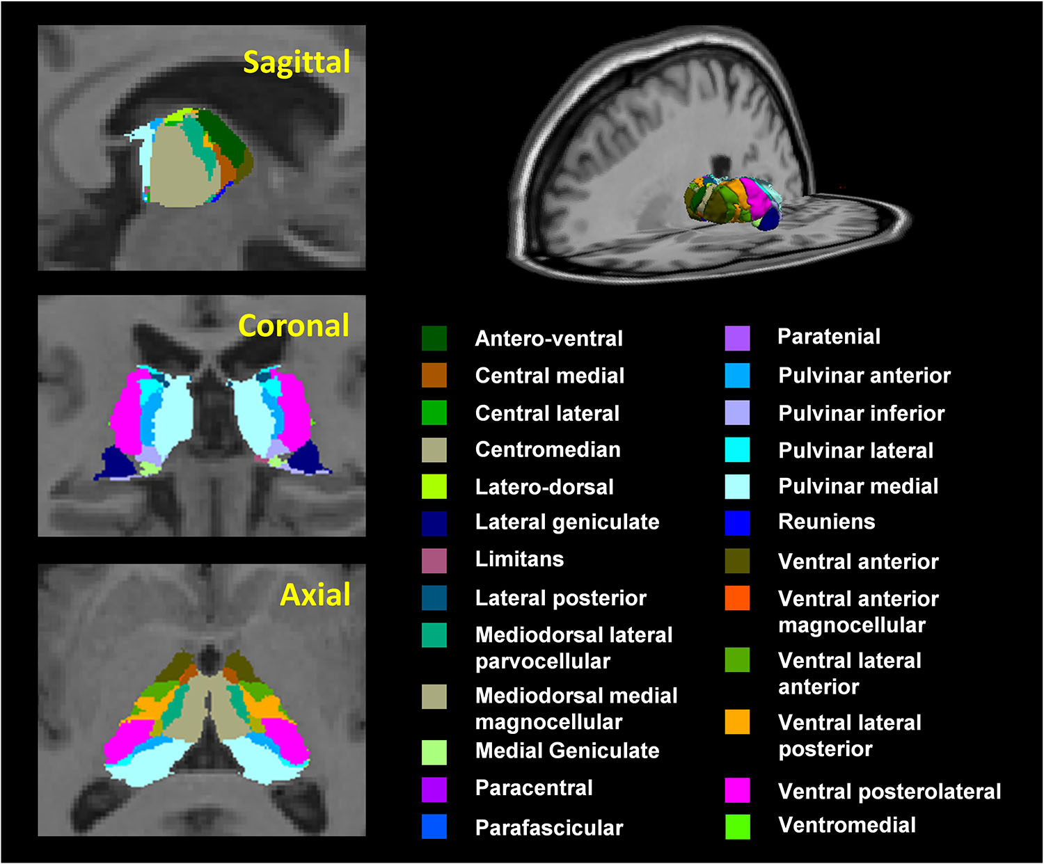
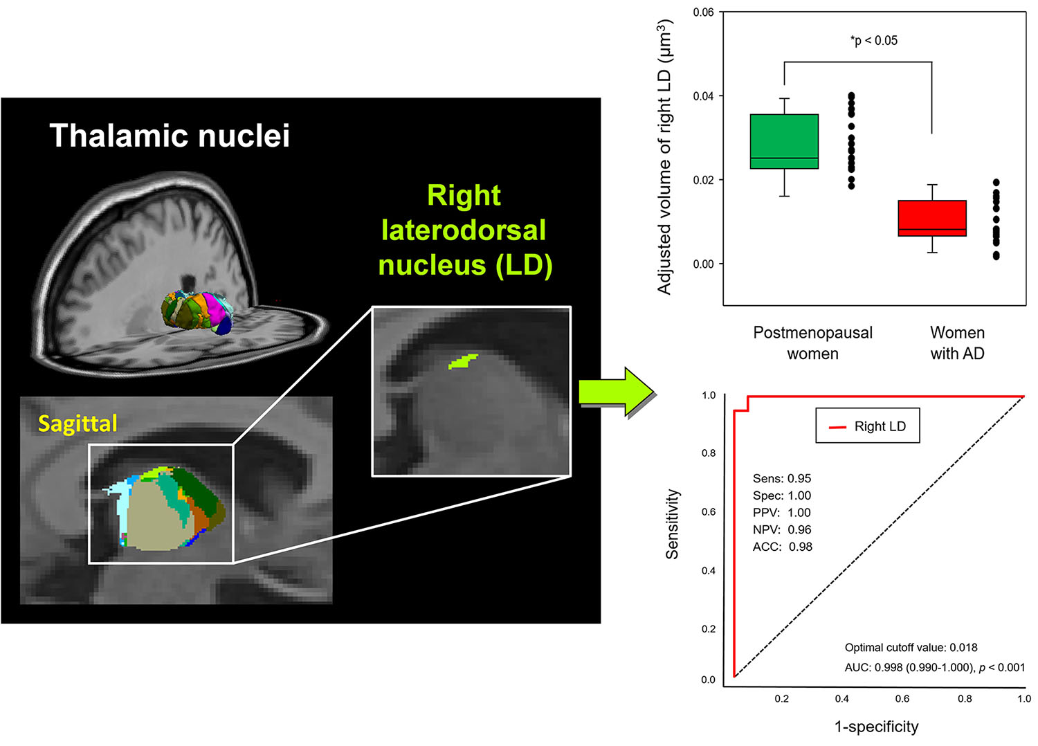
Twenty-one women with AD (mean age = 74.1 ± 8.3 years) and 21 postmenopausal women without AD (mean age = 55.2 ± 2.5 years) participated in this study. The symptom severity of AD was evaluated using the questionnaires of the mini-mental state examination (K-MMSE) and AD assessment scale−cognitive subscale (ADAS-Cog). Serum sex hormones including free testosterone (free-T) and estradiol (E2) were measured. Magnetic resonance imaging (MRI) scans were acquired using a 3.0 Tesla Magneton Tim Trio MR Scanner (Siemens Medical Solutions, Erlangen, Germany). T1 images were analyzed using SPM 12 software with difeomorphic anatomical registration through exponentiated Lie algebra (DARTEL) analysis. The thalamic subnuclei were calculated using the FreeSurfer v7.2 software. Twenty-five thalamic subnuclei (50 regions of interest (ROIs)) were extracted extracted from each hemisphere of the subject's T1 image (Fig. 1). Each thalamic subnucleus was used to evaluate the adjusted volume using the following equation: Adjusted volume (μm3) = (Each thalamic subnuclear volume (mm3) / Whole brain volume (mm3)) × 1000.Results
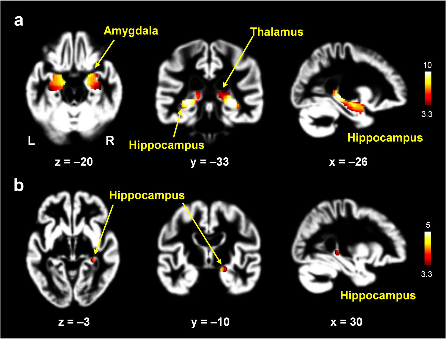
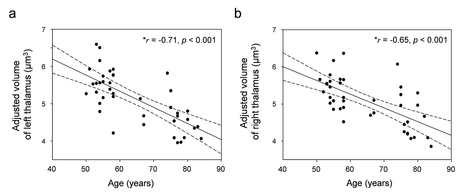
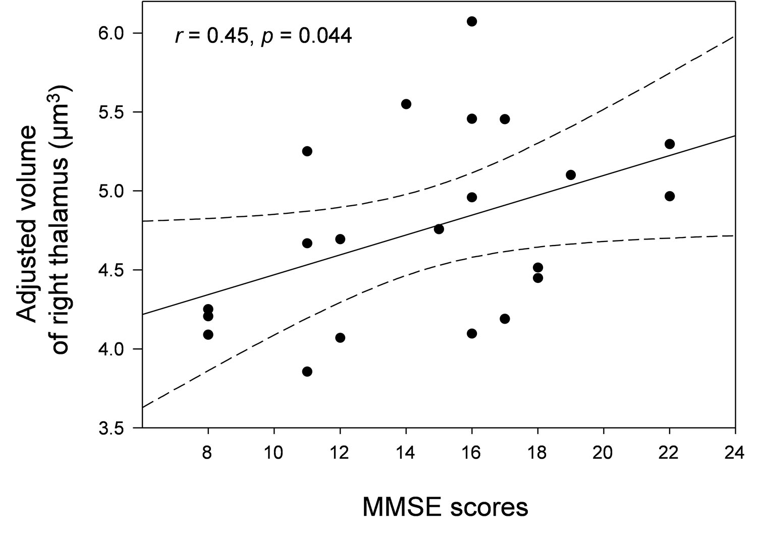
In the voxel-wise analysis, women with AD showed significantly lower gray matter volumes in the hippocampus, thalamus, and amygdala (p < 0.05, FWE-corrected; Fig. 2a). After adjusting for age, women with AD showed significantly lower gray matter volumes in the hippocampus (p < 0.05, FWE-corrected; Fig. 2b). Age was negatively correlated with the adjusted left (r = –0.71, p < 0.001) and right (r = –0.65, p < 0.001) thalamic volumes, respectively (Fig. 3). However, there were no significant correlations between the adjusted volumes of the left or right thalamic regions and the levels of estrogen or E2 in postmenopausal women (p > 0.05). The MMSE scores in women with AD were positively correlated with adjusted right thalamic volumes (r = 0.45, p = 0.044; Fig. 4). Although women with AD did not show volume atrophy in the thalamus after adjusting for age, the volume of the right laterodorsal thalamic nucleus was significantly decreased in women with AD compared to postmenopausal women (p < 0.05, Bonferroni-corrected; Fig. 5a,b). The sensitivity, specificity, positive predictive value (PPV), negative predictive value (NPV), accuracy, cutoff value, and area under the receiver operating characteristic (ROC) curve (AUC) of the adjusted volume of the right laterodorsal nucleus for diagnosing AD were 0.95, 1.00, 1.00, 0.96, 0.98, 0.018, and 0.998 (95% confidence interval: 0.990 – 1.000), respectively (Fig. 5c). The results indicated that right laterodorsal nucleus volume may be a predictive biomarker for AD. No significant differences were found in the volumes of the other 49 thalamic subnuclei ROIs between the two groups (all p > 0.05).Conclusion
This study compared differential thalamic subnuclear volumes between women with AD and postmenopausal women. Our findings suggest that reduced volume in both the right laterodorsal thalamic nucleus and right hippocampus could represent a key biomarker for predicting early stage of AD in postmenopausal women. These findings may be helpful for a better understanding of AD pathogenesis and also for providing an objective target for early interventions to prevent AD.Acknowledgements
This research was supported by the grants from the National Research Foundation funded by the Korea government (MSIT; 2021R1C1C2011748 and 2022R1A2C2007809).References
1. Barron AM, Pike CJ (2012) Sex hormones, aging, and Alzheimer's disease. Front Biosci (Elite Ed). 4:976-997.
2. Horstman AM, Dillon EL, Urban RJ, Sheffield-Moore M (2012) The role of androgens and estrogens on healthy aging and longevity. J Gerontol A Biol Sci Med Sci 67:1140-1152.
3. Subramaniapillai S, Almey A, Natasha RM, Einstein G (2021) Sex and gender differences in cognitive and brain reserve: Implications for Alzheimer's disease in women. Front Neuroendocrinol 60:100879.
4. Xiao Y, Wang J, Huang K, Gao L, Yao S (2022) Alzheimer's Disease Neuroimaging, I. Progressive structural and covariance connectivity abnormalities in patients with Alzheimer's disease. Front Aging Neurosci 14:1064667.
5. van de Mortel LA, Thomas RM, van Wingen GA, Initia AND (2021) Grey Matter Loss at Different Stages of Cognitive Decline: A Role for the Thalamus in Developing Alzheimer's Disease. Journal of Alzheimers Disease 83:705-720.
6. Kim GW, Kim BC, Park K, Jeong GW (2020) A pilot study of brain morphometry following donepezil treatment in mild cognitive impairment: volume changes of cortical/subcortical regions and hippocampal subfields. Sci Rep 10:10912.
Figures