3897
Investigation the relationship between cerebral small veins and microbleeds using 7T QSM in Alzheimer’s disease1Urmia University of Medical Science, Urmia, Iran (Islamic Republic of), 2Icahn School of Medicine at Mount Sinai, New York, NY, United States
Synopsis
Keywords: Alzheimer's Disease, Alzheimer's Disease
Motivation: Cerebral microbleeds (CMBs) are small hypointense lesions often associated with cerebral small vessel diseases like cerebral amyloid angiopathy and Alzheimer's disease (AD).
Goal(s): Our aim is to evaluate the feasibility of using ultra-high resolution QSM at 7T MRI to find a link between small veins and CMBs in AD.
Approach: We used 7T QSM to established a connection between CMBs and the venous vasculature to evaluate venous contributions to AD conditions.
Results: Our data support the notion that CMBs might not exclusively derive from arteries, but that venous contribution could play an important, yet not much explored, role in CMBs in AD cohorts.
Impact: Our results provide evidence of a potential connection between CMBs and small veins, suggesting a potential role for veins in AD.
Introduction
Cerebral microbleeds (CMBs) are small hypointense round lesions that indicate leakage of blood products from cerebral vessels damaged by β-amyloid (Aβ) and typically are detected by T2*-weighted GRE and susceptibility-weighted imaging (SWI) on MRI1,2. They are indicators of cerebral small vessel diseases like cerebral amyloid angiopathy and Alzheimer's disease (AD), primarily affecting cortical small arteries3. Notably, cortical and leptomeningeal venous Aβ accumulation has been observed in preclinical AD models4. Therefore, the possibility of small veins playing a role in the development of CMBs, attributed to venous pathology associated with cerebral small vessel diseases, is also worth considering. Results reported by Rotta et al. established a connection between cerebral CMBs and venous vasculature, suggesting that veins may play a role in cerebral small vessel disease5. This result was discerned through the utilization of the Quantitative Susceptibility Mapping (QSM) technique. QSM is an advanced MRI imaging technique used to quantify the magnetic susceptibility of tissues in the human body6. It is particularly valuable in neuroimaging to study various aspects of brain anatomy and function. In this study, we utilized the QSM technique to analyze venous structures at ultra-high resolution in a cohort of patients with AD and age-matched control subjects using 7T MRI. Our aim is to evaluate the feasibility of using ultra-high resolution QSM at 7T MRI to find a link between small veins and CMBs in individuals with AD.Methods
In-vivo human imaging was performed using a 7T MRI scanner (Siemens Healthineers, Erlangen, DE) with a Nova Medical 32Rx/1Tx head coil (Wilmington, Massachusetts, USA). Multi-echo magnitude and phase images were obtained using a 3D multi-echo GRE sequence (Echo number = 6, echo times (TE) =4 ms, DTE=4ms, voxels size = 0.3x0.3x1.5 mm3, TR= 32 ms, flip angle = 12°, and bw = 160 Hz/px). Generation of QSM requires several discrete steps. First, multi-echo phase images were combined supposing a linear weighted phase increment followed by Laplacian unwrapping7. Then, the background phase was removed using the variable-kernel sophisticated harmonic artifact reduction (VSHARP) for the phase data8. In the last step, QSM was calculated by dipolar inversion of the background corrected phase maps using the morphology-enabled dipole inversion (MEDI) algorithm9. A brain mask was created by using the BET tool of the FSL Software Library. We also obtained SWI images using the CLEAR SWI technique10. The same magnitude and phase images extracted from the multi-echo GRE sequence were used to post-process the SWI images. SWI postprocessing included Laplacian unwrapping, and magnitude homogeneity correction to avoid wrap-like artifacts and reduce signal dropouts and intensity variations.Results
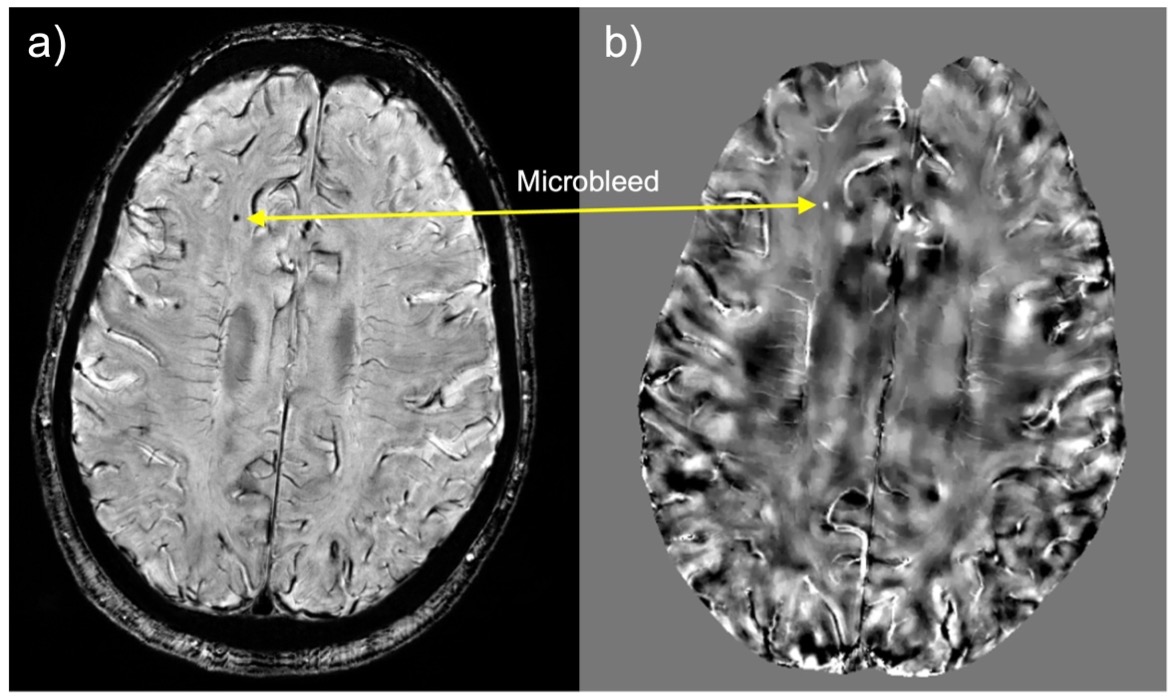
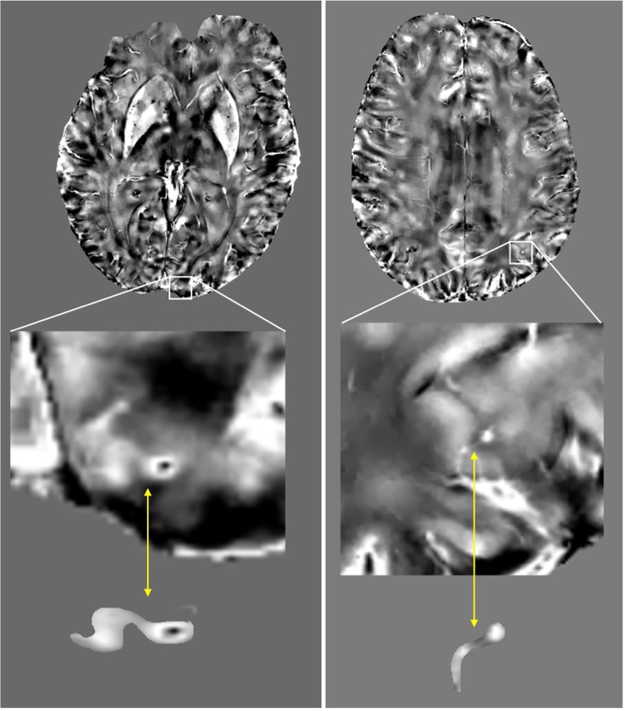
The images were analyzed by two neuroradiologists. CMBs were defined as hyperintense, round lesions in the QSM images. For verification, their appearance was matched on the SWI images, where CMBs are hypointense and associated with (Figure 1). CMBs with a direct link to a vein were assessed in the QSM sequence (Figure 2). They were defined as CMBs with small veins connection. This high-resolution 7T neuroimaging study investigated whether a spatial relationship between small veins and CMBs could be detected in a cohort of AD patients. QSM enabled the depiction of cerebral CMBs with a direct connection to a vein and their localization. Our data support the notion that CMBs might not exclusively derive from arteries, but that venous contribution could play an important, yet not much explored, role in CMBs in AD cohorts.Discussion
A key strength of our study lies in the utilization of high-resolution QSM at 7T MRI. High-resolution MRI enhances the visibility of small structures, and when maintained at the same resolution, imaging at a higher field MRI increases the signal-to-noise ratio. QSM, as a postprocessing technique, elevates CMB detection sensitivity while facilitating precise visualization of the brain's venous vasculature. Notably, the postprocessing algorithm ensures a high level of specificity for venous blood susceptibilities, minimizing the likelihood of measuring arterioles or slow-flow small arteries. Furthermore, by virtue of QSM's ability to eliminate the blooming effect, we can confidently rule out this artifact as a factor in the observed relationship between small veins and CMBs in AD cohorts.Conclusion
In this study we showed the feasibility of using 7T high-resolution QSM to investigate the spatial relation between cerebral small veins and CMBs in AD patients. These results provide evidence of a potential connection between CMBs and small veins, suggesting a potential role for veins in AD. To substantiate our findings, it is essential to conduct further investigations with larger study cohorts and incorporate pathological studies for confirmation.Acknowledgements
The authors would like to thank research coordinator Aislinn Diaz helping with the recruitment. This study was supported by a Developmental Project award from Mount Sinai ADRC (P30 AG066514) NIA/NIH, R21AG071179, and K01 AG075178-01 NIA/NIH grants.References
1. Greenberg SM, Vernooij MW, Cordonnier C, et al. Cerebral microbleeds: a guide to detection and interpretation. Lancet Neurol 2009;8:165–174.
2. Vernooij MW, Ikram MA, Wielopolski PA, Krestin GP, Breteler MM, van der Lugt A. Cerebral microbleeds: accelerated 3D T2*-weighted GRE MR imaging versus conventional 2D T2*-weighted GRE MR imaging for detection. Radiology. 2008 Jul;248(1):272-7. doi: 10.1148/radiol.2481071158. Epub 2008 May 19. PMID: 18490493.
3. Cordonnier C, van der Flier WM (2011) Brain microbleeds and Alzheimer’s disease: Innocent observation or key player? Brain 134, 335-344.
4. Keith J, Gao FQ, Noor R, Kiss A, Balasubramaniam G, Au K, Rogaeva E, Masellis M, Black SE. Collagenosis of the Deep Medullary Veins: An Underrecognized Pathologic Correlate of White Matter Hyperintensities and Periventricular Infarction? J Neuropathol Exp Neurol. 2017 Apr 1;76(4):299-312. doi: 10.1093/jnen/nlx009. PMID: 28431180.
5. Rotta J, Perosa V, Yakupov R, Kuijf HJ, Schreiber F, Dobisch L, Oltmer J, Assmann A, Speck O, Heinze HJ, Acosta-Cabronero J, Duzel E, Schreiber S. Detection of Cerebral Microbleeds With Venous Connection at 7-Tesla MRI. Neurology. 2021 Apr 20;96(16):e2048-e2057. doi: 10.1212/WNL.0000000000011790. Epub 2021 Mar 2. PMID: 33653897.
6. Liu C, Li W, Tong KA, Yeom KW, Kuzminski S. Susceptibility-weighted imaging and quantitative susceptibility mapping in the brain. J Magn Reson Imaging. 2015 Jul;42(1):23-41. doi: 10.1002/jmri.24768. Epub 2014 Oct 1. PMID: 25270052; PMCID: PMC4406874.
7. Li W, Wu B, Liu C. Quantitative susceptibility mapping of human brain reflects spatial variation in tissue composition. Neuroimage. 2011 Apr 15;55(4):1645-56. doi: 10.1016/j.neuroimage.2010.11.088. Epub 2011 Jan 9. PMID: 21224002; PMCID: PMC3062654.
8. Özbay PS, Deistung A, Feng X, Nanz D, Reichenbach JR, Schweser F. A comprehensive numerical analysis of background phase correction with V-SHARP. NMR Biomed. 2017 Apr;30(4):10.1002/nbm.3550. doi: 10.1002/nbm.3550. Epub 2016 Jun 3. PMID: 27259117; PMCID: PMC5136354.
9. Liu T, Liu J, de Rochefort L, Spincemaille P, Khalidov I, Ledoux JR, Wang Y. Morphology enabled dipole inversion (MEDI) from a single-angle acquisition: comparison with COSMOS in human brain imaging. Magn Reson Med. 2011 Sep;66(3):777-83. doi: 10.1002/mrm.22816. Epub 2011 Apr 4. PMID: 21465541.
10. Eckstein K, Bachrata B, Hangel G, Widhalm G, Enzinger C, Barth M, Trattnig S, Robinson SD. Improved susceptibility weighted imaging at ultra-high field using bipolar multi-echo acquisition and optimized image processing: CLEAR-SWI. Neuroimage. 2021 Aug 15;237:118175. doi: 10.1016/j.neuroimage.2021.118175. Epub 2021 May 15. PMID: 34000407; PMCID: PMC7612087.
Figures