3896
Neurovascular coupling dysfunction in type 2 diabetes patients1the First Affiliated Hospital of Dalian Medical University, Dalian, China
Synopsis
Keywords: Other Neurodegeneration, Diabetes, neurovascular coupling; fMRI; cerebral blood flow
Motivation: The neurovascular coupling is the basic structure of neurovascular unit. Various pathological injuries lead to abnormalities in neuronal activity and blood flow resulting in impaired function. There is still not much research on the coupling pattern.
Goal(s): We hypothesize that there exists an abnormal NVC pattern for T2DM.
Approach: We recruited 40 patients clinically diagnosed with T2DM, healthy controls and underwent BOLD fMRI scanning and image analysis.
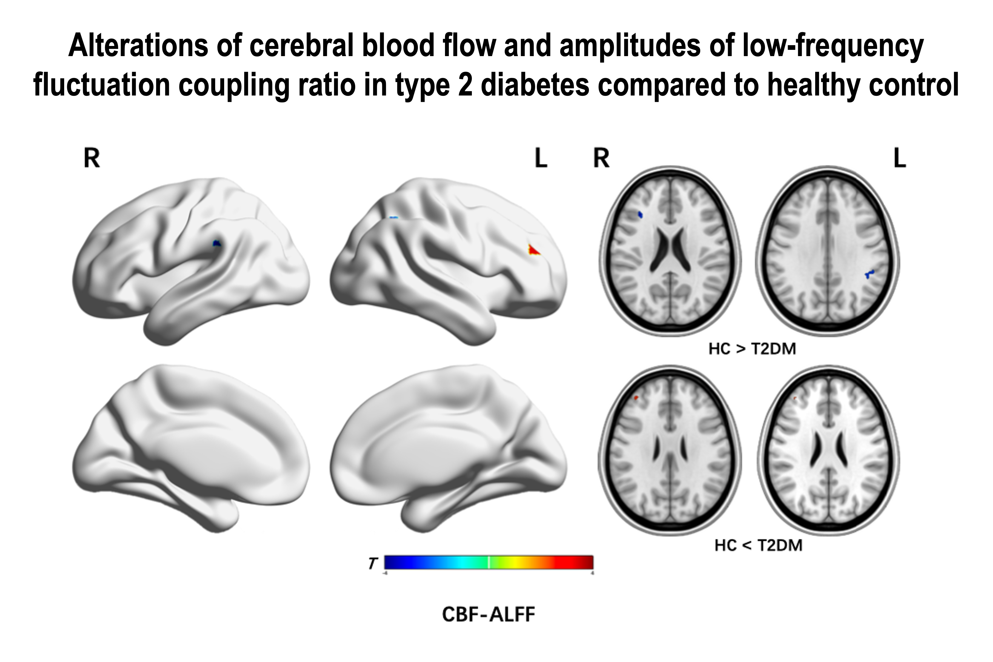
Results: Results found significant decreased CBF-ALFF ratio in supramarginal and frontal gyrus, and increased values in middle frontal gyrus of T2DM. The ALFF and CBF was found to be reduced or elevated in several brain regions.
Impact: In addition to the frontal and parietal gyrus, the cerebellum is also involved in the mechanisms of brain function impairment in T2DM. The different coupling results indicate the existence of mutually constraining balancing mechanism between neural activity and blood perfusion.
Introduction
The neurovascular coupling (NVC), consisting mainly of neurons, astrocytes and blood vessels, is the basic structure of the neurovascular unit. Increased neuronal activity often leads to increased cerebral blood flow (CBF) because of greater metabolic demands. However, various pathological injuries lead to abnormalities in neuronal activity and CBF resulting in impaired function of NVC. More studies have now revealed abnormal resting-state functional indicators and abnormal CBF in type 2 diabetes (T2DM). However, there is still not much research on the altered NVC pattern. We hypothesize that there exists an abnormal NVC pattern for T2DM. Therefore, we conducted this prospective study to analyze changes in the amplitudes of low-frequency fluctuation (ALFF), CBF and their coupling indicators (CBF-ALFF) to explore the mechanisms of cognitive impairment in T2DM.Methods
We recruited 40 patients clinically diagnosed with T2DM and 24 healthy controls (HC group). All participants were scanned using a Philips Ingenia CX 3.0T MR scanner (Philips Healthcare, Best, the Netherlands) with 32-channel head coil. Functional images were obtained using a gradient-echo planar sequence with 200 time points. Perfusion images were obtained using a 3D-pCASL technique. Functional data were analyzed using DPARSF on MATLAB. The first 10 points were discarded and then remaining data were slice-timing corrected, realigned, coregistered, and regressed to remove nuisance covariates, then normalized to the standard space and temporal bandpass filtered. Subjects with excessive head movement were discarded. The CBF maps were calculated and normalized. ALFF value was calculated and then ALFF and CBF values were standardized and smoothed. Finally, CBF-ALFF ratio of each voxel was computed. Two samples t-test and multiple comparison were implemented at grey matter level with gender, ages and education years as covariates. P < 0.05 was considered statistically significant.Results
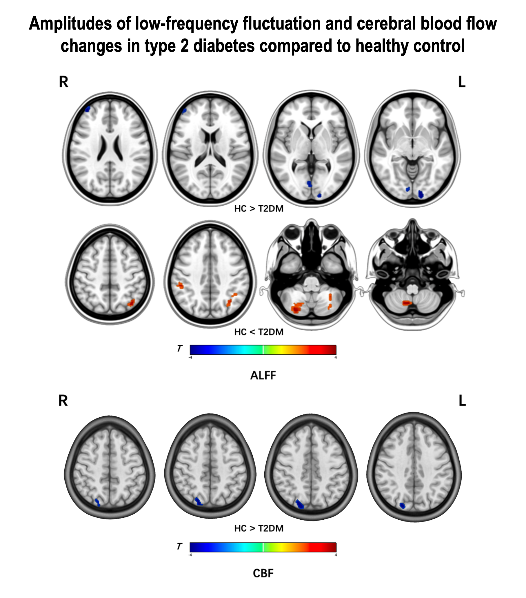
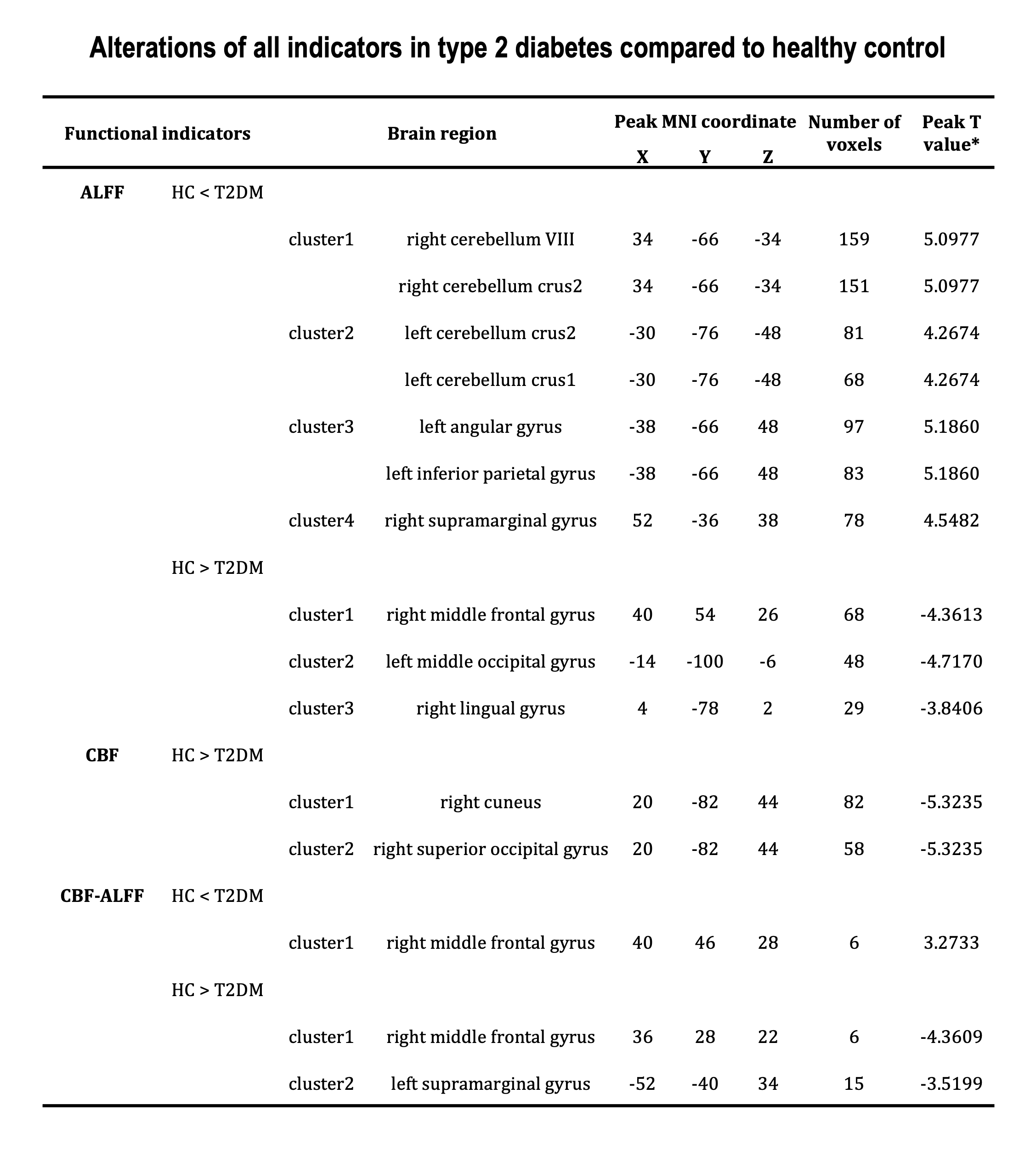
Compared with HC, we observed significant decreased CBF-ALFF ratio in left supramarginal gyrus and right middle frontal gyrus, and increased values in right middle frontal gyrus of T2DM group. The ALFF and CBF value was found to be reduced or elevated in several brain regions, including the lingual gyrus, cuneus, occipital lobe and cerebellum, in addition to the above-mentioned brain regions. (Gaussian random fields corrected, P < 0.05)Discussion
ALFF is thought to reflect spontaneous neural activity and has physiological significance. There is growing evidence that the cerebellum 1 and 2 plays a role in regulating cognition and emotion, attention processing, and decision making [1]. We found a greater number of regions with ALFF alterations, especially in cerebellum crus 1 and 2, suggesting that there are abnormalities of function. This finding is also consistent with other studies [2]. Decreased blood flow in occipital gyrus was also observed on CBF, which is similar to other studies [3]. The frontal lobes are involved in higher cognitive functions. Supramarginal gyrus belongs to the inferior parietal lobule and is an important component of the default mode and attention network [4]. The present study demonstrates the presence of damage to the neurovascular unit in these brain regions. But this result is not quite consistent with the results of the individual indicators. We hypothesize that it may be that the individual indicators are not sufficient to reflect the overall changes in the brain regions, suggesting that coupling can identify abnormal regions where ALFF and CBF are not altered. Moreover, it also confirms the existence of a mutually constraining balancing mechanism between neural activity and blood perfusion.Conclusion
Patients with T2DM have functional abnormalities and perfusion abnormalities in several brain regions compared to normal individuals, especially in the cerebellar region. Impaired neurovascular coupling was also observed in areas of higher cognitive function and in the default and attentional network brain regions. We hypothesize that this may be the result of a mutually constraining balancing mechanism between neural activity and blood perfusion. This provides some insight into advanced neurologic impairment in T2DM.Acknowledgements
No acknowledgement found.References
1. Sathyanesan A, Zhou J, Scafidi J, Heck DH, Sillitoe RV, Gallo V. Emerging connections between cerebellar development, behaviour and complex brain disorders. Nat Rev Neurosci. 2019;20(5):298-313. doi: 10.1038/s41583-019-0152-2
2. Zhang D, Qi F, Gao J, et al. Altered Cerebellar-Cerebral Circuits in Patients With Type 2 Diabetes Mellitus. Front Neurosci. 2020;14:571210. Published 2020 Sep 24. doi:10.3389/fnins.2020.571210
3. İlik İ, Arslan H, Yokuş A, Batur M, Üçler R, Akdeniz H. Cerebral perfusion in type 2 diabetes mellitus: A preliminary study with MR perfusion. Clin Neurol Neurosurg. 2023;231:107816. doi: 10.1016/j.clineuro.2023.107816
4. Hampel H. Amyloid-β and cognition in aging and Alzheimer's disease: molecular and neurophysiological mechanisms. J Alzheimers Dis. 2013;33 Suppl 1:S79-S86. doi: 10.3233/JAD-2012-129003
Figures