3895
Neurovascular coupling dysfunction of hippocampus-visual network in Post-Concussive Syndrome1Lanzhou University Second Hospital, LanZhou, China
Synopsis
Keywords: Other Neurodegeneration, Arterial spin labelling, post concussion syndrome
Motivation: The clinical relevance and modifications in perfusion and neural activity remain undetermined in Post-Concussive Syndrome.
Goal(s): To delineate changes in NVC during the pathophysiology of PCS, multimodal magnetic resonance imaging was performed on patients and healthy controls.
Approach: Five imaging-markers elucidates the relationship between neuronal activity and perfusion. Dynamic causal modeling evaluates associations with disease transitions, clinical variables, and information flow.
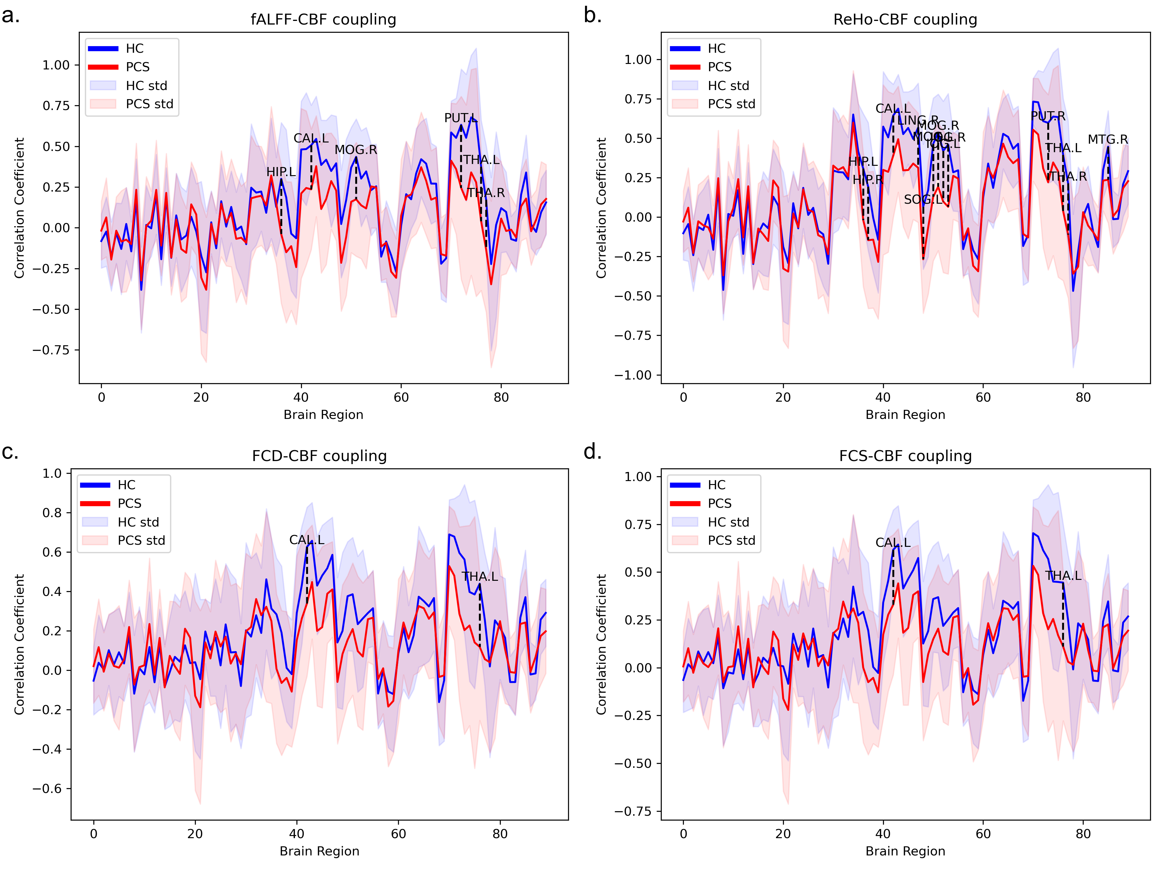
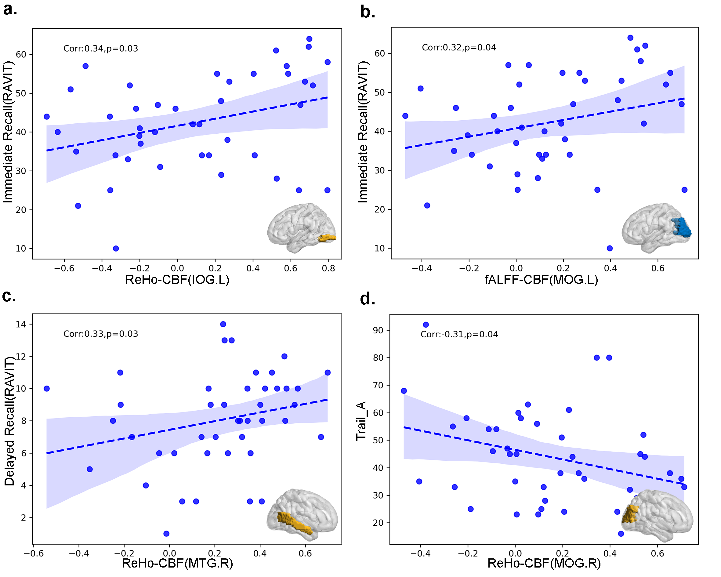
Results: The fALFF-CBF of the hippocampal was diminished, and the ReHo-CBF in the Temporal-Occipital cortex decreased, correlating with declining spatial processing and memory tasks.
Impact: The distinctive pattern of changes in neurovascular coupling in the hippocampal , temporal-occipital cortex provide fresh insights into PCS
Purpose
Post-Concussive Syndrome (PCS) is characterized by alterations in perfusion and neural activity, with changes in neurovascular coupling (NVC) playing a pivotal role in the evolution of post-injury sequelae and brain repair mechanisms following trauma. However, the clinical relevance and modifications in this coupling remain undetermined. To delineate changes in NVC during the pathophysiology of PCS, multimodal magnetic resonance imaging was performed on 43 patients, injured for over 4 weeks, and 35 healthy controls.Methods
Methods: For evaluations, five imaging markers - amplitude of low-frequency fluctuations (ALFF), fractional ALFF (fALFF), regional homogeneity (ReHo), functional connectivity strength (FCS), and functional connectivity density (FCD) were employed to quantify segregated and integrated neuronal activity. Cerebral blood flow (CBF) was utilized for vascular reactivity assessment. Pearson correlation coefficients were computed to elucidate the relationship between neuronal activity and cerebral perfusion. Furthermore, dynamic causal modeling was adopted to evaluate associations with disease transitions, clinical variables, and information flow.Results
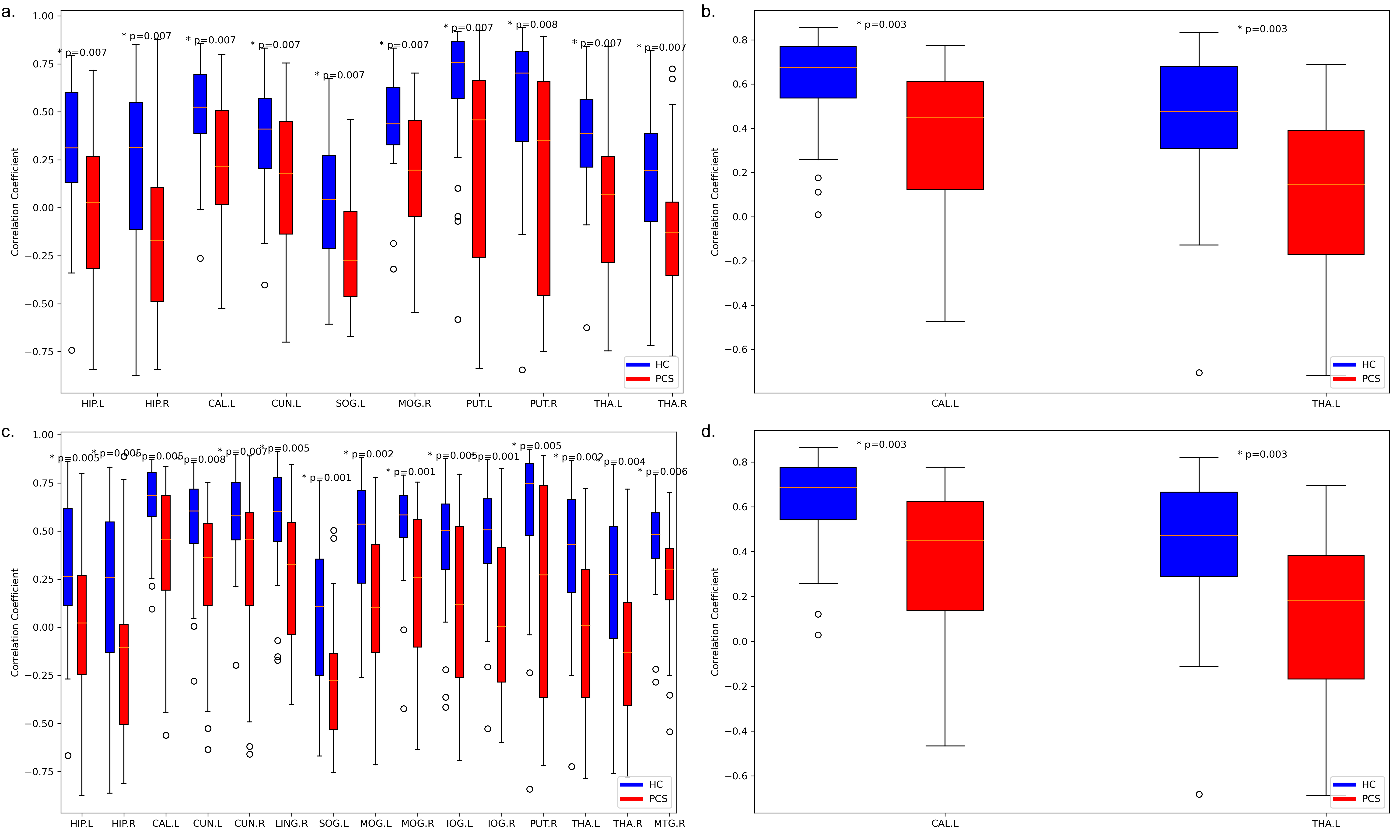
PCS patients exhibited impaired NVC. No significant discrepancies at the gray matter level were found for the aforementioned five neurovascular biomarkers. they were lower than those in HCs, indicating global decoupling in PCS. Although, neurovascular biomarkers in 15 distinct brain regions significantly diminished in PCS patients. The fALFF-CBF and ReHo-CBF in the left pericallosal fissure and right cuneus demonstrated negative correlations with Trial A of the digit test, while the fALFF-CBF in the right cuneus and the ReHo-CBF in the right wedge and cuneus were negatively correlated with the digit backward test (Trial B). The fALFF-CBF in the right cuneus had a positive correlation with orientation ability on the MocA scale, and the fALFF-CBF in the right medial temporal gyrus was positively correlated with immediate memory capabilities in the Rey Auditory Verbal Learning Test. Disease severity was negatively correlated with memory and executive function.The fALFF-CBF of the hippocampal cortex was diminished, and in the Visual Network (VN), the ReHo-CBF in the Temporal-Occipital cortex decreased, correlating with declining spatial processing and memory tasks.Discussion
Changes in NVC were explored by analyzing multi-parametric coupling between cerebral perfusion and corresponding intrinsic neural activity in PCS. The observed neurovascular unit damage in some brain areas may account for the reduced connections between vascular response and the neural activity in PCS, providing supporting evidence for the concept of disease-related decoupling.However, voxel-based analysis did not reveal significant differences. This may suggest that voxel-based analytical methods are inferior to ours in detecting brain injury-associated coupling, or they may not be appropriate for discerning impaired NVC. The decrease in the NVC coupling coefficient in the left pericallosal fissure, occipital lobe, and temporal lobe in the PCS group signals a decline in attention control, spatial processing, working memory, along with more delayed reaction times in decision-making. This suggests that PCS patients may exhibit persistent integration abnormalities in regional cerebral blood flow and neural activity post-trauma, leading to enduring behavioral and cognitive impairments. Insufficient blood supply per unit of functional segregation in the hippocampal cortex and temporal-occipital cortex may impact the functional activities of specific dorsal visual areas related to spatial vision in PCS, resulting in a decline in visual spatial processing functions and attention.Conclusion
The distinctive pattern of changes in neurovascular coupling in the hippocampal cortex and temporal-occipital cortex might provide fresh insights into understanding the pathophysiological mechanisms of PCS and its influence on cognitive functions. It may also aid in the development of more effective therapeutic strategies.Acknowledgements
No acknowledgement found.References
[1] Acosta CH, Clemons GA, Citadin CT, et al. A role for protein arginine methyltransferase 7 in repetitive and mild traumatic brain injury[J/OL]. Neurochem Int. 2023; 166:105524. DOI: 10.1016/j.neuint.2023.105524. [2] Li T, Liu T, Zhang J, et al. Neurovascular coupling dysfunction of visual network organization in Parkinson's disease[J]. Neurobiol Dis. 2023 Oct 12;188:106323. DOI: 10.1016/j.nbd.2023.106323. [3] Hu B, Yan LF, Sun Q,et al. Disturbed neurovascular coupling in type 2 diabetes mellitus patients: Evidence from a comprehensive fMRI analysis[J]. Neuroimage Clin. 2019;22:101802. DOI: 10.1016/j.nicl.2019.101802. [4] Iyer KK, Barlow KM, Brooks B, et al. Relating brain connectivity with persistent symptoms in pediatric concussion[J]. Ann Clin Transl Neurol. 2019; 6(5):954-961. DOI: 10.1002/acn3.764. [5] Amir J, Nair JKR, Del Carpio-O'Donovan R, et al. Atypical resting state functional connectivity in mild traumatic brain injury[J/OL]. Brain Behav. 2021; 11(8):e2261.[6]Barlow KM, Iyer K, Yan T, et al. Cerebral Blood Flow Predicts Recovery in Children with Persistent Post-Concussion Symptoms after Mild Traumatic Brain Injury[j]. J Neurotrauma. 2021 Aug 15;38(16):2275-2283. DOI: 10.1089/neu.2020.7566.Figures