3894
3D pCASL with dual PLDs could reflect cerebral blood flow regulation in patients with hydrocephalus1The Second Affiliated Hospital of Nanchang University, Nanchang, China, 2GE Healthcare, MR Research China, Beijing, China
Synopsis
Keywords: Other Neurodegeneration, Arterial spin labelling, Hydrocephalus, cerebral blood flow, cerebral blood flow regulation, hemodynamics
Motivation: It is unclear whether the 3D pCASL with dual PLDs can reflect blood flow (CBF) regulation in hydrocephalus patients.
Goal(s): To investigate hemodynamic characteristics in patients with hydrocephalus and whether ΔCBF can reflect cerebral regulation.
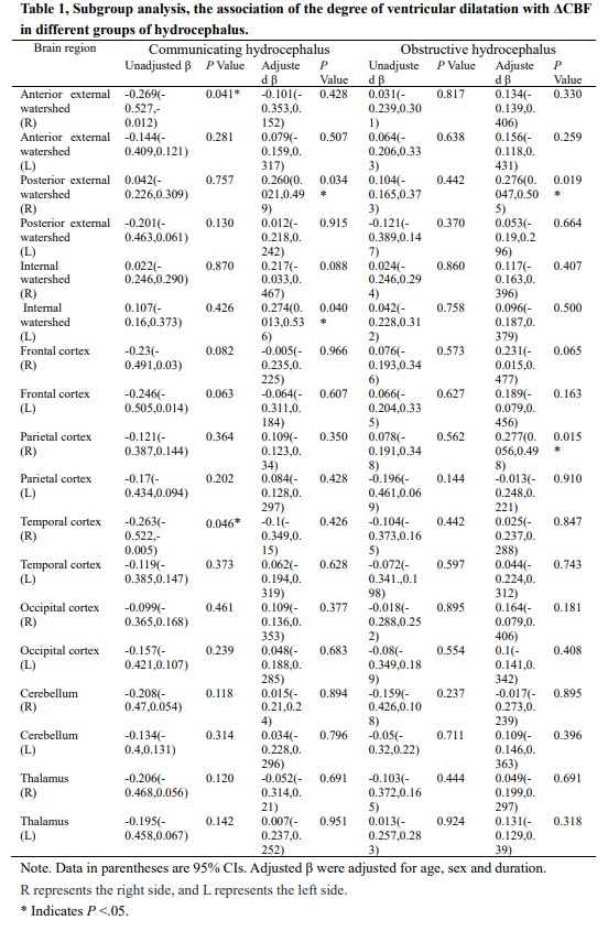
Approach: Patients with hydrocephalus and control subjects were retrospectively included. Regional CBF and ΔCBF were compared using covariance analyses. The relationship between ΔCBF and ventricular dilatation degree was investigated using linear regression analyses and interaction analysis.
Results: Compared to the control group, hydrocephalus patients showed larger ΔCBF in all brain regions except for the bilateral parietal cortex and cerebellum.
Impact: Patients with hydrocephalus initiate cerebral regulation to maintain CBF but require longer arterial transit times. The ability to regulate CBF in brain regions represented by the watershed is associated with the degree of ventricular dilation.
Introduction
Hydrocephalus is a pathological state that can be caused by a variety of reasons and mainly classified as obstructive hydrocephalus and communicating hydrocephalus. The physiological disorder of cerebrospinal fluid leads to abnormal ventricular expansion, usually accompanied by an increase in intracranial pressure1. Ventriculomegaly leads to the compression and extension of periventricular tissues, leading to ischemia, hypoxia, inflammation, and further leading to metabolic disorders and alterations in the permeability of the blood–brain barrier2.Research has identified that ventriculomegaly may directly reduce cerebral blood flow (CBF) through mechanical traction and vascular caliber reduction3. Previous studies used SPECT, CTP, and MR DSC to certify that the CBF of hydrocephalus patients was decreased, but these methods required the use of a radioactive tracer or contrast agent, SPECT, CTP exposed patients to ionizing radiation, and SPECT was expensive and had lower clinical universality4-6. These studies still leave a question, whether the brain regulate and maintain CBF in hydrocephalus patients experiencing hypoperfusion, ischemia, and hypoxia.
Three-dimensional pseudo-continuous arterial spin labeling (3D-pCASL) imaging is a non-invasive sequence for detecting CBF without contrast injection. CBF of different ages and diseases can be measured more accurately and reproducible by setting the suitable post-labeling delay time (PLD). Short PLD is sensitive to rapid blood flow, and long PLD may reflect delayed or compensated blood flow7. However, the method of single PLD obtains static information and cannot dynamically reflect the characteristics of CBF.
Therefore, we attempted to use the 3D-pCASL with dual PLDs to investigate hemodynamic characteristics in patients with hydrocephalus and whether ΔCBF can reflect cerebral regulation. The association between the ΔCBF and degree of ventricle dilation was also analyzed.
Material and Methods
Participants58 patients with communicating hydrocephalus, 57 patients with obstructive hydrocephalus, and 52 control subjects were included in the final analysis.
MRI protocol
Images were acquired on a 3.0T MR scanner (Discovery 750W, GE Healthcare, Waukesha, USA) with a 19-channel head and neck unit coil. MRI protocol included 3D FIESTA, Cube T2, DWI, MRA, T1WI, T2WI, FLAIR, as well as 3D pCASL with dual PLDs (1.5s and 2.5s).
Imaging processing
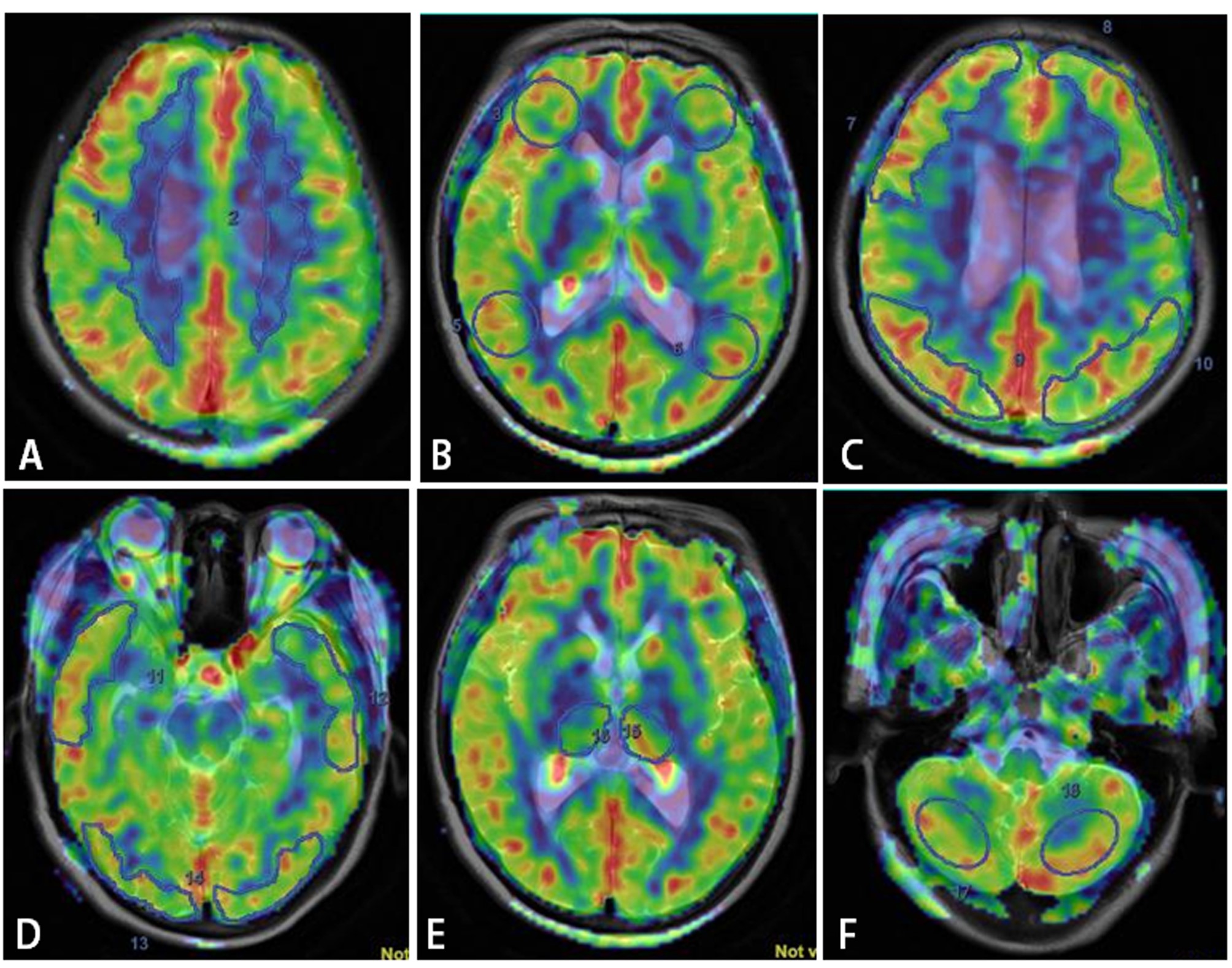
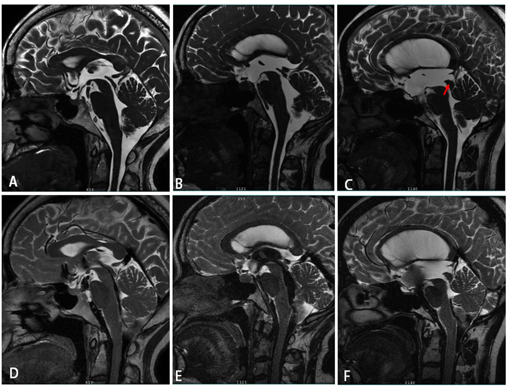
The 3D-pCASL was processed by the vendor-provided workstation (AW 4.7; GE Healthcare). The CBF maps derived from PLD 1.5s and 2.5s were co-registered and fused with T2WI or 3D T1 Bravo images. Manual corrections were performed when necessary. Then, region of interest (ROI) was manually delineated on the anatomical images to obtain the CBF values. The definition of ROIs used in this study was shown in Figure 1. Figure 2 showed representative images of healthy control, communicating and obstructive hydrocephalus to show the dilation of ventricle. The Evans’ index representing the degree of ventricular dilation was obtained by averaging the measurements from two neuroradiologists who were blind to patients’ information.
Statistical analysis
Differences in CBF and ΔCBF among groups were compared by using covariance analysis with post hoc LSD tests, correcting for the effect of age. Linear regression analyses were performed in communicating and obstructive hydrocephalus groups respectively, to assess the relationships between ΔCBF and ventricular dilatation after adjusting for age, sex, and duration. To further eliminate the effect of age on the CBF of hydrocephalus, the interaction between age and different hydrocephalus groups was analyzed by using the control group as a reference.
Results
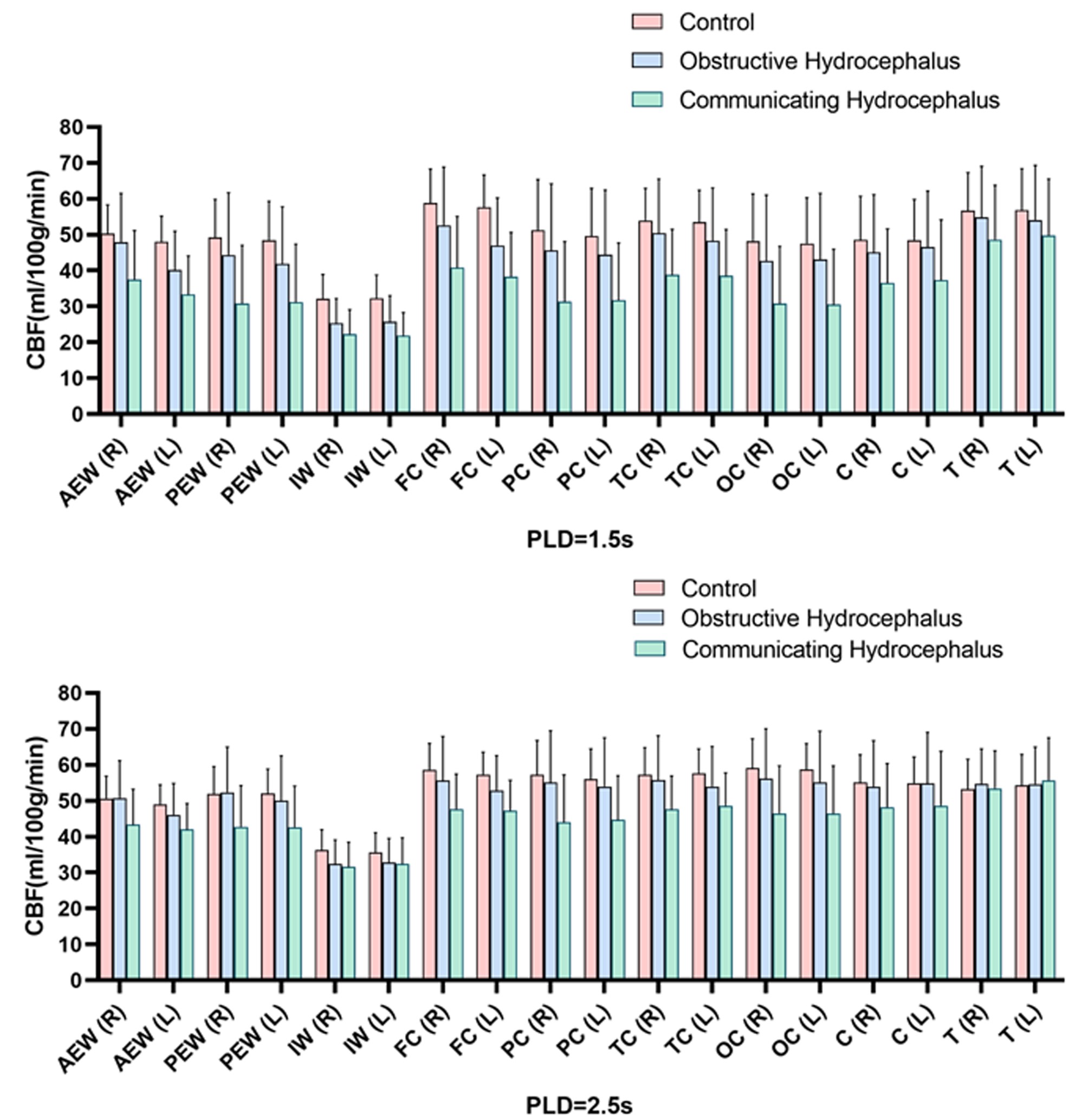
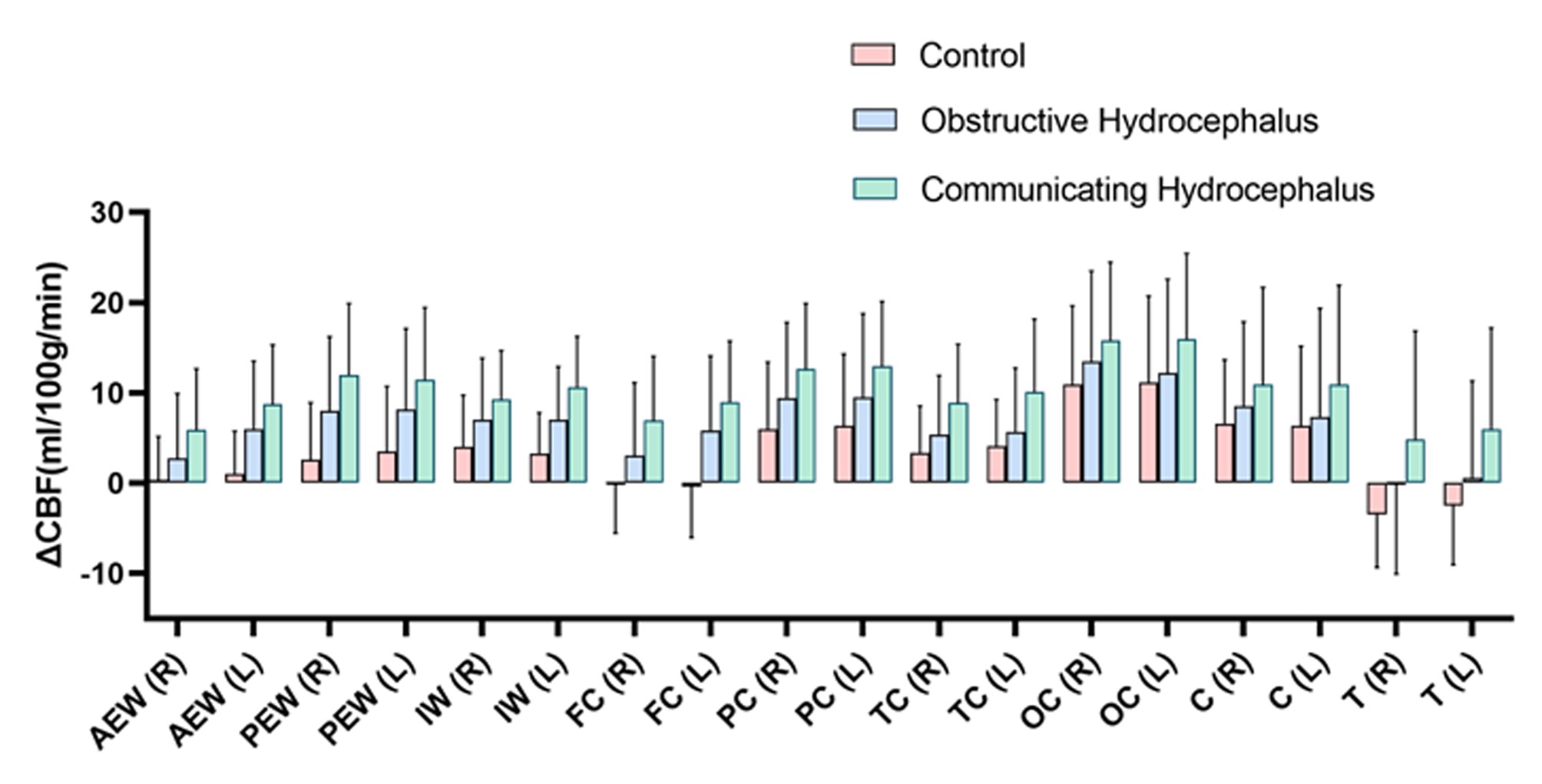
As shown in Figure 3, compared to healthy controls, both obstructive and communicating hydrocephalus patients presented decreased CBF in most brain regions for both PLD. Both hydrocephalus groups also showed larger ΔCBF in most brain regions (Figure 4). Furthermore, in the left medial watershed and left temporal cortex, communicating hydrocephalus exhibited greater ΔCBF than obstructive hydrocephalus. ΔCBF of the right posterior external watershed (adjusted β:0.276, P=0.019), right parietal cortex (adjusted β:0.277, P=0.015) in the obstructive hydrocephalus group while ΔCBF in the left internal watershed (adjusted β:0.274, P=0.04) in the communicating hydrocephalus group was associated with the degree of ventricular dilatation, respectively (Table 1).Discussion and Conclusions
This study investigated the hemodynamic alterations in hydrocephalus patients by 3D-pCASL with dual PLDs and its relationship with the degree of ventricular dilation in hydrocephalus. The results showed the CBF was decreased and the ΔCBF between the two PLDs in some brain regions were associated with ventricular dilatation in hydrocephalus patients. The ventriculomegaly may compress the brain parenchyma, causing vessels to initiate regulation and alter cerebral hemodynamics. Thus, the change in CBF may potentially reflects the cerebral self-regulation in the hypoperfusion pathology of hydrocephalus. In conclusion, this study provided insights into the hemodynamic regulation of hydrocephalus.Acknowledgements
No acknowledgement found.References
1. Kahle KT, Kulkarni AV, Limbrick DD, Jr., et al. Hydrocephalus in children. Lancet 2016; 387: 788-799.
2. McAllister JP, 2nd. Pathophysiology of congenital and neonatal hydrocephalus. Seminars in fetal & neonatal medicine 2012; 17: 285-294.
3. Del Bigio MR. Neuropathological changes caused by hydrocephalus. Acta Neuropathol 1993; 85: 573-585.
4. Takahashi R, Ishii K, Tokuda T, et al. Regional dissociation between the cerebral blood flow and gray matter density alterations in idiopathic normal pressure hydrocephalous: results from SINPHONI-2 study. Neuroradiology 2019; 61: 37-42.
5. van Asch CJ, van der Schaaf IC and Rinkel GJ. Acute hydrocephalus and cerebral perfusion after aneurysmal subarachnoid hemorrhage. AJNR Am J Neuroradiol 2010; 31: 67-70.
6. Graff-Radford NR, Rezai K, Godersky JC, et al. Regional cerebral blood flow in normal pressure hydrocephalus. Journal of neurology, neurosurgery, and psychiatry 1987; 50: 1589-1596.
7. Itagaki H, Kokubo Y, Kawanami K, et al. Arterial spin labeling magnetic resonance imaging at short post-labeling delay reflects cerebral perfusion pressure verified by oxygen-15-positron emission tomography in cerebrovascular steno-occlusive disease. Acta Radiol 2021; 62: 225-233.
Figures