3892
Age-Related Changes in Cerebral Blood Flow Values Measured Using Arterial Spin Labeling in Different Brain Regions of the Elderly1General Hospital of Ningxia Medical University, Yinchuan, China
Synopsis
Keywords: Aging, Aging
Motivation: In clinical work, there is an urgent need to understand the changes in CBF in different brain regions of normal elderly people to detect abnormalities.
Goal(s): to investigate and understand age-related changes in CBF values measured using ASL in different brain regions of the elderly
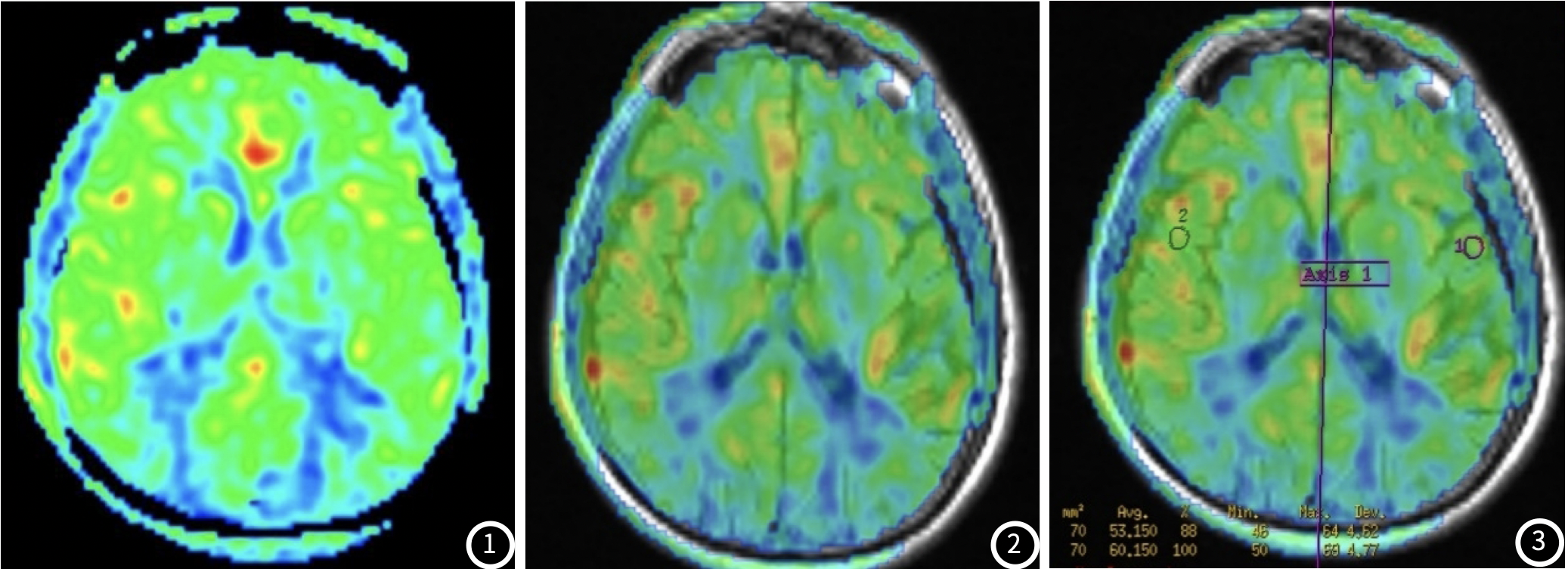
Approach: we recruited 344 healthy elderly participants and employed ASL magnetic resonance imaging to measure CBF values in multiple brain regions. We segmented the brain into distinct ROIs and quantified CBF values for each ROI.
Results: CBF reduction was manifested in some areas.Overall brain CBF decreases in both sexes begin at age 60 years
Impact: provides valuable insights into age-related alterations in CBF values measured using ASL in different brain regions of the elderly. Understanding these changes is crucial for developing strategies to support healthy brain aging and prevent cognitive decline in older populations
Introduction
Cerebral blood flow (CBF) is a vital parameter for maintaining optimal brain function, and its alteration is implicated in various age-related neurological conditions. Arterial spin labeling (ASL) offers a non-invasive means to quantify CBF in different brain regions, enabling the investigation of age-related changes and their potential implications on cognitive health in the elderly.Methods
In this cross-sectional study, we recruited 344 healthy elderly participants (217 males ,127 females ,age range: 40–89 years old) and employed ASL magnetic resonance imaging to measure CBF values in multiple brain regions. We segmented the brain into distinct regions of interest (ROIs) and quantified CBF values for each ROI. Statistical analyses, including regression models, were used to assess age-related changes in CBF.Results
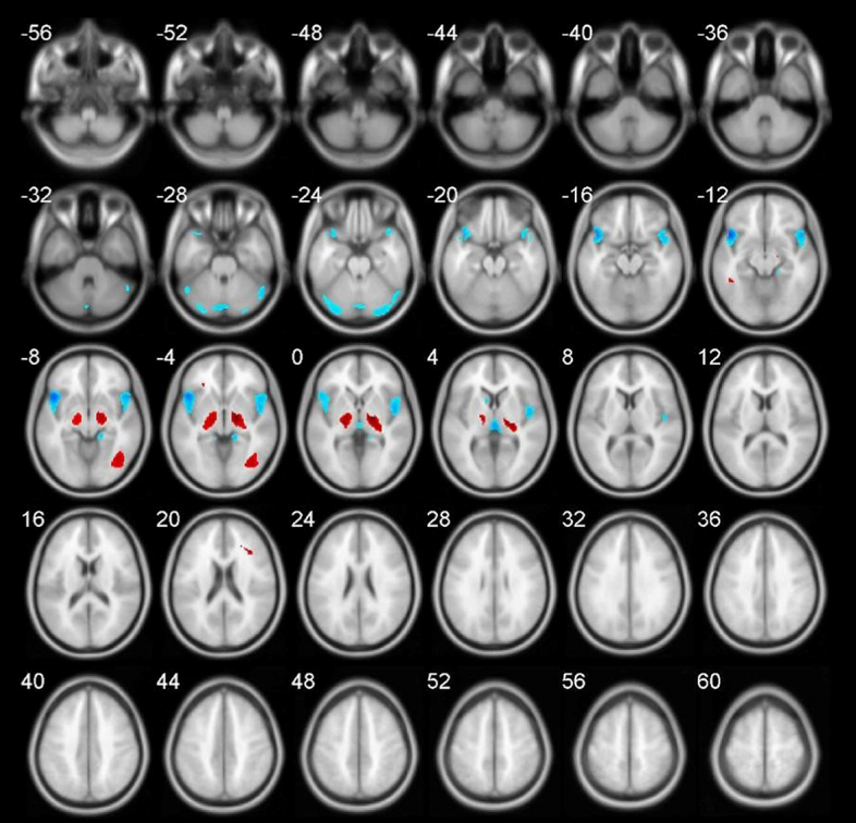
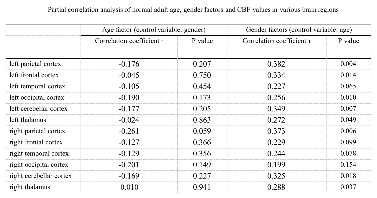
Compared with the baseline group, CBF reduction was manifested in bilateral frontal lobes, anterior cingulate gyrus, and bilateral temporal lobes, both in the entire cohort and in different gender groups. In men, areas of reduced CBF were the bilateral frontal lobes, caudate nucleus, and cingulate gyrus, and in women the left occipital lobe, cerebellum, and thalamus. However, overall brain CBF decreases in both sexes begin at age 60 years, and a further worsening decrease is observed only in men at age 70 years.Discussion
The observed age-related changes in CBF have important implications for understanding the aging brain's physiology and potential cognitive consequences. Altered CBF in specific brain regions may contribute to age-related cognitive decline and be associated with an increased risk of neurodegenerative diseases. Further research is needed to elucidate the underlying mechanisms and explore potential interventions to mitigate age-related CBF alterations and their associated cognitive effects.Conclusion
(1) After the age of 60, significantly decreased brain CBF were found in the bilateral frontal lobes, bilateral temporal lobes, and inferior cingulate gyrus; (2) We found specific brain CBF differences between genders, including the caudate nucleus in men area and the occipital lobe area in women; (3) The aging trends of men and women are different.Acknowledgements
This work is grateful for the support of Dr. Li Jian’s Ningxia Natural Foundation Project (No. 2023AAC03611).References
1.Pang H, Dang X, Ren Y, Zhuang D, Qiu T, Chen H, Zhang J, Ma N, Li G, Zhang J, Wu J, Feng X. 3D-ASL perfusion correlates with VEGF expression and overall survival in glioma patients: Comparison of quantitative perfusion and pathology on accurate spatial location-matched basis. J Magn Reson Imaging. 2019 Jul;50(1):209-220.
2.Zhuo Z, Su L, Duan Y, Huang J, Qiu X, Haller S, Li H, Zeng X, Liu Y. Different patterns of cerebral perfusion in SLE patients with and without neuropsychiatric manifestations. Hum Brain Mapp. 2020 Feb 15;41(3):755-766.
3.Yang Z, Rong Y, Cao Z, Wu Y, Zhao X, Xie Q, Luo M, Liu Y. Microstructural and Cerebral Blood Flow Abnormalities in Subjective Cognitive Decline Plus: Diffusional Kurtosis Imaging and Three-Dimensional Arterial Spin Labeling Study. Front Aging Neurosci. 2021 Feb 1;13:625843.
Figures