3891
Sex- and age-related changes in cerebral microvasculature can be detected with vascular architecture mapping1Department of Neurology, Heidelberg University Hospital, Heidelberg, Germany, 2Department of Diagnostic and Interventional Radiology, Heidelberg University Hospital, Heidelberg, Germany, 3Department of Neuroradiology, Heidelberg University Hospital, Heidelberg, Germany, 4Department of Radiology, German Cancer Research Center, Heidelberg, Germany, 5Clinical Cooperation Unit Neurooncology, German Cancer Research Center, Heidelberg, Germany
Synopsis
Keywords: Aging, Microstructure
Motivation: While vessel architecture mapping (VAM) is an emerging quantitative MR imaging technique that can characterize cerebral blood vessel microstructure in vivo based on dynamic changes in gradient-echo and spin-echo relaxation rates during contrast agent administration, no study has examined how age-related morphological changes affect VAM parameters.
Goal(s): Our goal was to assess region-specific age- and sex-related changes in cerebral microvasculature with VAM.
Approach: We applied high-resolution VAM on the healthy contralateral hemisphere of 72 age-matched women and men with stable low-grade brain tumors.
Results: We could show that microvascular morphology and aging-related remodeling differ between sexes, particularly in thalamus, insular cortex, and putamen.
Impact: This is the first study to characterize age- and sex-specific changes in cerebral microvascular architecture across different anatomical regions using vascular architecture mapping. Results may be of particular importance for future studies on sex-specific diagnostics and prevention of cerebrovascular disease.
Introduction
Histological studies indicate that the morphology and function of cerebral microvasculature change with aging, such as a decrease in capillary density, or a dilatation of arterioles.1,2 These microvascular alterations play a crucial role in both cerebrovascular disease, i.e. lacunar infarctions or small vessel disease, as well as in therapy approaches for malign tumors, e.g. glioblastoma.3,4 Previous studies further indicate that age-related changes differ between sexes, as well as between different brain regions.5 Our goal was to map and evaluate age-related changes in human microvasculature in vivo by using whole-brain vascular architecture mapping (VAM), a combined spin- and gradient echo echo-planar imaging (SAGE-EPI) sequence. VAM parameters that characterize microvascular properties such as vessel caliber, vessel type or blood flow, can be derived from time-parametrised vortex curves of dynamic changes in relaxation rates R2* and R2 during contrast agent bolus passage.6,7Methods
We examined 40 women and 32 men from an institutional database of patients with suspected unifocal low-grade glioma, each stable for at least two years and with no prior history of chemo- or radiotherapy, on the non-tumor hemisphere. Subjects were matched for age [range: 20-70 years] and BMI. Exlusion criteria were any diagnosis of cerebrovascular or cardiovascular disease, arterial hypertension, hypercholesterinemia, diabetes mellitus or smoking. VAM was conducted at 3 Tesla (Prisma, Siemens) using a multiband SAGE-EPI sequence with parallel imaging acceleration (parallel imaging factor=3) and 20-channel head receive radio-frequency coil. 60 SAGE-EPI readouts with 24 slices each were recorded during administration of contrast agent (0.1mmol/kg bodyweight gadoterate meglumine at a rate of 4ml/s) followed by a 20ml saline bolus. Detailed sequence parameters were TE(GE/SE)=22/90ms, TR=1.5s, acquisition time=90s, multiband factor=2. After motion correction in SPM12.0 (Matlab R2020a), changes in relaxation rates were calculated GE and SE signals with $$$ ΔR(t)=-\frac{1}{TE} ln(\frac{S(t)}{S_0}) $$$. We corrected for contrast agent leakage effects as in 8. VAM parameters were obtained from the resulting vortex curve: the signed area of the curve as vessel type indicator (VTI) and its slope as caliber gradient indicator (GCI) as described before.9 Calculation of vessel size index (VSI) and microvessel density Q was based on 6. We only analyzed volume-of-interests (VOIs) from the healthy hemispheres contralateral to the suspected lesion, including inidividual cortical grey matter (cGM) and supratentorial white matter (WM) VOIs that were identified using FMRIB’s automated segmentation tool in FSL10 on isotropic T1-weighted images and co-registered to VAM parameter maps. Additionally, VOIs for putamen, thalamus, caudate nucleus (CN) and insula were analyzed using a digital human brain atlas in SPM (AAL3).11 T-tests were conducted between age-matched men and women. All correlations with age were adjusted for BMI.Results
Higher age was associated with less negative or even positive values for parameter VTI, corresponding to a shift in dominant vessel types with aging, from capillaries to an arteriole-dominated profile, particularly noticeable in insula (r=0.30, p=0.010) and thalamus (r=0.24, p=0.048). Additionally, aging correlated with an increase in microvessel caliber as measured by both parameters CGI and VSI, in insula (CGI: r=0.25, p=0.03, VSI: r=0.27, p=0.022), thalamus (CGI: r=0.38, p=0.001, VSI: 0.35, p=0.003) and CN (CGI: r=0.37, p=0.002, VSI: r=0.31, p=0.009), see Fig. 1. Compared to age-matched men, women had smaller VSI in insula, thalamus and putamen, while no significant difference was found for the other brain regions. Further, women exhibited a higher microvessel density (Q) than men across all analyzed grey matter areas, but not in white matter, see Fig. 2.Discussion
In line with previous studies, we found an increase in vessel diameter and regional changes of dominant vessel types with age. While VSI has been shown to be increased in patients with vascular dementia,12 we could show that subclinical changes in vessel caliber occur even in the normal aging process. Furthermore, sex-specific differences in vessel diameter and microvessel density matches epidemiological data that women have lower incidence of vascular dementia across all age groups.13Conclusions
Using in vivo high-resolution vascular architecture mapping, we could show that microvascular morphology and aging-related remodeling differs between women and men. These results are particularly relevant to future studies on sex-specific diagnostics and prevention of cerebrovascular disease. Future research should consider race-related differences in microvasculature changes as well.Acknowledgements
This study was supported by a grant from the Deutsche Forschungsgemeinschaft. A. Hohmann was supported by the Rahel-Goitein-Straus-Program from the medical faculty of Heidelberg University.
References
1. Berthiaume, A.-A. et al. Pericyte remodeling is deficient in the aged brain and contributes to impaired capillary flow and structure. Nat Commun 13, 5912 (2022).
2. Xu, X. et al. Age-related Impairment of Vascular Structure and Functions. Aging Dis 8, 590–610 (2017).
3. Caplan, L. R. Lacunar infarction and small vessel disease: pathology and pathophysiology. J Stroke 17, 2–6 (2015).
4. Das, S. & Marsden, P. A. Angiogenesis in glioblastoma. N Engl J Med 369, 1561–1563 (2013). 5. Huxley, V. H. & Kemp, S. S. Sex-Specific Characteristics of the Microcirculation. Adv Exp Med Biol 1065, 307–328 (2018).
6. Kiselev, V. G., Strecker, R., Ziyeh, S., Speck, O. & Hennig, J. Vessel size imaging in humans. Magn Reson Med 53, 553–563 (2005).
7. Emblem, K. E. et al. Vessel architectural imaging identifies cancer patient responders to anti-angiogenic therapy. Nat Med 19, 1178–1183 (2013).
8. Boxerman, J. L., Schmainda, K. M. & Weisskoff, R. M. Relative cerebral blood volume maps corrected for contrast agent extravasation significantly correlate with glioma tumor grade, whereas uncorrected maps do not. AJNR Am J Neuroradiol 27, 859–867 (2006).
9. Zhang, K. et al. Vessel architecture imaging using multiband gradient-echo/spin-echo EPI. PLoS ONE 14, e0220939 (2019).
10. Zhang, Y., Brady, M. & Smith, S. Segmentation of brain MR images through a hidden Markov random field model and the expectation-maximization algorithm. IEEE Trans Med Imaging 20, 45–57 (2001).
11. Rolls, E. T., Huang, C.-C., Lin, C.-P., Feng, J. & Joliot, M. Automated anatomical labelling atlas 3. Neuroimage 206, 116189 (2020).
12. Choi, H.-I., Ryu, C.-W., Kim, S., Rhee, H. Y. & Jahng, G.-H. Changes in Microvascular Morphology in Subcortical Vascular Dementia: A Study of Vessel Size Magnetic Resonance Imaging. Front Neurol 11, 545450 (2020).
13. Ruitenberg, A., Ott, A., van Swieten, J. C., Hofman, A. & Breteler, M. M. Incidence of dementia: does gender make a difference? Neurobiol Aging 22, 575–580 (2001).
Figures
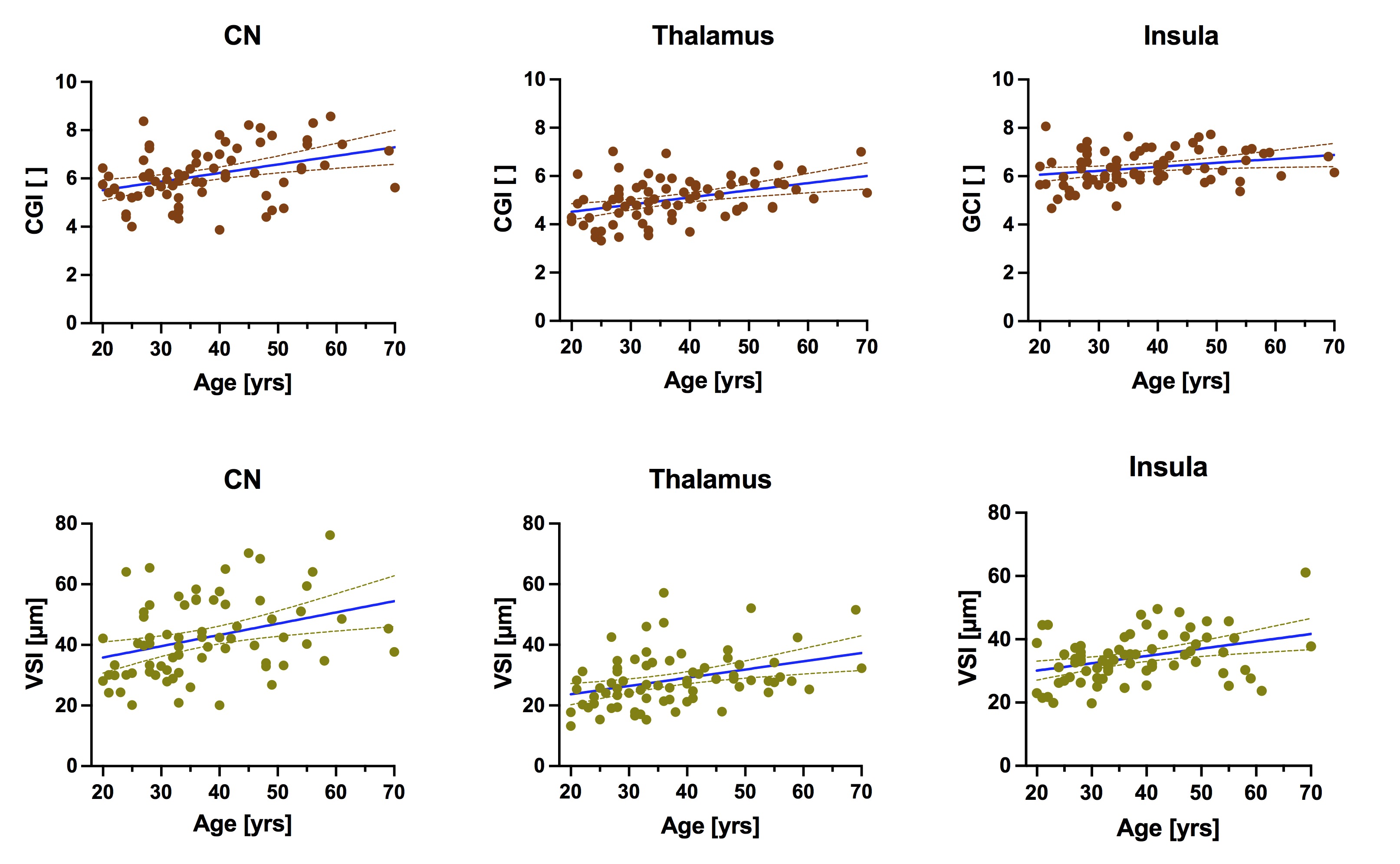
Fig. 1 Aging-related increase in microvessel caliber.
Measured with parameter caliber gradient indicator (CGI), above and vessel size index (VSI), below. Mean values for caudate nucleus (CN), thalamus and insular cortex for all subjects (n=72). Line of best fit for simple linear regression (blue), with 95% confidence intervals (dashed lines).
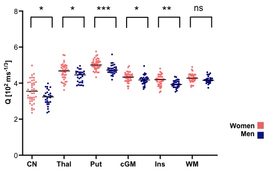
Fig. 2: Sex-specific values for microvessel density (Q) in different brain regions.
Mean values per VOI for women and men. Line indicates median value. Asterisks show p-values for unpaired t-tests between women (n=42) and men (n=30). * p<0.05,** p<0.01, *** p<0.001. ns: not significant. [CN: caudate nucleus, Thal: thalamus, Put: putamen, cGM: cortical grey matter, Ins: insular cortex, WM: white matter]