3888
ASL-derived cerebrovascular brain-age improves associations with cognitive decline1Radiology & Nuclear Medicine,Amsterdam University Medical Centers, Vrije Universiteit, Amsterdam, Netherlands, 2Amsterdam Neuroscience, Brain Imaging, Amsterdam, Netherlands, 3Alzheimer Center Amsterdam, Neurology, Vrije Universiteit Amsterdam, Amsterdam, Netherlands, 4Amsterdam Neuroscience, Neurodegeneration, Amsterdam, Netherlands, 5Neurochemistry lab, Department of Clinical Chemistry, Amsterdam Neuroscience, Amsterdam UMC, Amsterdam, Netherlands, 6Department of Physics and Computational Radiology, Division of Radiology and Nuclear Medicine, Oslo University Hospital, Oslo, Norway, 7Norwegian Centre for Mental Disorders Research (NORMENT), Oslo University Hospital, Oslo, Norway, 8Department of Psychology, University of Oslo, Oslo, Norway, 9Department of Psychiatric Research, Diakonhjemmet Hospital, Oslo, Norway, 10KG Jebsen Centre for Neurodevelopmental Disorders, University of Oslo, Oslo, Norway, 11Dementia Research Centre, Queen Square Institute of Neurology, UCL, London, United Kingdom, 12Centre for Medical Imaging Computing, Computer Science, UCL, London, United Kingdom, 13Epidemiology and Data Science, Vrije Universiteit Amsterdam, Amsterdam UMC location VUmc, Amsterdam, Netherlands, 14Queen Square Institute of Neurology and Centre for Medical Image Computing, UCL, London, United Kingdom, 15Helmholtz-Zentrum Dresden-Rossendorf, Institute of Radiopharmaceutical Cancer Research, Dresden, Germany
Synopsis
Keywords: Aging, Aging
Motivation: Structural brain ageing models are associated with cognitive decline, and the addition of arterial spin labelling (ASL)-derived improved brain-age estimation accuracy, but the relation between cerebrovascular ageing and cognitive decline is not yet fully understood.
Goal(s): To assess the contribution of ASL in the relationship between brain-age estimates and cognitive decline.
Approach: Brain-age estimation accuracy and linear relationships with composite cognitive scores were compared between structural-only (T1w and FLAIR), ASL-only, and structural+ASL models.
Results: Combined structural and ASL brain-age models showed the highest accuracy and increased effect sizes with composite cognitive scores, however, ASL-only models showed unexpected relationships.
Impact: Combined structural-ASL brain-age models might present a surrogate biomarker in an earlier stage of cognitive decline, aiding in diagnosis and treatment monitoring. Possible mediation effects of ASL on the association of structural decline with cognitive domains should be investigated further.
Introduction
Accelerated brain ageing is associated with cognitive decline and dementia1. The biological brain age can be estimated using structural brain MRI and machine learning methods, which subtracted with chronological age results in the brain-predicted age difference (brain-PAD). However, structural MRI is limited, mostly sensitive to late irreversible structural changes, and there is increasing evidence that accelerated cerebrovascular compromise is an important factor for cognitive dysfunction2.The addition of arterial spin labelling (ASL) perfusion scans has been shown to improve brain-PAD accuracy in healthy ageing and brain-PAD-based classification of healthy controls and Alzheimer’s Disease (AD)3,4. However, the added value of cerebrovascular ageing, its underlying mechanisms, and their relation to (domain-specific) cognitive decline are not yet fully understood. Furthermore, there is evidence that multi-sequence brain-PAD approaches offer different insights into pathological developments5.
In this study, we assessed the relationship between brain-PAD and 1) stages of cognitive decline, and 2) global cognitive scores and domain-specific cognitive tests. We compared brain-PAD results between structural-only (T1w and FLAIR), ASL-only, and structural+ASL models.
Methods
Methods-study design/dataHealthy controls (HC, n=1107, Table 1) were drawn from the StrokeMRI and TOP datasets6. From the Amsterdam Dementia Cohort7 (ADC; n=213), patients with Subjective Cognitive Decline (SCD), Mild Cognitive Impairment (MCI), and probable Alzheimer’s Disease (AD) were selected (Table 1). All ASL datasets were acquired with the same 3D spiral-FSE PCASL protocol (3T GE MR750, post-labelling delay=2025ms, labelling duration=1450ms). ASL post-processing was performed with ExploreASL8. Cognitive tests (ADC only) were z-scored and averaged per domain (Table 1)9. Features10 were created by extracting the following imaging derivatives: GM and WM volumetrics (T1w), WMH volumetrics (FLAIR), and vascular territory-based CBF and spatial coefficient of variation (sCoV; ASL, Table 1).
Brain-age estimation
The three brain-age estimation models (T1w+FLAIR+ASL, T1w+FLAIR, or ASL-only) were trained on the HC dataset, and model performance was assessed with five-fold cross-validation using the mean absolute error (MAE).
Brain-age predictions
For all three models, brain-age estimates were subtracted from chronological age to determine the Brain-PAD in the ADC dataset. Differences in imaging derivatives between HC and ADC were assessed using a t-test. Brain-PAD group differences were assessed per model, using one-way ANOVA with Tukey’s Honest Significant Difference post-hoc test. Relationships between cognitive performance, per domain, and brain-PADs, per model, were assessed using linear regression.
Results
All imaging measures differed (p<0.01) between HC and ADC (Table 1).All cognitive composite scores differed between the diagnostic groups (p<0.05) except for the attention & executive function and visuospatial function between SCD and MCI (Table 1). The T1w+FLAIR+ASL model performed best (MAE=7.8 years), and model performance did not differ (p>0.05) between diagnostic groups (all models, Figure 1).
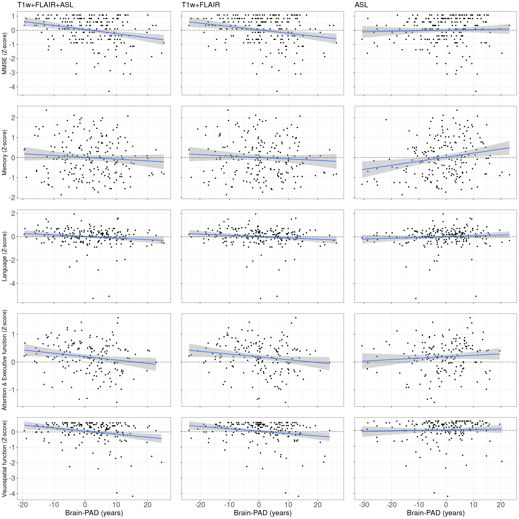
Brain-PADs differed (p<0.05) between SCD and AD (all models) and between SCD and MCI (ASL model, Figure 1). The T1w+FLAIR+ASL brain-PADs was associated with MMSE (ß=-0.031,p<0.001), language (ß=-0.013,p=0.04), attention & executive (ß=-0.012,p=0.02) and visuospatial (ß=-0.02,p<0.001) domains (Table 2, Figure 2). Brain-PAD from the T1w+FLAIR model was associated with MMSE (ß=-0.021,p<0.001), attention & executive (ß=-0.010,p=0.03) and visuospatial (ß=-0.020,p=0.006) domains (Table 2, Figure 2). The ASL only brain-PAD was associated with the memory domain (ß=0.20,p=0.01; Table 2, Figure 2).
Discussion
Our brain-PADs findings are in agreement with previous studies, with SCD being similar to HC, and MCI and AD groups showing increased predicted brain age gaps1. The addition of ASL to T1w+FLAIR Brain-age improved sensitivity to global and domain-specific cognitive decline.Only the ASL-only model was associated with cognitive staging and memory, which is unexpected as structural derivatives such as hippocampal volume usually show stronger effects11. Also unexpectedly, a lower ASL-only brain-AD (i.e., a younger/healthier brain) was related with poorer cognition. Perhaps, ASL derivatives were more sensitive to early-stage cognitive decline, and the observed relationships might be explained by early-stage compensatory perfusion effects that have been described in initial stages of AD-related cognitive decline, or because ADC contains many young-onset AD patients.
Conclusion
ASL improved the performance of structural brain-age models both in relation with AD staging and domain-specific cognitive tests. Further studies in various stages of AD and with larger clinical datasets are required to assess if and how ASL mediates the association of structural changes with cognitive domains.Acknowledgements
We acknowledge the following grants: the Dutch Heart Foundation 2020 T049—Mathijs B. J. Dijsselhof, Jan Petr, and Henk J. M. M. Mutsaerts—the Eurostars-2 joint programme with co-funding from the European Union Horizon 2020 research and innovation programme (ASPIRE E!113701), provided by the Netherlands Enterprise Agency (RvO) — Jan Petr, and Henk J. M. M. Mutsaerts—and the EU Joint Program for Neurodegenerative Disease Research, provided by the Netherlands Organisation for Health Research and Development and Alzheimer Nederland DEBBIE JPND2020-568-106—Jan Petr, Henk J. M. M. Mutsaerts. Lars T. Westlye is supported by The Research Council of Norway (273345, 298646, 300767), the South-Eastern Norway Regional Health Authority (2018076, 2019101), the European Research Council under the European Union's Horizon 2020 research and Innovation program (802998). Frederik Barkhof is supported by the NIHR Biomedical Research Centre at UCLH.References
1. Franke, K. & Gaser, C. Ten Years of BrainAGE as a Neuroimaging Biomarker of Brain Aging: What Insights Have We Gained? Front. Neurol. 10, 789 (2019).2. Iadecola, C. & Gottesman, R. F. Neurovascular and Cognitive Dysfunction in Hypertension. Circ. Res. 124, 1025–1044 (2019).
3. Dijsselhof, M. B. J. et al. The value of arterial spin labelling perfusion MRI in brain age prediction. Hum. Brain Mapp. 44, 2754–2766 (2023).
4. Rokicki, J. et al. Multimodal imaging improves brain age prediction and reveals distinct abnormalities in patients with psychiatric and neurological disorders. Hum. Brain Mapp. 42, 1714–1726 (2021).
5. Jirsaraie, R. J. et al. A systematic review of multimodal brain age studies: Uncovering a divergence between model accuracy and utility. Patterns (N Y) 4, 100712 (2023).
6. Richard, G. et al. Assessing distinct patterns of cognitive aging using tissue-specific brain age prediction based on diffusion tensor imaging and brain morphometry. PeerJ 6, e5908 (2018).
7. van der Flier, W. M. & Scheltens, P. Amsterdam Dementia Cohort: Performing Research to Optimize Care. J. Alzheimers. Dis. 62, 1091–1111 (2018).
8. Mutsaerts, H. J. M. M. et al. ExploreASL: An image processing pipeline for multi-center ASL perfusion MRI studies. Neuroimage 219, 117031 (2020).
9. Scheltens, N. M. E. et al. Cognitive subtypes of probable Alzheimer’s disease robustly identified in four cohorts. Alzheimers. Dement. 13, 1226–1236 (2017).
10. Dijsselhof et al. Cerebrovascular Brainage. ISMRM Perfusion Workshop.
11. Roux, C. M., Leger, M. & Freret, T. Memory Disorders Related to Hippocampal Function: The Interest of 5-HT4Rs Targeting. Int. J. Mol. Sci. 22, (2021).
Figures
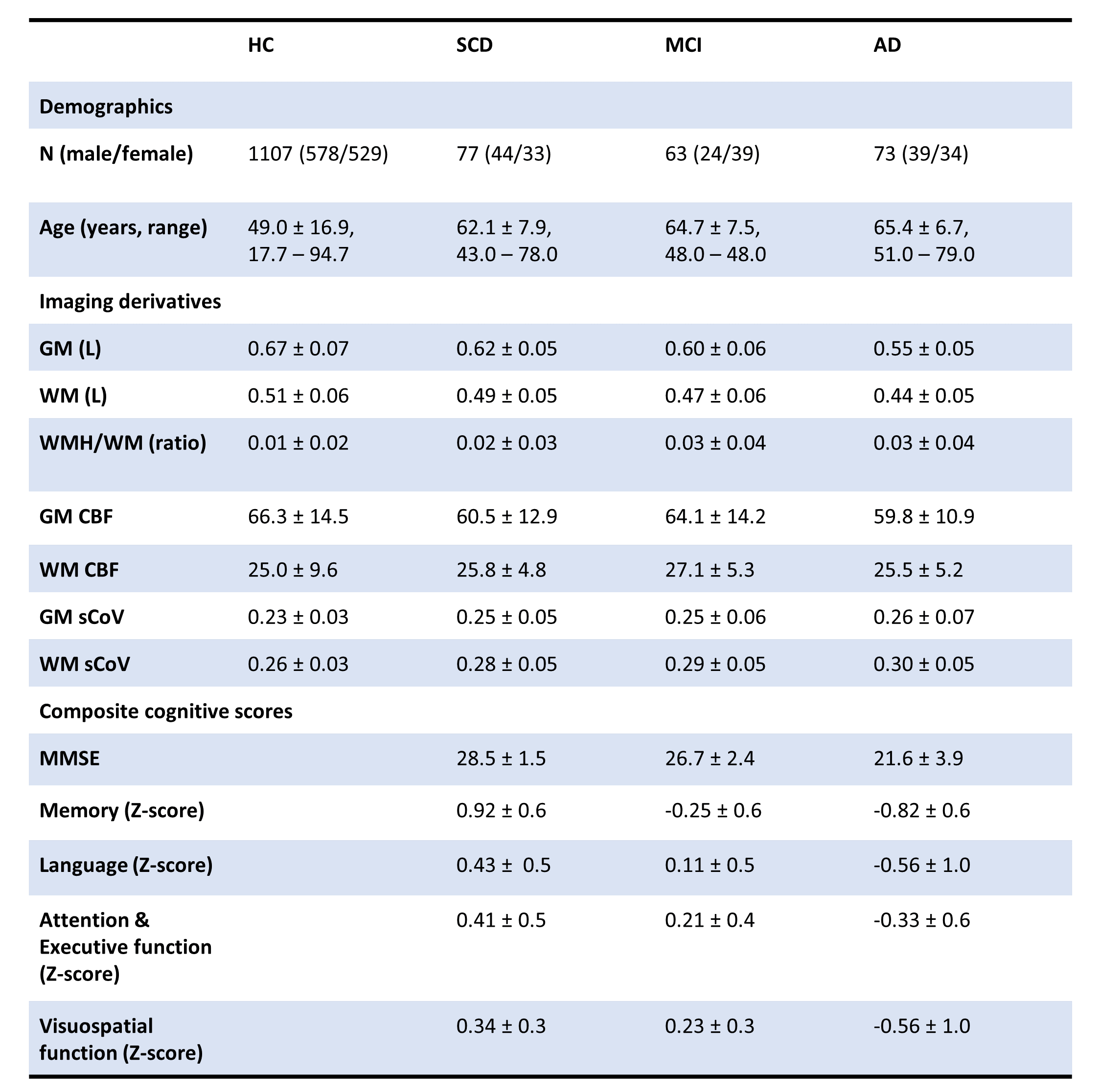
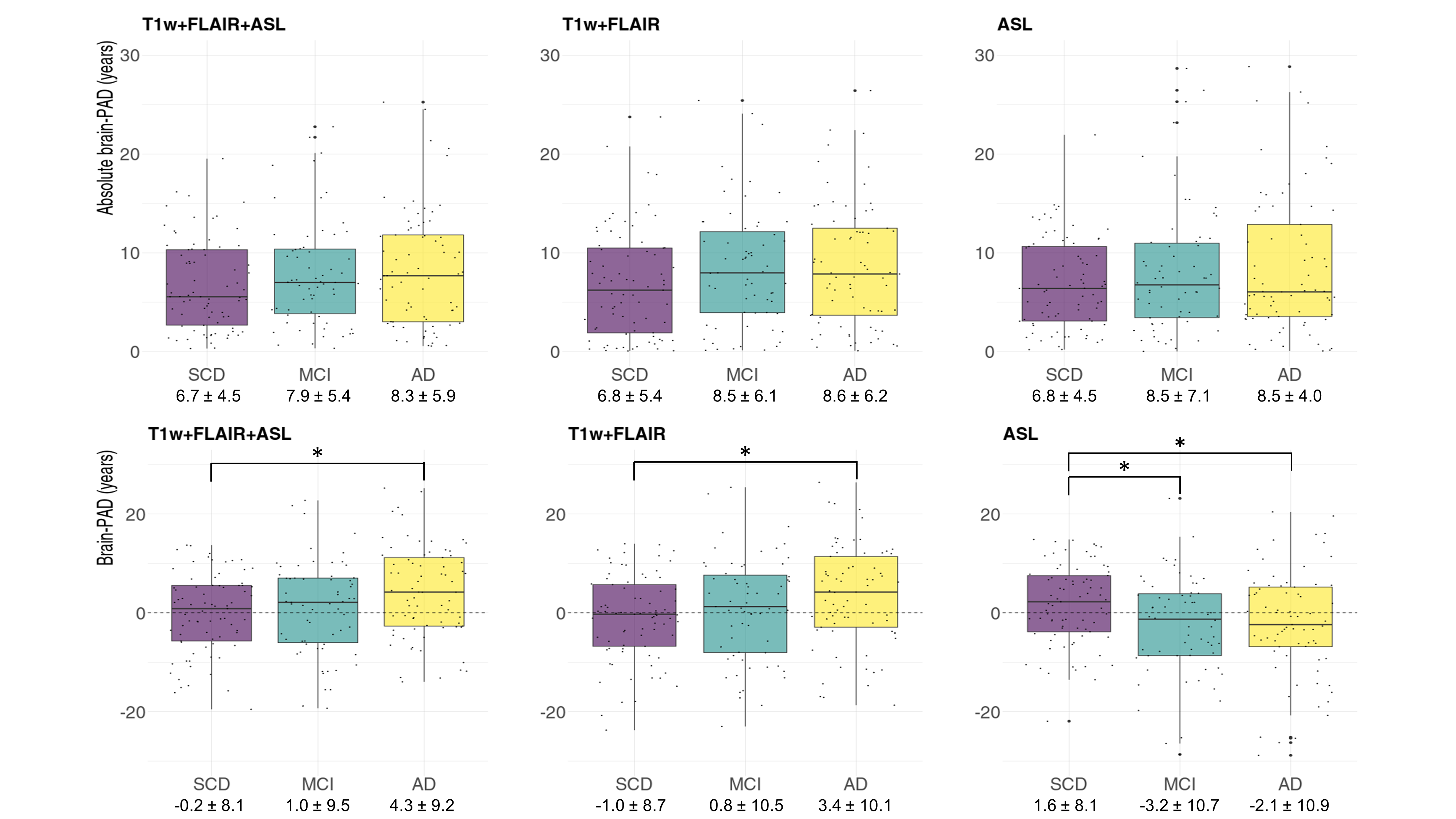
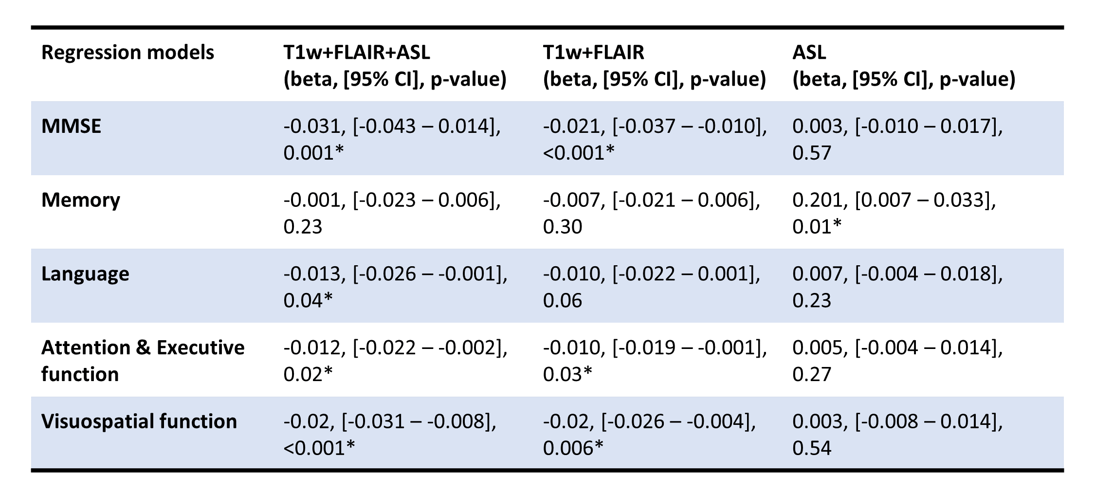
Table 2: Linear regression analyses between global and domain-specific cognitive composites and brain-PADs per model. The asterisks denote statistical significance. ASL = arterial spin labeling; brain-PAD = brain-predicted age difference. MMSE = mini-mental state examination.