3886
Assessment of Pulsatility Index of Lenticulostriate Arteries using 2D PC-MRI with Dual-VENC in elderly adults at 7T: A Preliminary Study1Department of Radiology, Northwestern University, Chicago, IL, United States, 2Department of Biomedical Engineering, Northwestern University, Chicago, IL, United States, 3Department of Neurology, University of Southern California, Los Angeles, CA, United States
Synopsis
Keywords: Aging, High-Field MRI
Motivation: LSA plays an important role in the pathology of small vessel disease. Directly assessing LSA pulsatility may offer valuable insight into SVD pathology.
Goal(s): To assess the utility of 7T dual-VENC PC-MRI for the LSA pulsatility assessment in elderly adults by investigating its association with age and cognitive impairment.
Approach: LSA pulsatility was assessed using 7T PC-MRI using both dual- and single-VENC on 25 elderly participants. Cognitive tests including MMSE and MOCA were conducted.
Results: Our results showed that the LSA pulsatility was significantly correlated with upstream major arteries and increased LSA pulsatility was associated with aging, education level, and cognitive impairment.
Impact: Increased LSA pulsatility measured by dual-VENC PC-MRI shows a strong association with aging, education, and cognitive impairment. Compared to single-VENC, dual-VENC PC-MRI enhanced the detection and characterization of LSAs, providing a promising imaging tool for investigating vascular pathology of SVD.
Introduction
Small vessel disease (SVD) is characterized by pathological changes in cerebral perforating arteries within the basal ganglia, which serves as a common cause of both stroke and vascular dementia1. Arterial pulsatility, quantified by the pulsatility index (PI), is a well-established marker of vascular dysfunction. Elevated arterial pulsatility can lead to the transmission of excessive pulsatile energy into downstream microvasculature and tissue, resulting in endothelial dysfunction, injury, and vascular inflammation2. Therefore, directly assessing the pulsatility of perforating arteries offers valuable insight into the contribution of vascular dysfunction to the pathophysiology of SVD. Our recent 7T test-retest work has shown improved detection of LSA perforators and enhanced reliability of PI measurements using submillimeter-resolute dual-VENC phase-contrast MRI (PC-MRI) in comparison to the conventional PC-MRI with a single-VENC3. This study aims to evaluate the utility of 7T dual-VENC PC-MRI in the LSA pulsatility assessment by investigating its association with age and cognitive functions within an aged cohort.Methods
Study Participants and Cognitive AssessmentsTwenty-five elderly participants (13 female: 71 ± 9.2 years) were enrolled in the study after providing written informed consent. 23 out of 25 provided information about their education level. Cognitive assessments were conducted including 19 undergoing the Mini-Mental State Exam (MMSE) and 23 taking Montreal Cognitive Assessment (MoCA). Additionally, 2D PC-MRI scans on M1 segment of middle cerebral artery (MCA) and internal carotid artery (ICA) were performed on 13 of the participants.
MRI experiments
All MRI experiments were performed on a Siemens 7T MAGNETOM Terra MRI system using an 8-transmit/32-receive head coil. Retrospectively gated single-slice 2D PC-MRI sequences with VENCs of 20 cm/s and 40 cm/s were acquired (FOV = 180x200 mm2, slice thickness = 2 mm, voxel size = 0.2x0.2x2.0 mm3, flip angle = 50°, TE/TR= 9.72/95ms, 20 phases across a cardiac cycle). A 3D high-resolution TOF was performed on each subject prior to PC-MRI scans to locate the perforating arteries.
Image processing and statistical analysis
LSA masks were blindly generated for single-VENC of 40cm/s and dual-VENC data, respectively. Background phase correction and phase unwrapping were done before calculating the blood velocity and PI. In the dual-VENC image processing, high VENC phase maps were used to unaliase the phase wrapping in low VENC phase maps to ensure a proper velocity profile. Pearson correlation was used to evaluate the association between PI and age, education level, and cognitive measurements. A linear mixed-effects model was used to describe the linear relationship between the PI and cognitive measurements while accounting for the effects of age, gender, and education.
Results and Discussion
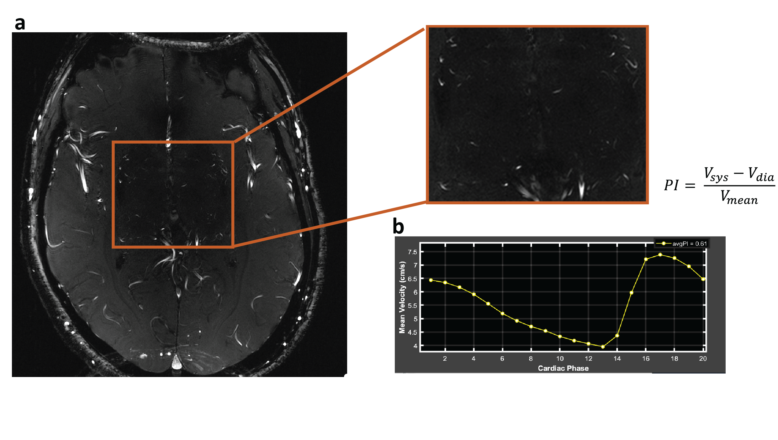
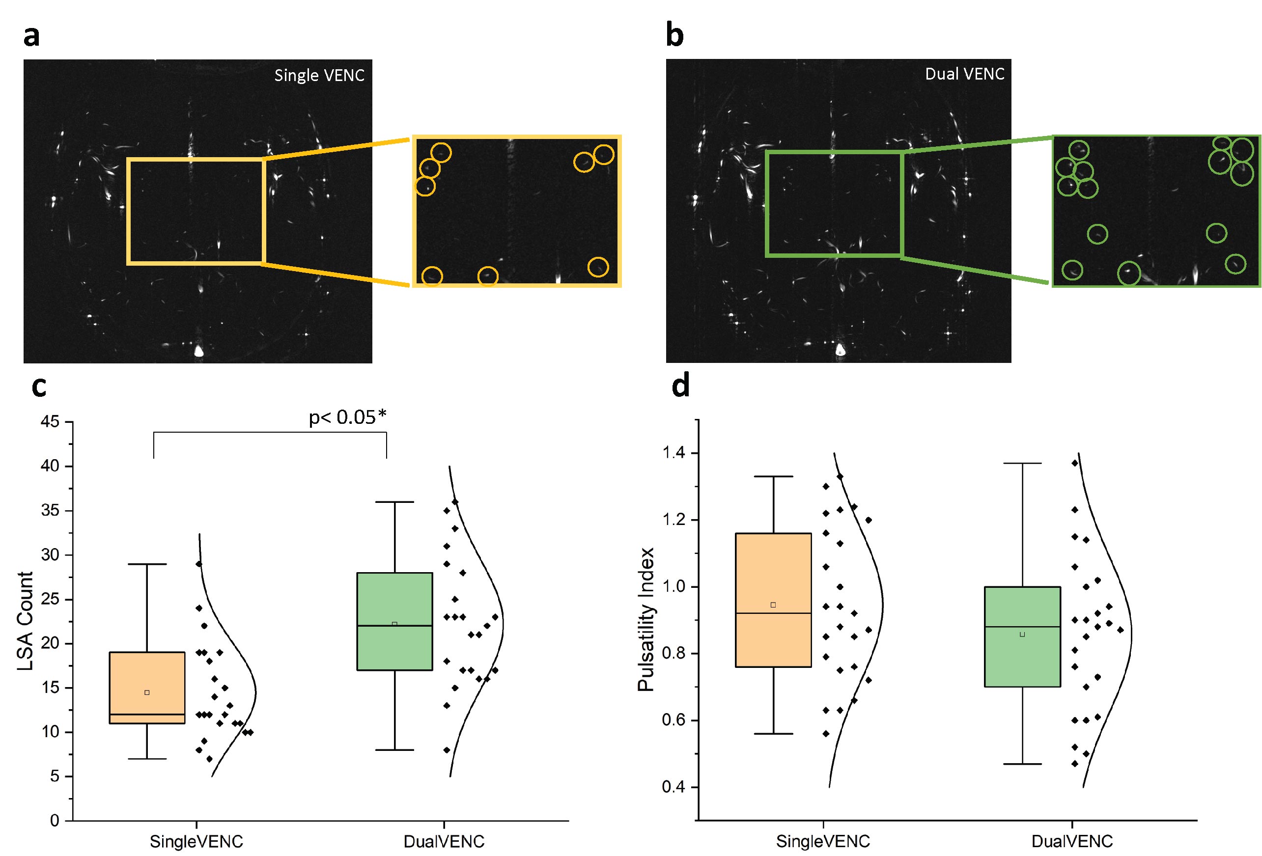
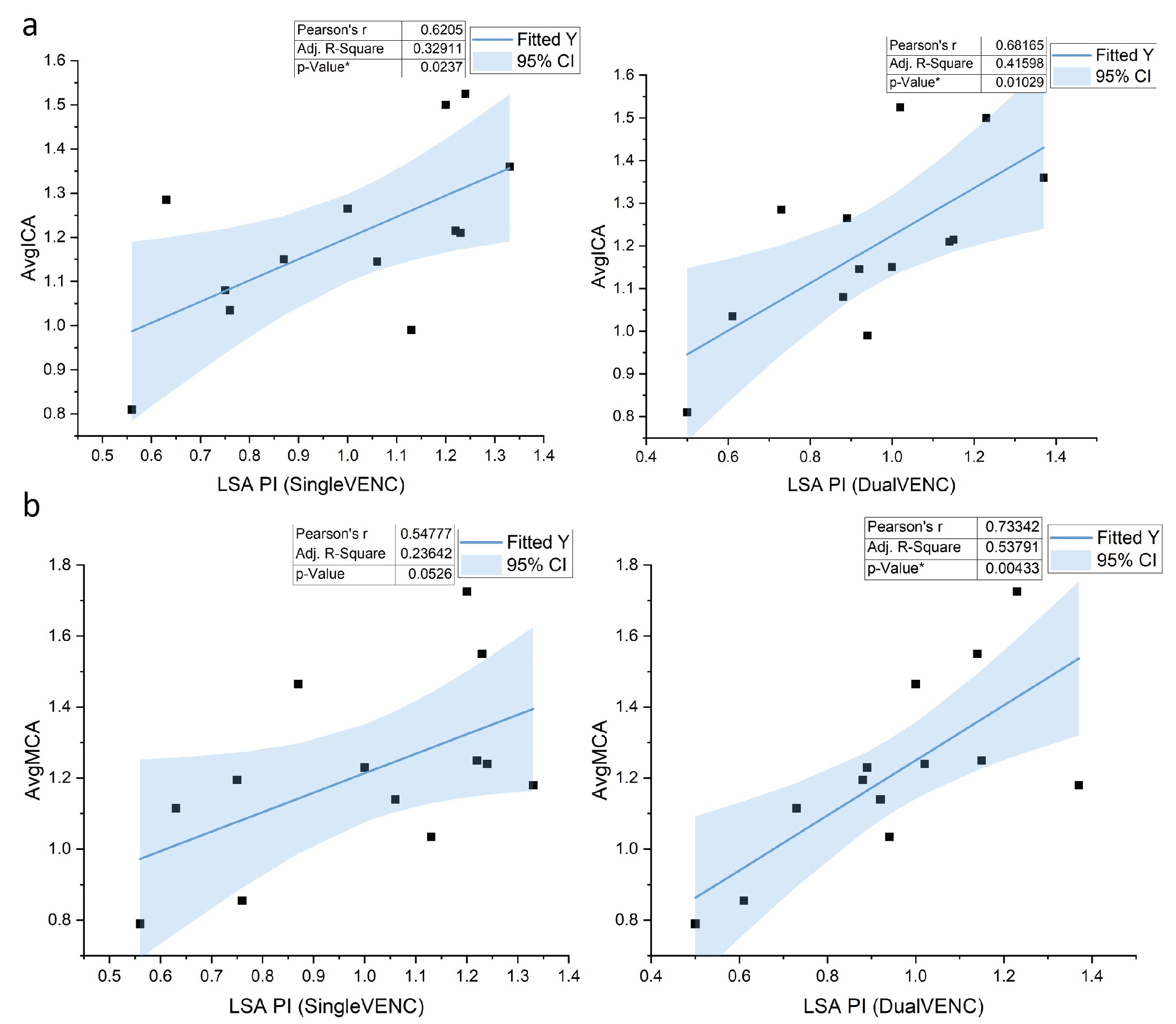
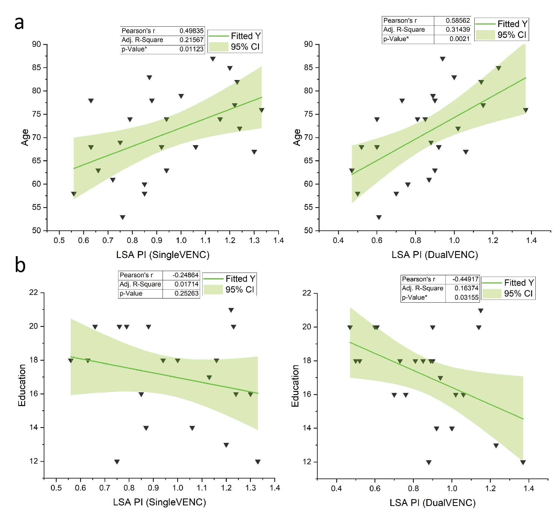
Flow velocity curves and PI values were successfully extracted and calculated from all 25 participants. An example is illustrated in Figure 1. Consistent with our previous findings in healthy young participants3, significantly more LSAs were detected using dual-VENC (p<0.001) compared to single-VENC of 40cm/s (Figure 2). No significant difference (p=0.19) was found in the PI values between single VENC (PI=0.94+/-0.23) and dual VENCs (PI=0.86+/-0.23) (Figure 2). The LSA PI values measured by both single and dual-VENC highly correlated with the PI values obtained from the proximal vessel segments including the ICA and the M1 of MCA (Figure 3). This finding further validates the reliability of the LSA PI measurement using PC-MRI with both single and dual-VENC. Moreover, the PI values with dual-VENC showed a slightly stronger correlation with those of ICA and MCA compared to those with single VENC (p=0.01 vs p=0.024, p=0.004 vs 0.053).Consistent with the previous findings with single-VENC PC-MRI 3, LSA PI showed a significant correlation with age for both single and dual-VENC (p=0.01, 0.002 respectively). In addition, LSA PI measured by dual VENC showed a significant correlation with the education level (p=0.03), while no significant association was found with single VENC (p=0.25).
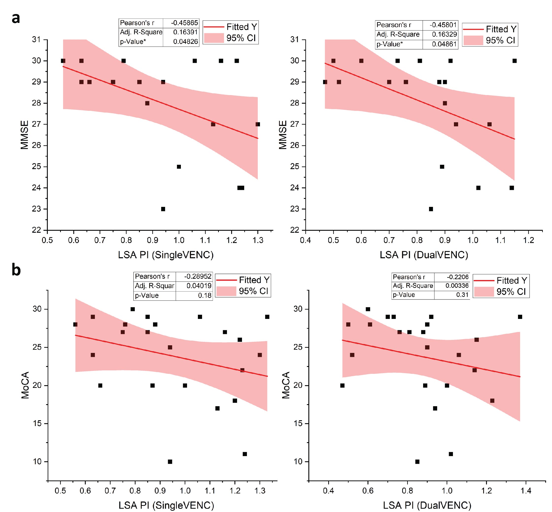
Increased PI was strongly associated with lower MMSE scores with (p = 0.028) and without correcting for age, gender, and educational level (p=0.048). A similar trend was observed between PI and MOCA scores, although there was no significance (p=0.18, 0.31). These findings suggest a strong association between LSA dysfunction and cognitive performance.
Conclusion
This study has demonstrated that elevated LSA pulsatility is associated with aging, education, and cognitive function. These findings indicate that LSA dysfunction may play an important role in aging and cognitive decline in elderly adults. Dual-VENC detects more LSAs compared to the single-VENC, which could provide more reliable measures from both larger and smaller LSAs, compared to single-VENC. The advantages of PC-MRI with dual-VENC will be further evaluated in a larger cohort in future work.Acknowledgements
This work was partly supported by National Institute of Health (NIH) grants R01NS118019, RF1AG072490, and BrightFocus Foundation A20201411SReferences
1. Wardlaw, Joanna M., Colin Smith, and Martin Dichgans. "Small vessel disease: mechanisms and clinical implications." The Lancet Neurology 18.7 (2019): 684-696.
2. Geurts, Lennart J., et al. "Higher pulsatility in cerebral perforating arteries in patients with small vessel disease related stroke, a 7T MRI study." Stroke 50.1 (2019): 62-68.
3. Tang,J et al. " Comparison of arterial pulsatility of cerebral perforating arteries between dual-VENC and single-VENC phase-contrast MRI at 7T." ISMRM Proceedings, 2023.
Figures