3884
Characterizing the age-related changes of hippocampal arterial transit time and perfusion across adult lifespan1Bernard and Irene Schwartz Center for Biomedical Imaging, Department of Radiology, New York University Grossman School of Medicine, New York, NY, United States, 2Center for Advanced Imaging Innovation and Research (CAI2R), Department of Radiology, New York University Grossman School of Medicine, New York, NY, United States, 3Vilcek Institute of Graduate Biomedical Sciences, New York University Grossman School of Medicine, New York, NY, United States, 4Department of Neurology, New York University Grossman School of Medicine, New York, NY, United States
Synopsis
Keywords: Aging, Perfusion, Aging brains
Motivation: Age-related changes of arterial transit time (ATT) and perfusion (CBF) measurement in hippocampus subfields is still under-investigated.
Goal(s): To quantitatively characterize the age-related hippocampal perfusion changes in a subfield-specific manner to better understand its involvement in neurodegenerative changes and dementia.
Approach: T1-weighted images and mbPCASL data from the Human Connectome Project-Aging (HCA) was analyzed to obtain subfield-specific measurements of ATT and CBF in hippocampus.
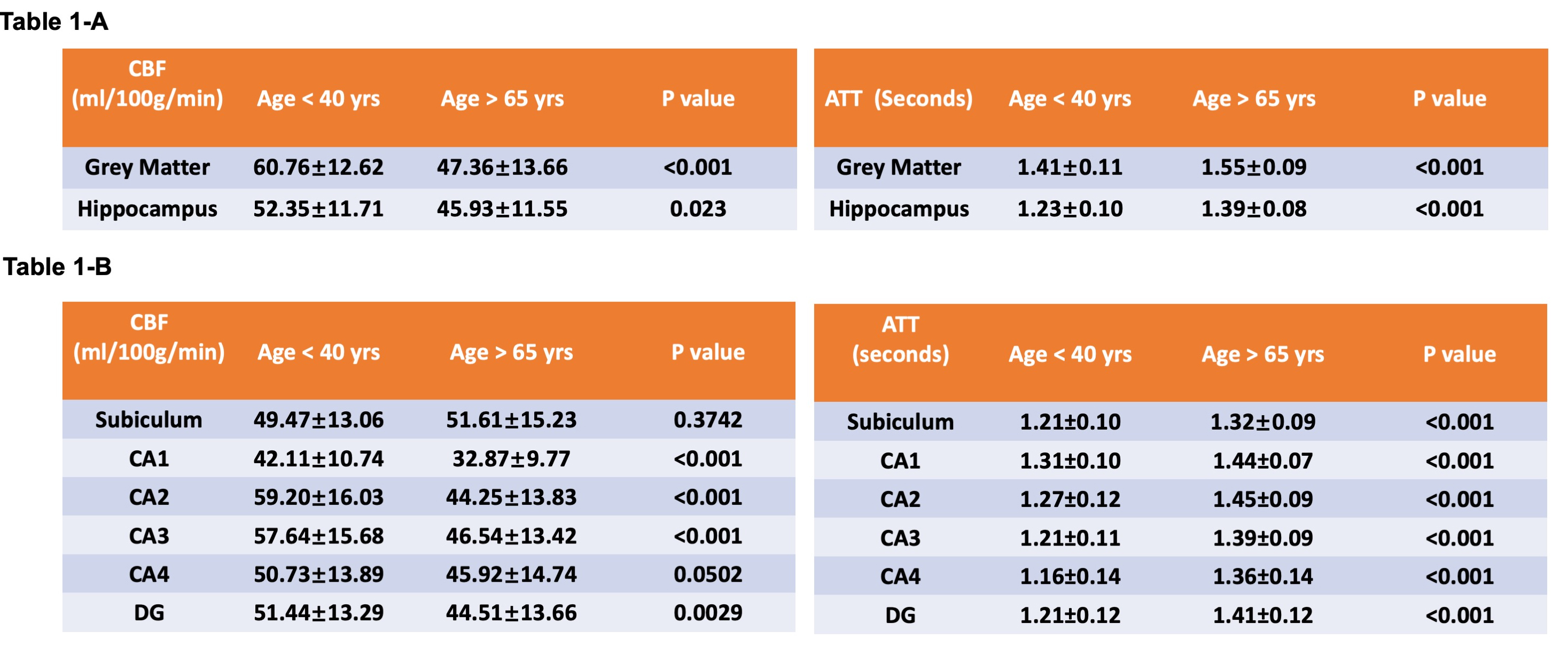
Results: The lowest perfusion measurement was observed in CA1 region across all age groups. Age trajectories of CBF and ATT were demonstrated in different subfields with female showing a more significant decrease of hippocampal perfusion.
Impact: Using Human Connectome Project–Aging (HCA) dataset, this study revealed age-related subfield-specific changes in hippocampal ATT and CBF across the adult normative lifespan, including subiculum, CA1-CA4 and the dentate gyrus.
Introduction
The hippocampus, a complex structure situated in the medial temporal lobe, plays a pivotal role in various cognitive functions in human brain, including memory encoding and learning1,2. Advancing age is by far the most prevalent risk factor contributing to hippocampal atrophy and age-related neurodegenerative diseases such as Alzheimer’s disease or dementia. Traditionally, the assessment of hippocampal degeneration in aging populations primarily relies on volumetric analysis, which serves as the most common biomarker3,4. However, this method can only identify structural changes in the hippocampus, representing a late and irreversible stage without functional aspects of hippocampal alterations. There is a need for novel biomarkers capable of detecting early hippocampal changes, enabling the development of preventative strategies. Biomarkers focusing on functional aspects, such as perfusion5 and oxygen utilization6, have shown promise in this regard. In this study, we leveraged arterial spin labeling (ASL) data from 541 subjects in the Human Connectome Project – Aging (HCP-A) dataset, spanning the adult lifespan. Our objective was to characterize the perfusion changes in the hippocampus during normal aging7,8, with the goal of elucidating age-related vascular functional changes. This exploration can contribute valuable insight into understanding atypical such as Alzheimer’s Disease (AD) and AD-related dementia.Materials and methods
T1-weighted and arterial spin labeling (ASL) data of 541 subjects from Human connectome Project Aging (HCP-Aging) was processed and analyzed. The T1-MPRAGE data was acquired with the following parameters: voxel size = 0.8x0.8x0.8mm, TE=1.8/3.6/5.4/7.2ms, TR/TI=2500/1000ms. The segmentation of hippocampus subfields, including subiculum, CA1-4 and dentate gyrus was performed using Hippfold toolbox9, employing T1-MPRAGE images as input data. Cerebral blood flow (CBF) and arterial transit time (ATT) in hippocampal subfields were unfolded and projected into the hippounfolded space to acquire subfield-specific values. For the ASL data, a multiple post labelling delay (PLD) and a simultaneous multi-slice acquisition (SMS) pseudo-continuous ASL (pCASL) protocol were employed with following parameters: labelling duration of 1500 ms, PLDs: 200 ms, 700ms, 1200ms, 1700ms and 2200ms, each PLD was repeated 6, 6, 6, 10, and 15 times, respectively, and multiband factor: 6. The voxel size was 2.5mm isotropic with a total of 60 slices. The mbPCASL data was preprocessed using hcpasl minimal processing pipeline (https://github.com/physimals/hcp-asl) to yield CBF and ATT maps10. Statistical analysis was performed in GraphPad Prism and Matlab with p value < 0.05 as statistically significant.Results
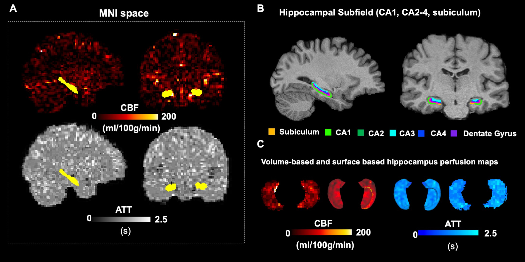
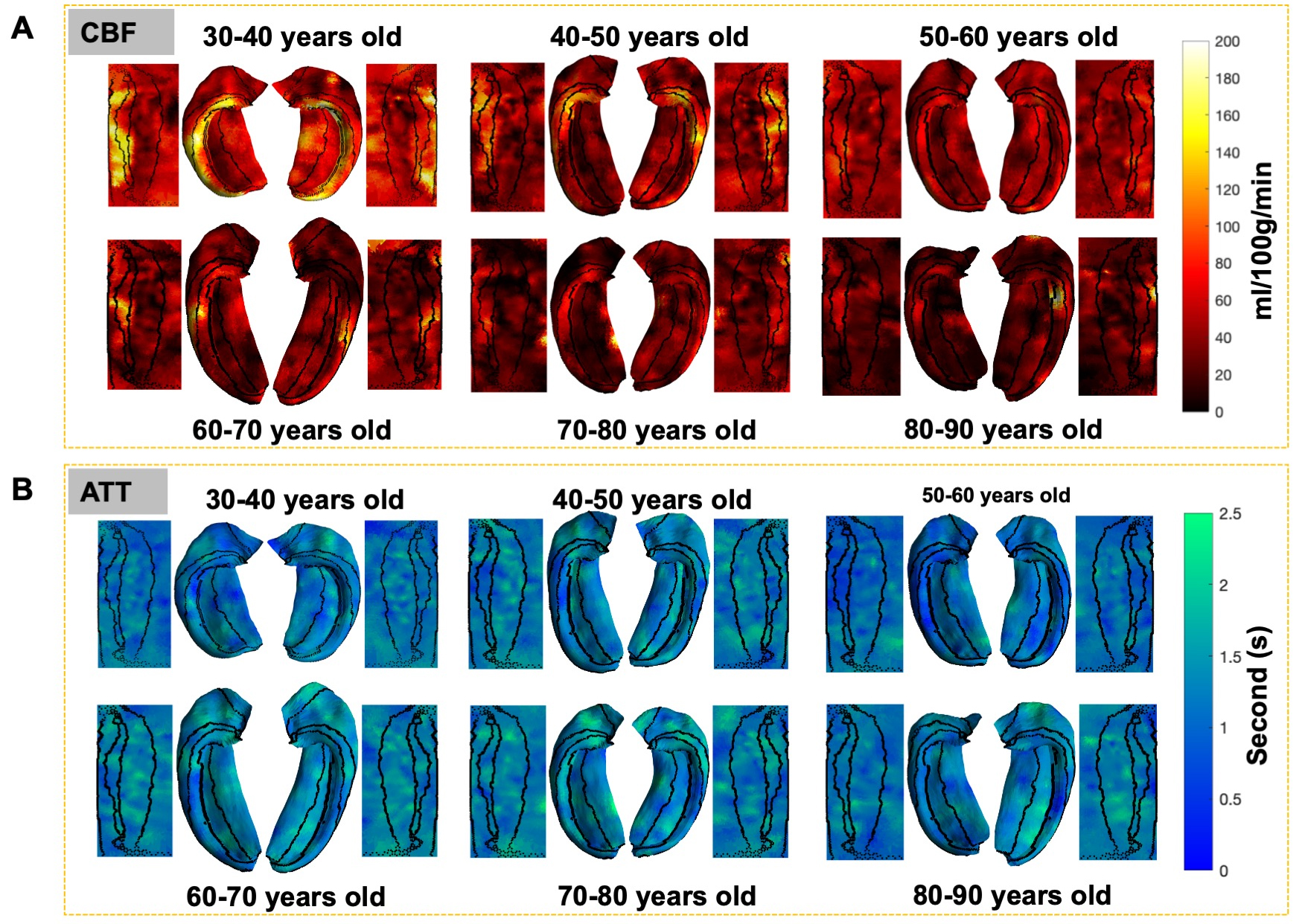
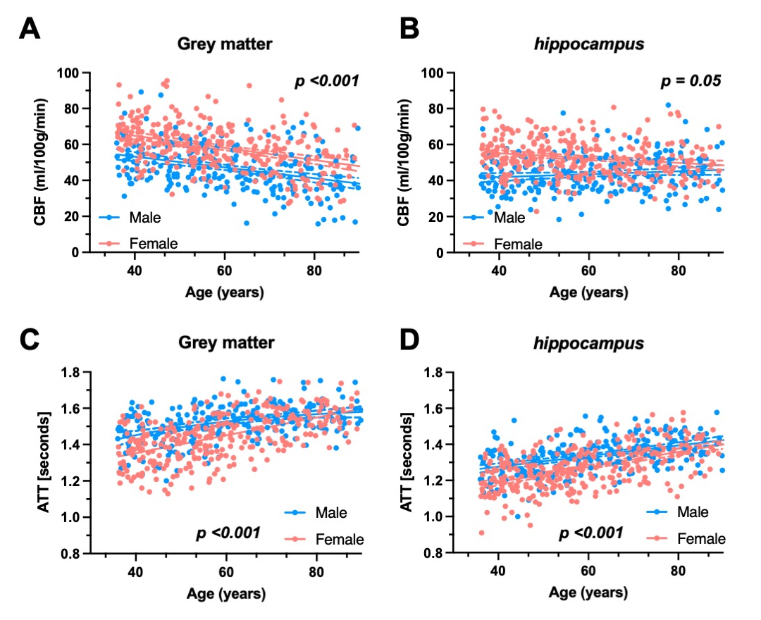
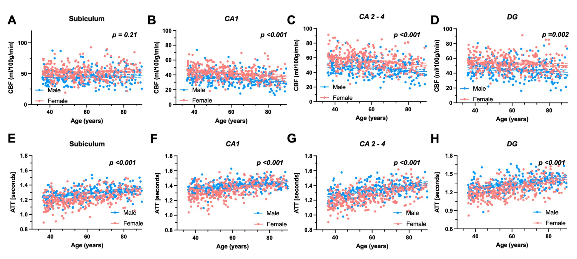
Figure 1A showed the representative CBF and ATT maps in MNI space, with overlays of hippocampus masks for whole hippocampal perfusion measurements. In Figure 1B, subfield masks on T1-MPRAGE data are displayed. The hippocampal CBF and ATT are visualized in both volume-based and surface-based renderings, each highlighting the spatial distribution differently. For subfield-specific analysis, Figure 2 showcases representative CBF and ATT maps in Hippunfold space. Figure 3 illustrated a comparison of total hippocampal CBF and ATT with grey matter. Notably, higher CBF is observed in the grey matter, and it exhibits a steeper age-related decline (Y=-0.35*Age+74.2) compared to the hippocampus (Y=-0.06*Age+52.3). Moreover, within the hippocampus, females (Y=-0.13*Age+60.4) demonstrate a more significant decline in CBF with age compared to males (Y = 0.08*Age+38.7). Lastly, the scatter plots of CBF and ATT are presented in Figure 4 to provide comprehensive view of age-related changes in hippocampal subfields (Subiculum, CA1-4 and Dentate Gyrus) of CBF and ATT.Conclusion and discussion
Several key findings have been revealed in this study. Firstly, we compared the ATT and CBF of the entire hippocampus with whole brain grey matter. Our results corroborate previous reports, indicating female populations tend to exhibit higher blood flow than their male counterparts11. Secondly, we compared CBF and ATT values across distinct hippocampus subfields in two groups: the younger group (< 40 years) and the elderly group (> 65 years). Our findings indicate that the CA1 exhibits the lowest perfusion relative to other hippocampal subfields, aligning with prior research that observed diminished CBF5 and vascular density12 in CA1 region using different methods. Finally, our study demonstrates that the hippocampal ATT is more sensitive to age than CBF, suggesting ATT may serve as a more sensitive indicator for characterizing age-related perfusion changes within the hippocampus. In conclusion, our investigation has evaluated age-related variations in hippocampal CBF and ATT across different subfields, which may provide valuable insights into vascular perfusion changes occurring in the aging hippocampus, serving as a vital reference point for future research on dementia.Acknowledgements
This work was supported by the National Institute on Aging of the National Institutes of Health under Award Number U01AG052564 and by funds provided by the McDonnell Center for Systems Neuroscience at Washington University in St. Louis. The HCP-Aging 2.0 Release data used in this report came from DOI: 10.15154/1520707. This work was performed under the rubric of the Center for Advanced Imaging Innovation and Research (CAI2R, www.cai2r.net), an NIBIB National Center for Biomedical Imaging and Bioengineering (NIH P41 EB017183).References
1. Bettio, Luis EB, Luckshi Rajendran, and Joana Gil-Mohapel. "The effects of aging in the hippocampus and cognitive decline." Neuroscience & Biobehavioral Reviews 79 (2017): 66-86.
2. Fox, Nick C., and Jonathan M. Schott. "Imaging cerebral atrophy: normal ageing to Alzheimer's disease." The Lancet 363.9406 (2004): 392-394.
3. Bussy, Aurelie, et al. "Hippocampal subfield volumes across the healthy lifespan and the effects of MR sequence on estimates." NeuroImage 233 (2021): 117931.
4. Bussy, Aurelie, et al. "Hippocampal shape across the healthy lifespan and its relationship with cognition." Neurobiology of Aging 106 (2021): 153-168.
5. Haast, Roy AM, et al. "Novel insights into hippocampal perfusion using high-resolution, multi-modal 7T MRI." bioRxiv (2023).
6. Jiang, Dengrong, et al. "MRI assessment of cerebral oxygen extraction fraction in the medial temporal lobe." NeuroImage 266 (2023): 119829.
7. Bookheimer, Susan Y., et al. "The lifespan human connectome project in aging: an overview." Neuroimage 185 (2019): 335-348.
8. Li, Xiufeng, et al. "Pushing the limits of asl imaging for the lifespan human connectome projects." International Society for Magnetic Resonance Medicine .
9. DeKraker, Jordan, et al. "Automated hippocampal unfolding for morphometry and subfield segmentation with HippUnfold." Elife 11 (2022): e77945.
10. McConnell, Flora Kennedy, et al. "The minimal processing pipeline for arterial spin labeling data from the Human Connectome Project Lifespan studies of Aging and Development." International Society for Magnetic Resonance Medicine.
11. Ibaraki, Masanobu, et al. "Interindividual variations of cerebral blood flow, oxygen delivery, and metabolism in relation to hemoglobin concentration measured by positron emission tomography in humans." Journal of Cerebral Blood Flow & Metabolism 30.7 (2010): 1296-1305.
12. Buch, Sagar, et al. "Vascular mapping of the human hippocampus using Ferumoxytol-enhanced MRI." Neuroimage 250 (2022): 118957.
Figures