3882
Relationship between cardiovascular outcomes, hippocampal vascularization and hippocampal volume1University of Pittsburgh, Pittsburgh, PA, United States
Synopsis
Keywords: Aging, Neurodegeneration
Motivation: Studies have shown a correlation between cardiovascular health and the size of the hippocampus. However, how cardiovascular conditions impact blood flow and vessel health in the brain, and how this potentially affects hippocampus, is still not fully understand
Goal(s): We tested whether hippocampal vascularization statistically mediates the association of multiple cardiovascular risk factors and hippocampal volumes in each hemisphere.
Approach: Hippocampal vascularization was segmented from T1-weighted MPRAGE images at 7T. Mediation analysis was performed to test the relationship between various cardiovascular variables and hippocampal volume via hippocampal vascularization
Results: Hippocampal vascularization statistically mediates the association between cardiovascular health and hippocampal volume
Impact: understanding the exact causal pathways between cardiovascular outcomes, hippocampal vascularization, and hippocampal volume is crucial for establish novel strategies for potentially mitigating the risk of cognitive decline and cerebrovascular diseases
INTRODUCTION
Cardiovascular risk factors are associated with poor cerebrovascular health, brain volume loss, and cognitive decline in aging adults1. The integrity of blood vessels supplying the hippocampus is crucial for maintaining its structural integrity and function, and alterations in these vessels may contribute to hippocampal volume reduction. The brain is functionally lateralized and there is differential blood supply to the left and right hippocampus. Thus, alterations in blood flow due to cardiovascular risk factors might relate to each hemisphere differently. Here, we developed a method to segment blood vessels from T1-weighted images. Since head coil is used for RF excitation/inversion at 7T, this enhances the inflow of unsaturated fresh blood into the slices, increasing the signal intensity of blood vessels. We quantified hippocampal vascularization as the volume of blood vessels connected to the hippocampus, and we tested whether hippocampal vascularization statistically mediates the association of multiple cardiovascular risk factors and hippocampal volumes in each hemisphere.METHODS
145 participants (30-59 years old (45.2±8.4), Fe/male= 85/60) were studied at 7T. 20 cardiovascular variables were examined, including root mean square of successive differences (RMSSD), stroke volume, cardiac output (CO), pulse wave velocity (PWV), mean, standard deviation and coefficient of beat to beat variation of systolic and diastolic blood pressure, power/variance of low (.04 - .149 Hz) and high frequency (.15 - .40 Hz) blood pressure variability (BPV), peak low and high frequency power, resting flow and maximum flow and % increase of venous occlusion plethysmography (VOP), mean heart rate, baroreflex sensitivity (BRS) and Framingham risk score (FRS).Anatomical 3D-T1-weighted images (MPRAGE, TR/TE = 3000/1.96ms, TI = 1.2s, voxel-size = 0.7 mm isotropic) were acquired. The anatomical brain parcellations including left and right hippocampus for the brain volume measurement were obtained from FreeSurfer. After signal intensity normalization, vessel segmentation was performed by the Frangi vesselness filter, which estimate the likelihood of tube-like structures2. The vessels connected to segmented hippocampus volume were identified in the left and right hemispheres. Voxels with this identified vessel were calculated as the vascularization (i.e., vessel volume).A path analysis was conducted using the Mediation Toolbox3. The null hypothesis of zero indirect, direct, and total effects was tested based on the 95% bootstrap confidence intervals with 50,000 iterations between various cardiovascular outcomes and left and right hippocampal volumes via left and right vascularization, respectively, with age and sex as covariables.RESULTS
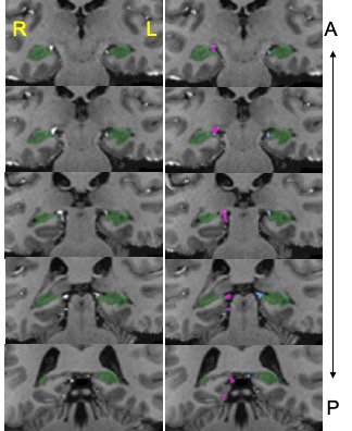
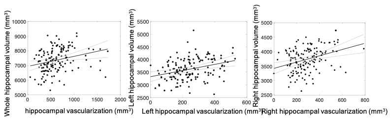
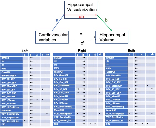
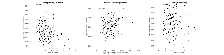
Right hippocampal volume was significantly larger than left hippocampal volume (3739±414 vs. 3623±421 mm3, p < 0.001), The vessels connected to hippocampus were successfully identified (Fig. 1). The vascularization of right hippocampus is larger than that of left hippocampus (291±131 vs. 246±115 mm3, p < 0.001). The hippocampal vascularization was highly associated with the hippocampal volume, adjusting for age and sex (Fig. 2, p < 0.01). Fig.3 shows relationships between various cardiovascular variables and (left and/or right) hippocampal volumes that were mediated by left and/or right hippocampal vascularization. Among various cardiovascular variables, resting flow of VOP and standard deviation of DBP showed a mediated relationship with hippocampal volume through hippocampal vascularization, but did not have a direct relationship with hippocampal volume. Fig. 4 demonstrates one example from the relationship between standard deviation of DBP, right hippocampal vascularization and right hippocampal volume.DISCUSSION
Some cardiovascular variables relate to hippocampal vascularization, which in this study may be related to changes in blood velocity. Slower or lesser blood supply, which is associated with cardiovascular health, may be a substrate for tissue damage, which may be associated with reduced hippocampal volume.CONCLUSION
Development of cerebrovascular segmentation on high-resolution T1weighted image at 7T without dedicated vascular imaging (e.g. angiography) allows for estimation of hippocampal vascularization. Hippocampal vascularization mediated the association between peripheral blood flow, a measure of blood pressure variability, and hippocampal volume, with more consistent effects for the right hippocampus. Improving one aspect may positively impact the others. Thus, understanding the exact causal pathways between cardiovascular outcomes, hippocampal vascularization, and hippocampal volume is crucial for establish novel strategies for potentially mitigating the risk of cognitive decline and cerebrovascular diseasesAcknowledgements
This work was supported by the NIH grant: P01HL040962References
1. Song R, Xu H, Dintica CS, Pan KY, Qi X, Buchman AS, Bennett DA, Xu W. Associations Between Cardiovascular Risk, Structural Brain Changes, and Cognitive Decline. J Am Coll Cardiol. 2020 May 26;75(20):2525-2534.
2. Frangi AF et al., Mutiscale Vessel Enhancement Filtering. Medical Image Computing and Computer-Assisted Intervention. 1998
3. Tor Wager. MediationToolbox (https://github.com/canlab/MediationToolbox), GitHub. Retrieved November 8, 2023.
Figures