3881
Changes of auditory γ-aminobutyric acid and cerebral blood flow in presbycusis and their relationship with cognitive impairment1Department of Biomedical Engineering, School of Life Sciences, Tiangong University, Tianjin, China, 2Russell H. Morgan Department of Radiology and Radiological Science, The Johns Hopkins University School of Medicine, Baltimore, MD, United States, 3Philips Healthcare, Shanghai, China, 4Department of Radiology, Shandong Provincial Hospital Affiliated to Shandong First Medical University, Jinan, Shandong, China
Synopsis
Keywords: Aging, Brain
Motivation: Studies have shown that presbycusis is associated with cognitive impairment.
Goal(s): To study the pathophysiological mechanism of presbycusis and its potential association with cognitive impairment.
Approach: To study the levels of auditory γ-aminobutyric acid (GABA) and glutamate (Glu), cerebral blood flow (CBF) and their relationship with auditory and cognitive function in patients with presbycusis and healthy controls.
Results: The results showed that the decrease of auditory GABA, Glu levels and CBF in presbycusis was related to the decrease of speech perception or cognitive impairment, and mediated the association between hearing loss and impaired information processing speed in presbycusis.
Impact: The decrease of GABA and CBF mediates the correlation between hearing loss and impaired information processing speed in presbycusis, which helps us to reveal the relationship between hearing loss and cognitive impairment in presbycusis.
Introduction
Presbycusis (PC) is characterized by bilateral symmetric progressive neurological hearing loss1. Numerous studies have demonstrated that presbycusis is closely related to cognitive impairment2,3,4. However, the pathological and physiological mechanisms of presbycusis and its association with cognitive impairment are still poorly understood. Previous magnetic resonance imaging (MRI) studies have suggested cognitive impairment in presbycusis patients is related to the auditory cortex and cognitive-related cortex5,6,7. A study using PCASL observed significantly reduced CBF levels in the right primary auditory cortex of patients with presbycusis, which were associated with hearing loss8. However, the study did not continue to explore whether CBF values in other brain regions decreased, nor did it investigate the relationship between CBF decline and cognitive impairment. A study using MEGA-PRESS found decreased auditory GABA levels in presbycusis patients, which was closely associated with hearing loss9. Another MEGA-PRESS study found that decreased GABA levels were related to decreased executive function in patients with presbycusis10. Notably, a study of healthy subjects found a positive correlation between baseline GABA and cerebral blood flow (CBF) in the visual cortex11. Thus, aberrant GABA and Glu levels in presbycusis patients measured by MRS are likely related to alterations in CBF. In summary, we hypothesized that abnormal metabolite levels and neurotransmitter concentrations are associated with cognitive impairment, and studied the auditory GABA and Glu levels, CBF and their relationship with auditory and cognitive function in presbycusis patients and healthy controls.Methods
One hundred and twenty-four patients with presbycusis and ninety-one normal hearing (NH) controls were recruited for this research. We evaluated their hearing levels at the 0.5, 1, 2, and 4 kHz thresholds using the pure tone average (PTA) method10. To assess auditory information processing speed and executive function, the Trail-Making Test(TMT) Part A(TMT-A) and Part B(TMT-B)12 were administered13 . CBF data was gathered by PCASL. GABA-edited data was gathered through a macromolecules (MM)-suppressed MEGA-PRESS sequence utilizing a symmetrical-suppression method to suppress the MM signal10,13., and the PRESS sequence was used to collect the Glu data.Results
No statistically significant difference was found in fitting error or CRLB between groups within the bilateral auditory regions (all p > 0.05, Table 1). Compared with the NH group, presbycusis patients had decreased GABA levels in bilateral auditory region (left: 1.10 ± 0.22 i.u. vs 0.98 ± 0.32 i.u., p < 0.001, right: 1.36 ± 0.30 i.u. vs 1.17 ± 0.27 i.u., p < 0.001). Furthermore, decreased Glu levels were found in bilateral auditory regions in patients with presbycusis (left: 4.71 ± 0.69 i.u. vs. 4.39 ± 0.71 i.u., p = 0.001, right: 4.54 ± 0.62 vs 4.19 ± 0.99 i.u., p = 0.002, Table 1).Compared with NH group, CBF in bilateral insula, precuneus, central operculum and medial frontal gyrus of presbycusis patients decreased significantly. At the same time, CBF was also significantly decreased in the right auditory cortex, anterior cingulate gyrus, superior occipital gyrus, inferior parietal lobule, calcarine fissure and surrounding cortex, angular gyrus, and left caudate nucleus, thalamus, superior frontal gyrus, superior parietal gyrus, middle cingulate cortex ( FDR correction, p < 0.05; clustering defined threshold, P = 0.001), as shown in Table 2 and Fig. 1.
The serial mediation model revealed a significant total effect (c = 0.6528, p = 0.0162), suggesting that PTA predicts TMT-A scores in presbycusis patients (Fig. 2). In the path analysis, PTA was negatively related to GABA levels in the right auditory region (a1 = 0.0092, p = 0.0001) and negatively related to SPG CBF (a2 = -0.0785, p = 0.3441). The right auditory GABA levels were positively related to AN-DMN FC (a3 = 8.3799, p = 0.0094) and negatively related to TMT-A scores (b1 = -43.505, p = 0.0001). The SPG CBF was negatively related to TMT-A scores (b2 = 0.9134, p = 0.0025). After controlling for GABA levels and SPG CBF, the direct effect of PTA on TMT-A scores was no longer statistically significant (c’ = 0.2504,p=0.3483). The total indirect effect was significant (a1×b1+ a2×b2 + a1×a3×b2 = 0.4024, 95%CI[0.0816,0.7652]), with a significant serial indirect effect observed from PTA via GABA levels and SPG CBF to TMT-A scores (a1×a3×b2 = 0.0706, 95%CI[-0.1818,-0.0133]).
Conclusion
This study shows that hearing loss in presbycusis patients can lead to decreased auditory GABA, Glu levels and CBF levels, and this phenomenon is related to decreased speech perception or cognitive impairment. Importantly, decreased auditory GABA levels and decreased CBF levels mediate the association between hearing loss and impaired information processing speed in presbycusis patients.Acknowledgements
Over the course of my researching and writing this paper, I would like to express my thanks to all those who have helped me. A special acknowledgement should be shown to Doctor Yao Wang and Doctor Fei Gao, who gave me kind encouragement and useful instruction all through my writing. Sincere gratitude should also go to all my partners, friends and family who have greatly helped me in my study as well as in my life.References
1. Guan B, Xu Y, Chen Y C, et al. Reorganized Brain Functional Network Topology in Presbycusis[J]. Frontiers in Aging Neuroscience, 2022, 14: 905487.2. Lin F R, Ferrucci L, Metter E J, et al. Hearing loss and cognition in the Baltimore Longitudinal Study of Aging[J]. Neuropsychology, 2011, 25(6): 763.
3. Lin F R, Metter E J, O’Brien R J, et al. Hearing loss and incident dementia[J]. Archives of neurology, 2011, 68(2): 214-220.
4. Gates G A, Mills J H. Presbycusis[J]. The lancet, 2005, 366(9491): 1111-1120.
5. Delano P H, Belkhiria C, Vergara R C, et al. Reduced suprathreshold auditory nerve responses are associated with slower processing speed and thinner temporal and parietal cortex in presbycusis[J]. PLoS One, 2020, 15(5): e0233224.
6. Ren F, Ma W, Li M, et al. Gray matter atrophy is associated with cognitive impairment in patients with presbycusis: a comprehensive morphometric study[J]. Frontiers in neuroscience, 2018, 12: 744.
7. Chen Y C, Yong W, Xing C, et al. Directed functional connectivity of the hippocampus in patients with presbycusis[J]. Brain Imaging and Behavior, 2020, 14: 917-926.
8. Ponticorvo S, Manara R, Pfeuffer J, Cappiello A, Cuoco S, Pellecchia MT, Saponiero R, Troisi D, Cassandro C, John M, Scarpa A, Cassandro E, Di Salle F, Esposito F. Cortical pattern of reduced perfusion in hearing loss revealed by ASL-MRI. Hum Brain Mapp. 2019 Jun 1;40(8):2475-2487.
9. Gao, F., Wang, G., Ma, W., Ren, F., Li, M., Dong, Y., Liu, C., Liu, B., Bai, X., Zhao, B., Edden, R.A., 2015. Decreased auditory GABA+ concentrations in presbycusis demonstrated by edited magnetic resonance spectroscopy. Neuroimage 106, 311–316.
10. Li N, Ma W, Ren F, et al. Neurochemical and functional reorganization of the cognitive-ear link underlies cognitive impairment in presbycusis[J]. NeuroImage, 2023, 268: 119861.
11. Donahue M J, Near J, Blicher J U, et al. Baseline GABA concentration and fMRI response[J]. Neuroimage, 2010, 53(2): 392-398.
12. Wang K, Xu C, Smith A, et al. Genome-wide association study identified INSC gene associated with Trail Making Test Part A and Alzheimer's disease related cognitive phenotypes[J]. Progress in Neuro-Psychopharmacology and Biological Psychiatry, 2021, 111: 110393.
13. Harris A D, Puts N A J, Barker P B, et al. Spectral‐editing measurements of GABA in the human brain with and without macromolecule suppression[J]. Magnetic resonance in medicine, 2015, 74(6): 1523-1529.
Figures
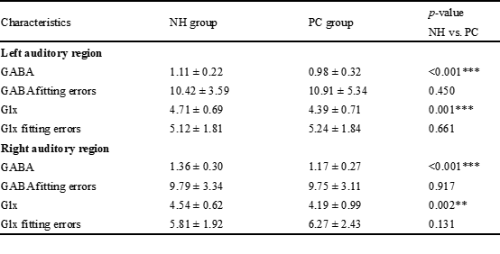
Data are presented as means ± standard deviations. MRS, magnetic resonance spectroscopy; NH, normal hearing; PC, Presbycusis; dB/HL, decibel hearing level; GABA, gamma-aminobutyric acid; Glx, glutamate-glutamine complex.
** p < 0.01. *** p < 0.001.
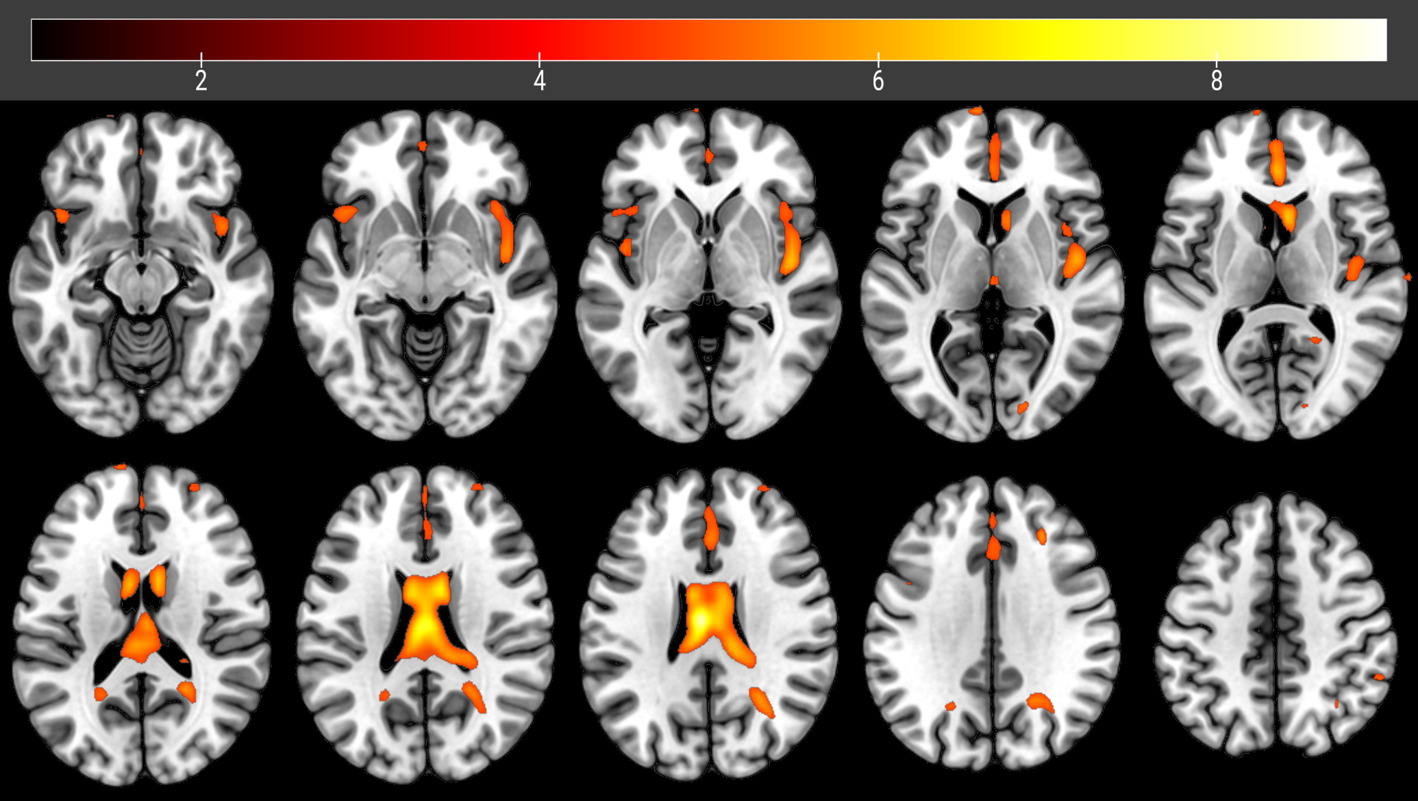
Fig. 1. The brain regions with significant intergroup differences of the CBF. The voxel-based analysis demonstrates the survived clusters between the NH group and presbycusis group. These clusters are referred to multiple comparisons correction using the FDR rate (a cluster-defining threshold of P = 0.001 and a corrected cluster significance of P < 0.05). CBF, cerebral blood flow; NH, normal hearing; FDR, False Discovery Rate correction.
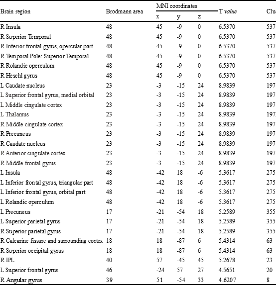
Table 2 Brain regions with significant differences in the CBF between the PC group and NH group.
FDR corrected p < 0.001. CBF, cerebral blood flow; NH, normal hearing; PC, Presbycusis; MNI, Montreal Neurological Institute; IPL, Inferior parietal, but supramarginal and angular gyri; FDR, False Discovery Rate correction; L, left; R, right.
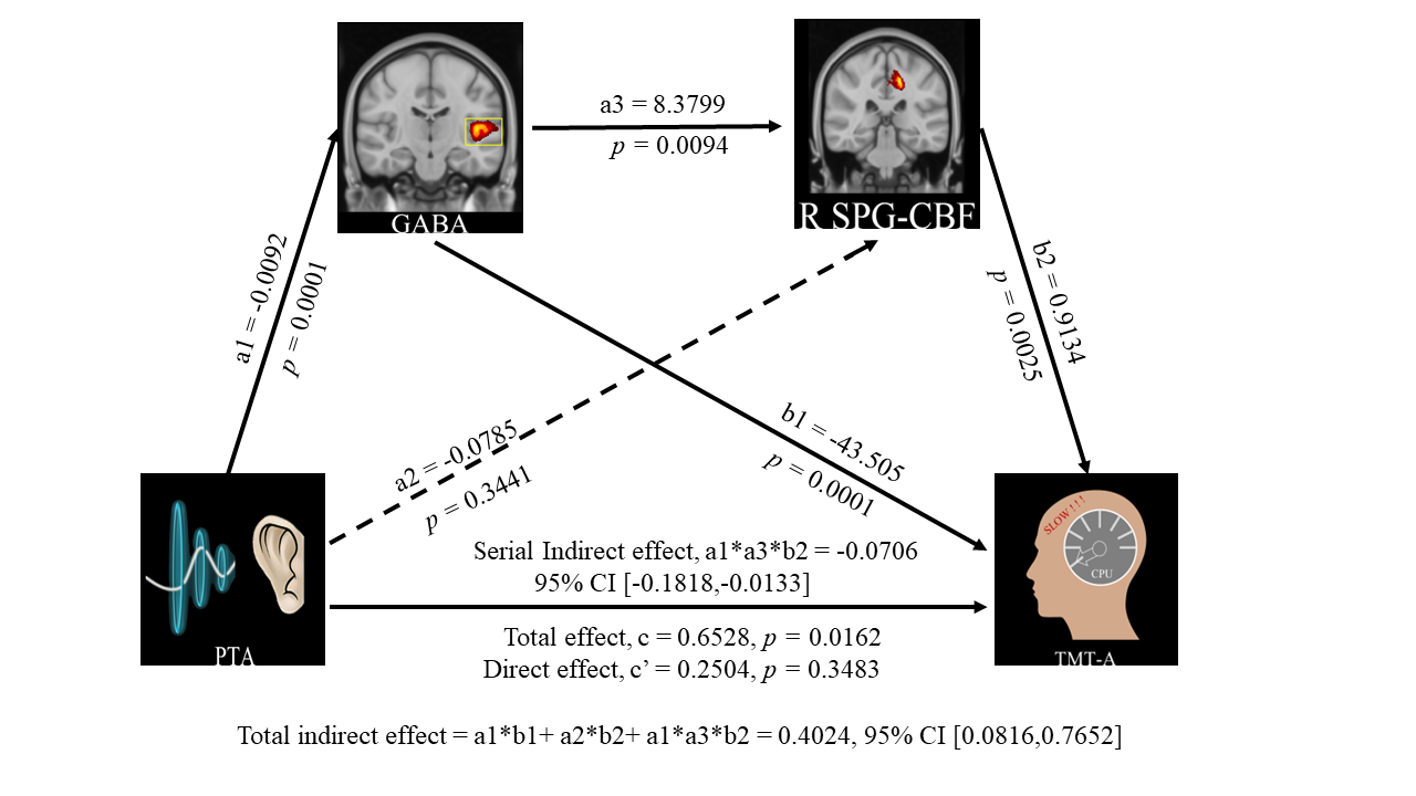
Fig. 2. A serial mediation model including the right auditory GABA levels and SPG CBF as mediators of the associations between hearing loss and cognitive impairment in presbycusis group. PTA, pure tone average; GABA, gamma-aminobutyric acid; CBF, cerebral blood flow; SPG, superior parietal gyrus; TMT-A, Trail-Making Test-A.