3879
Association Between Body Mass Index and Brain Aging in Adults: A 16-Year Population-Based Cohort and Mendelian Randomization Study1Beijing Friendship Hospital, Capital Medical University, Beijing, China
Synopsis
Keywords: Aging, Aging, Cohort study; Body mass index; Obesity; Neuroimaging; Mendelian randomization
Motivation: The causal relationship between BMI and brain health remains unclear.
Goal(s): This study aimed to demonstrate the effect of cumulative BMI on neuroimaging features in adults of different ages and verify the causal relationship.
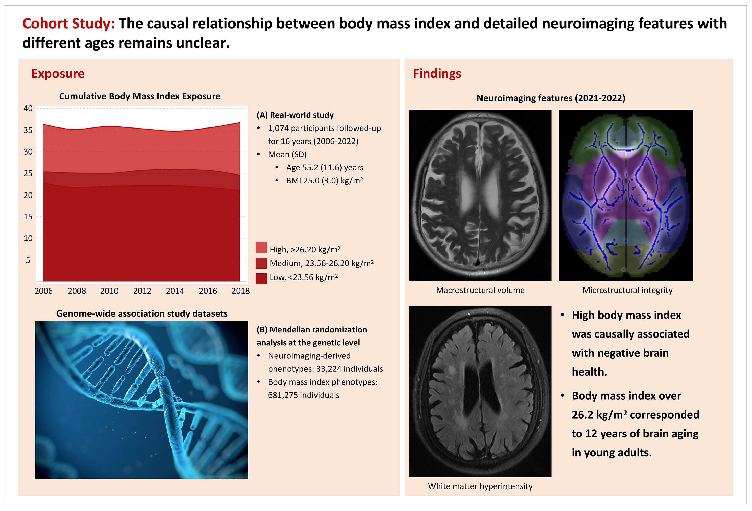
Approach: This study was based on the KaiLuan Study that began in 2006. We also performed two-sample Mendelian randomization analysis using genetic data from 681,275 individuals.
Results: For adults aged under 45 years but BMI > 26.2 kg/m2 corresponded to 12.0 years of brain aging. Genetic analysis indicated causal relationships among high BMI, smaller volume of the cerebral parenchyma, and higher fractional anisotropy in projection fibers.
Impact: High BMI is causally associated with smaller brain volume and abnormal microstructural integrity in projection fibers, especially in young adults. These findings provide a basis for future brain health promotion and disease prevention strategies.
Background
High body mass index (BMI), a modifiable factor associated with poor cardiovascular health, is linked to brain health, but the causal relationship between BMI and brain health remains unclear. This study aimed to demonstrate the effect of cumulative BMI on neuroimaging features in adults of different ages and verify the causal relationship.Methods
This study was based on the KaiLuan Study, a multicenter, long-term follow-up, community-based longitudinal cohort study of the adult population that began in 2006. The study included participants who visited the hospital at least 3 times and underwent brain MRI examination, with no evidence of dementia or mental disorders. Exclusion criteria were incomplete or poor-quality neuroimaging data and diagnosed cancer. We modeled the trajectories of BMI over 16 years to evaluate cumulative exposure. Multimodality neuroimaging data were collected using 3.0-T MRI, starting in 2020, for volumetric measurements of the brain structure, white matter hyperintensity (WMH), and skeletonized white matter tract at the voxel level. We performed two-sample Mendelian randomization analysis using genetic data from 681,275 individuals to analyze the causal relationship between BMI and neuroimaging features.Results
In the population-based longitudinal study, clinical and neuroimaging data were obtained from 1,074 adults (aged 25–83 years). High BMI was associated with a wide range of negative brain health effects. For adults aged under 45 years, the differences in cerebral parenchyma volume between those with BMI > 26.2 kg/m2 and those with normal BMI corresponded to 12.0 years (95% confidence interval [CI], 3.0 to 20.0) of brain aging. The volumetric results corresponded to -17.9 ml (95% CI, -29.8 to -4.5). Differences in WMH were statistically significant for participants aged over 60 years, with a 6.0-ml (95% CI, 1.5 to 10.5) larger volume. Genetic analysis indicated causal relationships among high BMI, smaller volume of the cerebral parenchyma and gray matter, and higher fractional anisotropy in projection fibers.Conclusions
High BMI is causally associated with smaller brain volume and abnormal microstructural integrity in projection fibers, especially in young adults. These findings provide a basis for future brain health promotion and disease prevention strategies.Acknowledgements
The authors would like to thank all the involved study investigators, clinicians, nurses, and technicians for dedicating their time and skills to the completion of this study.References
1.Hamer M, Batty GD. Association of body mass index and waist-to-hip ratio with brain structure: UK Biobank study. Neurology. 2019;92(6):e594-e600
2.Zhang Y, Pletcher MJ, Vittinghoff E, et al. Association between cumulative low-density lipoprotein cholesterol exposure during young adulthood and middle age and risk of cardiovascular events. JAMA Cardiol. 2021;6(12):1406
3.Smith SM, Douaud G, Chen W, et al. An expanded set of genome-wide association studies of brain imaging phenotypes in UK Biobank. Nat Neurosci. 2021;24:737-745
4.Han YP, Tang X, Han M, et al. Relationship between obesity and structural brain abnormality: Accumulated evidence from observational studies. Ageing Res Rev. 2021;71:101445
Figures
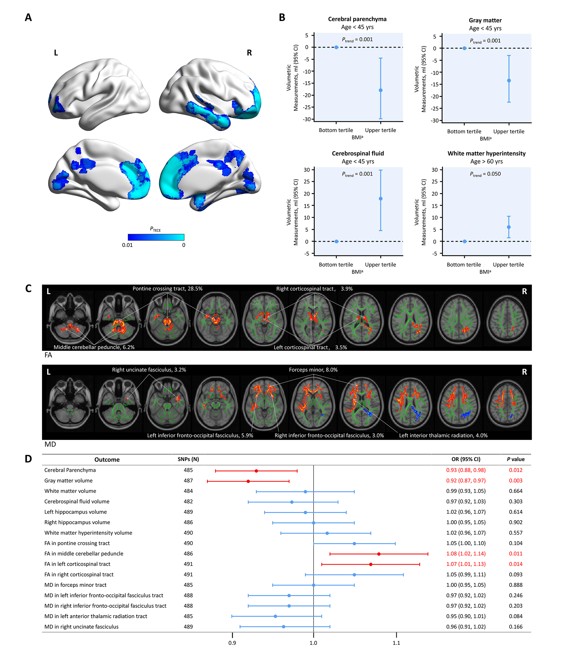
Association of high BMI with neuroimaging features.
A. High BMI is associated with smaller gray matter volume
B. Association of BMI with volume of cerebral parenchyma, gray matter, and white matter hyperintensity in different age groups
C. Association of BMI with microstructural integrity