3877
Deciphering the Mediator Role of White Matter Function in Age-Related Cognitive Decline1Vanderbilt University Institute of Imaging Science, Vanderbilt University Medical Center, Nashville, TN, United States, 2Department of Radiology and Radiological Sciences, Vanderbilt University Medical Center, Nashville, TN, United States, 3Shandong Medical Imaging Research Institute, Shandong University, Jinan, China, 4Department of Electrical and Computer Engineering, Vanderbilt University, Nashville, TN, United States, 5Department of Biomedical Engineering, Vanderbilt University, Nashville, TN, United States, 6Department of Computer Science, Vanderbilt University, Nashville, TN, United States
Synopsis
Keywords: Aging, Aging, fMRI, White matter, Resting state, Cognition
Motivation: Unraveling how age-related changes in brain structure and function affect cognitive functions.
Goal(s): To determine the mediation role of white matter functional metrics in cognitive decline with aging.
Approach: Employing fMRI, graph theory, and mediation analysis to assess how the effect of changes in WM BOLD activity with age influence or reflect cognitive performance.
Results: Identified significant white-matter mediators linking age to cognitive performance.
Impact: Contributes a fresh perspective to our understanding of the functional architecture of the aging brain.
Introduction
Aging usually leads to declines in several cognitive functions, which are often accompanied by changes in brain structure and functional architecture, such as reduced volumes and altered patterns of blood oxygenation level-dependent (BOLD) signals detectable by fMRI. While extensive research has been conducted on BOLD signals in gray matter (GM), their presence and significance in white matter (WM) are usually disregarded. Recent studies confirm that BOLD signals in WM are reliably detectable, they vary with neural activity, and change with aging (1). In exploring the link between aging, brain function, and cognitive ability, mediation analysis can reveal functional and/or structural features of the brain that channel the influence of age on cognition. However, no previous studies have considered the role of WM functional activity as a mediator in the age-cognition relationship, though degenerative changes in WM in aging are known to correlate with decline of mental functions. Our study targets this gap by identifying WM regions and functions that change with age and how these alterations affect cognitive function.Methods
We analyzed data from 688 healthy adults (ages 36-100) from the HCP-A database (2). Participants, representing typically aging subjects underwent a multi-modal imaging protocol. We focused on resting-state and structural fMRI data from the first-session scan, acquired on Siemens 3T Prisma scanners. Participants also completed a battery of cognitive and sensory performance assessments, including tests for episodic memory (Picture Sequence Memory test, PSM), cognitive flexibility (Dimensional Change Card Sort, DCCS), attention and inhibitory capability (Flanker), processing speed (Pattern Completion Processing Speed, PCPS), working memory (List Sorting Working Memory, LSW), and visual acuity, along with an HCP-derived Cognition Composite Score. Our preprocessing opts for minimally preprocessed images. T1-weighted images underwent non-linear alignment to MNI space. fMRI data were corrected for motion and physiological noise using RETROICOR, distortion from susceptibility effects, and filtered to relevant frequency bands. We constructed a rigorous WM mask for focused analysis. After that, the images within WM mask were spatially smoothed with a 4-mm FWHM. Employing Independent Component Analysis (ICA), we segmented WM into spatially independent components (ICs) and constructed a graph representing their functional connectivity, modeling WM as a complex network (3). We derived graph-theoretical metrics, including clustering coefficients, efficiency, and strength, to quantify regional functional properties and used mediation analysis (4) to explore how aging affects cognitive abilities through changes in those functional metrics of specific WM regions.Results
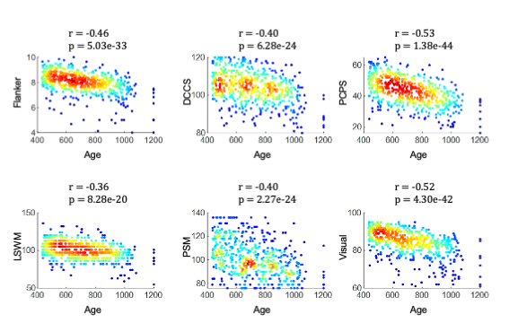
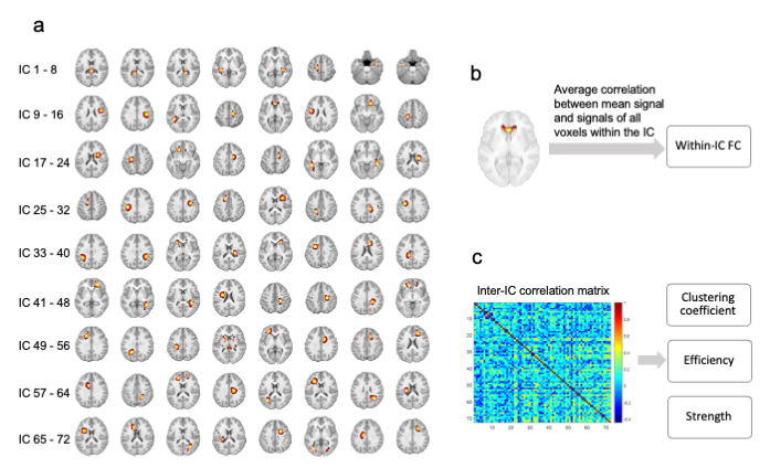
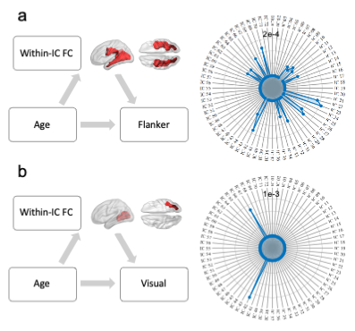
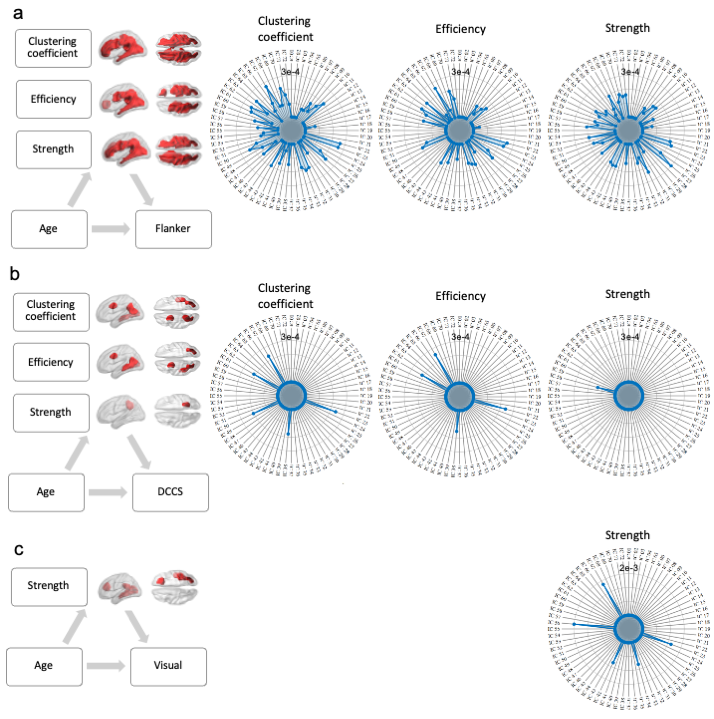
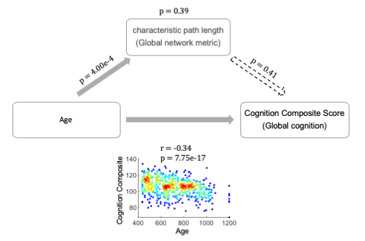
Figure 1 shows negative correlations between age and various clinical scores. As age increased, cognitive and sensory abilities typically decreased, with processing speed (PCPS) and visual acuity being most affected. The spatial maps of the 72 ICs in WM, as depicted in Figure 2a, elucidate spatially distinct clusters in the brain, often with bilateral symmetry. The ICs capture diverse areas of the brain and identify distinct patterns of intrinsic BOLD signal fluctuations. The within- and inter-IC relationships, as shown in Figures 2b and 2c provided insight into the intricate patterns of the brain. Mediation analysis revealed that the integrity of specific brain networks influenced the relationship between age and certain cognitive functions. As shown in Figure 3, the within-IC FC was a significant mediator between age and performance on the Flanker task, with the temporal lobe playing a crucial role. Similar mediation effects were found for visual acuity, with significant roles for ICs in the temporal and occipital regions. When examining network metrics including clustering coefficient, efficiency, and strength, these were found to mediate the link between age and performance on behavioral scores. As shown in Figure 4, wide-spread WM regions show significant mediation, with temporal and frontal regions being particularly influential in the connection between age and Flanker. In contrast, temporal areas show strong mediating effects on the relationship between age and DCCS. Moreover, occipital and frontal WM regions significantly mediate the age effects on visual ability. Lastly, Figure 5 shows that while aging correlated with a decline in cognitive composite scores, the expected mediation effect of the characteristic path length, a global network metric, on this relationship was not confirmed, suggesting that the architectural configuration of particular regions is more crucial in mediating the effects of age on specific behavioral outcomes, rather than a broad global metric influencing overall cognitive decline.Conclusion
Our study elucidates the aging brain's impact on cognition, showing that alterations of BOLD signals within white matter (WM) mediate cognitive decline. It highlights that aging affects cognitive functions variably, but is linked to specific WM changes. This contributes to understanding the aging brain's functional architecture and may guide interpretation of other functional studies.Acknowledgements
This work was supported by the National Institutes of Health (NIH) grants RF1 MH123201 (JG and BL), R01 NS113832 (JG), R01 NS129855 (ZD), and K01 EB032898 (KS). Research reported in this publication was supported by the National Institute On Aging of the National Institutes of Health under Award Number U01AG052564 and by funds provided by the McDonnell Center for Systems Neuroscience at Washington University in St. Louis. The HCP-Aging 2.0 Release data used in this report came from DOI: 10.15154/1520707.References
(1) Li M, Gao Y, Lawless RD, Xu L, Zhao Y, Schilling KG, Ding Z, Anderson AW, Landman BA, Gore JC. 2023. Changes in white matter functional networks across late adulthood. Frontiers in Aging Neuroscience. 15.
(2) Bookheimer SY, Salat DH, Terpstra M, Ances BM, Barch DM, Buckner RL, Burgess GC, Curtiss SW, Diaz-Santos M, Elam JS, Fischl B, Greve DN, Hagy HA, Harms MP, Hatch OM, Hedden T, Hodge C, Japardi KC, Kuhn TP, Ly TK, Smith SM, Somerville LH, Uğurbil K, van der Kouwe A, Van Essen D, Woods RP, Yacoub E. 2019. The Lifespan Human Connectome Project in Aging: An overview. NeuroImage. 185:335–348.
(3) Calhoun VD, Adali T, Pearlson GD, Pekar JJ. 2001. A method for making group inferences from functional MRI data using independent component analysis. Human Brain Mapping. 14:140–151.
(4) Wager TD, Waugh CE, Lindquist M, Noll DC, Fredrickson BL, Taylor SF. 2009. Brain mediators of cardiovascular responses to social threat: Part I: Reciprocal dorsal and ventral sub-regions of the medial prefrontal cortex and heart-rate reactivity. NeuroImage, Brain Body Medicine. 47:821–835.
Figures