3876
Healthy aging in human thalamic nuclei: an evaluation of volumetric atrophy and kurtosis microstructural metrics1Center for Mind/Brain Sciences - CIMeC, University of Trento, Rovereto, Italy, 2Data Science for Health, Fondazione Bruno Kessler, Trento, Italy, 3Champalimaud Foundation, Lisbon, Portugal
Synopsis
Keywords: Aging, Aging, Human thalamus, Other diffusion techniques
Motivation: Volumetric and microstructural characterization of thalamic nuclei is currently meeting growing interest in neuroimaging. Thalamic nuclei microstructural features, especially, may be used as biomarkers to study changes in both normal aging and degenerative diseases.
Goal(s): Quantify age-related differences in volumetry and microstructure of thalamic nuclei
Approach: We used 3T structural MRI (volumetry) and Diffusion Kurtosis Imaging (microstructure)
Results: Our main result is that thalamic volumetric atrophy effects related to healthy aging are significant and stronger than those given by microstructure estimates.
Impact: Macroscopic atrophy is more sensitive to healthy aging differences relative to microstructure effects derived from Diffusion Kurtosis Imaging.
Introduction
The thalamus is a subcortical structure composed of distinct grey matter nuclei involved in sensorimotor, attention, and memory functions1. Research evidenced differential age-related volume reductions depending on thalamic territories2,3, and volumetric changes become more variable as subjects age4,5. Diffusion Tensor Imaging (DTI) studies evidenced lower fractional anisotropy (FA) and higher mean diffusivity (MD) in thalamic regions during aging6,7,8. Findings suggest that, generally, anterolateral territories are more affected2,6. Diffusion Kurtosis Imaging (DKI) is an extension of DTI that accounts for non-Gaussian water displacement in biological tissues, providing more accurate characterization of tissue microstructure9. DKI evidence currently available for healthy aging10 is limited to the study of thalamus as a unitary structure. In the present study, we evaluate the effects of healthy aging on volume and microstructure of individual thalamic nuclei.Methods
Acquisition: 15 young (mean age (years,sd)=25.7(4.4); 7 males) and 7 elderly (mean age (years,sd)=71.1(3.6); 4 males) healthy volunteers underwent 3T MRI (MAGNETOM Prisma, Siemens Healthcare, Erlangen, Germany, 64-channel head-neck RF receive coil), to acquire 3D T1-weighted (T1w) multi-echoMPRAGE (TR=2530 ms, TE1-4=1.69/3.55/5.41/7.27 ms, TI=1100 ms, flip angle=7°, 1mm-isotropic), and dMRI (2mm-isotropic, TE/TR=76/4200 ms, shells: b={0,700,1000,2850} s/mm2, 32/64/64 directions) data.Image processing: T1w images were processed through FastSurfer’s recon-surf, a whole-brain segmentation tool alternative to FreeSurfer11. Thalamic nuclei were segmented using FreeSurfer’s thalamic segmentation algorithm12 to obtain nuclei volumetric data. The following thalamic nuclei were considered: medial pulvinar (PuM), ventroposterolateral (VPL), ventrolateral anterior (VLa) and posterior (VLp), mediodorsal medial (MDm) and lateral (MDl), ventral anterior (VA), centromedian (CM), and lateral geniculate (LGN). dMRI data were denoised13, corrected for Gibbs-ringing14, eddy currents, head motion, and geometric distortions in FSL (v.6.0.3)15, and for bias field in MRtrix (v.3.0.2)16. DKI was fitted and FA, MD, and MK maps derived17. DKI data was spatially smoothed using a Gaussian kernel with FWHM=1.259,18,19 in DIPY (v.1.4.1). To align thalamic nuclei segmentations with microstructural maps, linear registrations were computed in ANTs16 between each participant's first b=0 volume and T1w image, applied to thalamic segmentations, and the median value across each thalamic nucleus was extracted for each DKI metric.
Statistical analysis: a cross-sectional design was used. Wilcoxon rank-sum tests were used to investigate group differences in volumes and microstructural metrics, considering nuclei of both hemispheres separately. Subjects’ thalamic volumes were corrected for total intracranial volume (ICV). P-values were adjusted for False Discovery Rate for both volumetric and microstructural analyses.
Results
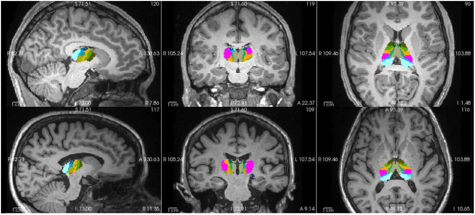
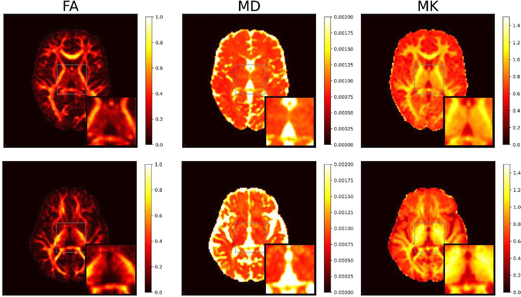
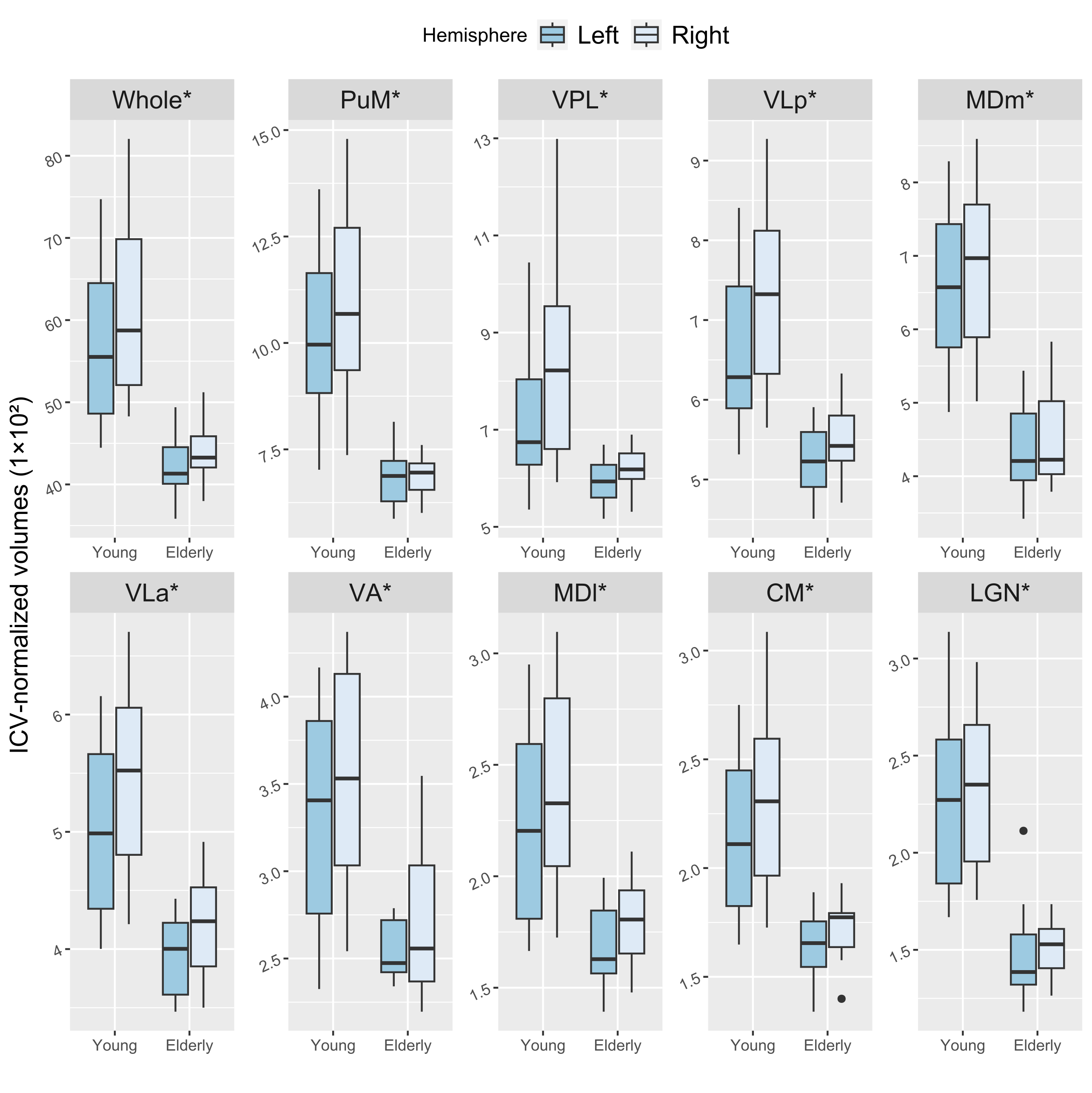
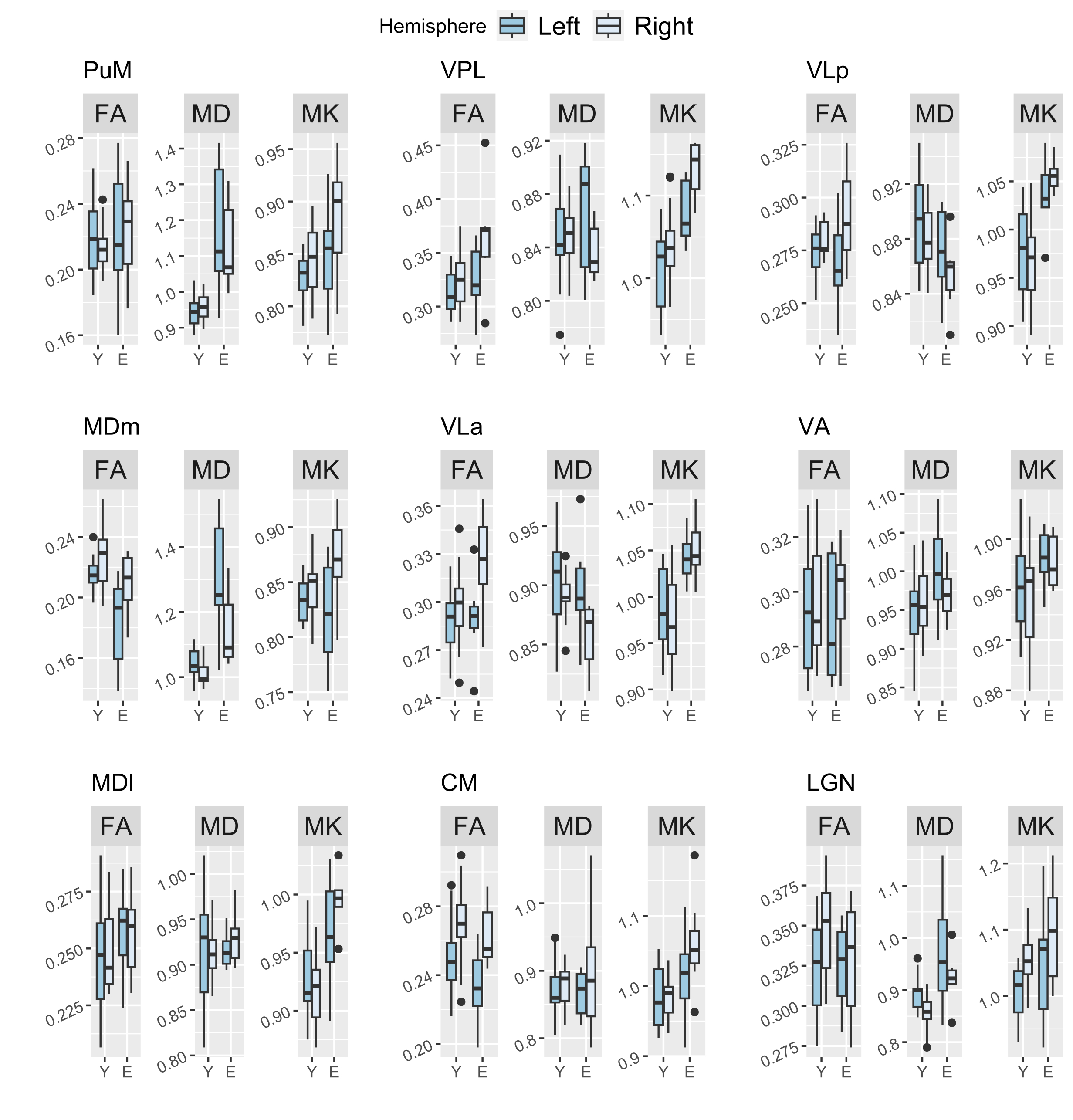
Thalamic parcellations (Fig. 1) and DKI-derived FA, MD, and MK maps (Fig. 2) are shown for representative young and elderly subjects. Figure 3 shows distributions of thalamic volumes in the two groups. Wilcoxon tests yielded significant volume differences in all considered thalamic nuclei. Figure 4 shows distributions of DKI-derived microstructural estimates. Comparisons for FA, MD, and MK were non-significant.Discussion
In agreement with previous studies, we found that whole thalamus and thalamic nuclei volumes are reduced in elderly as compared to younger subjects2,3,4,5. Previous DTI evidence showed that thalamic anterolateral regions are more associated with age-related microstructural changes2,6,7,8. We did not find any significant difference in the considered nuclei using DKI. Our preliminary results suggest that age-related volume reductions in thalamic nuclei might not necessarily coincide with changes in microstructural organization, not even in nuclei known to play a role in attention and memory2,20. There is evidence that volumes of dentate gyrus and lateral ventricles are good biomarkers for the assessment of the disease progression in mild cognitive impairment, while tissue microstructure plays little role21. Our preliminary results suggest that, similarly, thalamic nuclei volumes could better highlight group differences as opposed to DKI microstructural estimates. Nevertheless, trends in nuclei close to the lateral ventricles (e.g., MDm and PuM) suggest higher MD in elderly subjects, as expected from previous DTI studies6,7,8, as well as higher MK in sensorimotor nuclei VPL, VLa, and VLp. Although non-significant, these preliminary results need further evaluation with a larger sample size.Conclusions
We compared volume and microstructure of thalamic nuclei in young vs. elderly subjects with a cross-sectional design. Our results indicate age-related volume reductions, which do not accompany microstructural changes in any of the considered thalamic nuclei.Acknowledgements
This work was supported by funding from the Municipality of the City of Rovereto (Trento, Italy), for the project “Advanced neuroimaging to study aging”.References
1. Sherman, S. M., & Guillery, R. W. (2006). Exploring the thalamus and its role in cortical function. MIT press.
2. Fama, R., & Sullivan, E. V. (2015). Thalamic structures and associated cognitive functions: Relations with age and aging. Neuroscience & Biobehavioral Reviews, 54, 29-37.
3. Hughes, E. J., Bond, J., Svrckova, P., Makropoulos, A., Ball, G., Sharp, D. J., ... & Counsell, S. J. (2012). Regional changes in thalamic shape and volume with increasing age. Neuroimage, 63(3), 1134-1142.
4. Walhovd, K. B., Fjell, A. M., Reinvang, I., Lundervold, A., Dale, A. M., Eilertsen, D. E., ... & Fischl, B. (2005). Effects of age on volumes of cortex, white matter and subcortical structures. Neurobiology of aging, 26(9), 1261-1270.
5. Walhovd, K. B., Westlye, L. T., Amlien, I., Espeseth, T., Reinvang, I., Raz, N., ... & Fjell, A. M. (2011). Consistent neuroanatomical age-related volume differences across multiple samples. Neurobiology of aging, 32(5), 916-932.
6. Ota, M., Obata, T., Akine, Y., Ito, H., Matsumoto, R., Ikehira, H., ... & Suhara, T. (2007). Laterality and aging of thalamic subregions measured by diffusion tensor imaging. Neuroreport, 18(10), 1071-1075.
7. Abe, O., Yamasue, H., Aoki, S., Suga, M., Yamada, H., Kasai, K., ... & Ohtomo, K. (2008). Aging in the CNS: comparison of gray/white matter volume and diffusion tensor data. Neurobiology of aging, 29(1), 102-116.
8. Cherubini, A., Péran, P., Caltagirone, C., Sabatini, U., & Spalletta, G. (2009). Aging of subcortical nuclei: microstructural, mineralization and atrophy modifications measured in vivo using MRI. Neuroimage, 48(1), 29-36.
9. Jensen, J. H., Helpern, J. A., Ramani, A., Lu, H., & Kaczynski, K. (2005). Diffusional kurtosis imaging: the quantification of non‐gaussian water diffusion by means of magnetic resonance imaging. Magnetic Resonance in Medicine: An Official Journal of the International Society for Magnetic Resonance in Medicine, 53(6), 1432-1440.
10. Gong, N. J., Wong, C. S., Chan, C. C., Leung, L. M., & Chu, Y. C. (2014). Aging in deep gray matter and white matter revealed by diffusional kurtosis imaging. Neurobiology of aging, 35(10), 2203-2216.
11. Henschel, L., Conjeti, S., Estrada, S., Diers, K., Fischl, B., & Reuter, M. (2020). Fastsurfer-a fast and accurate deep learning based neuroimaging pipeline. NeuroImage, 219, 117012.
12. Iglesias, J. E., Insausti, R., Lerma-Usabiaga, G., Bocchetta, M., Van Leemput, K., Greve, D. N., ... & Alzheimer's Disease Neuroimaging Initiative. (2018). A probabilistic atlas of the human thalamic nuclei combining ex vivo MRI and histology. Neuroimage, 183, 314-326.
13. Veraart, J., Novikov, D. S., Christiaens, D., Ades-Aron, B., Sijbers, J., & Fieremans, E. (2016). Denoising of diffusion MRI using random matrix theory. Neuroimage, 142, 394-406.
14. Kellner, E., Dhital, B., Kiselev, V.G., Reisert, M. (2016). Gibbs‐ringing artifact removal based on local subvoxel‐shifts. Magnetic Resonance in Medicine, 76(5), 1574-1581.
15. Andersson, J. L., & Sotiropoulos, S. N. (2016). An integrated approach to correction for off-resonance effects and subject movement in diffusion MR imaging. Neuroimage, 125, 1063-1078.
16. Tustison, N. J., Avants, B. B., Cook, P. A., Zheng, Y., Egan, A., Yushkevich, P. A., & Gee, J. C. (2010). N4ITK: improved N3 bias correction. IEEE transactions on medical imaging, 29(6), 1310-1320.
17. Henriques, R. N., Correia, M. M., Marrale, M., Huber, E., Kruper, J., Koudoro, S., ... & Rokem, A. (2021). Diffusional kurtosis imaging in the diffusion imaging in python project. Frontiers in Human Neuroscience, 15, 675433.
18. Diffusion in Python (DIPY) documentation: https://dipy.org/documentation/1.1.1./examples_built/reconst_dki/#references
19. Jones, D. K., & Cercignani, M. (2010). Twenty‐five pitfalls in the analysis of diffusion MRI data. NMR in Biomedicine, 23(7), 803-820.
20. Kensinger, E. A. (2009). Cognition in aging and age-related disease. In L. R. Squire (Ed.), Encyclopedia of neuroscience (pp. 1055–1061). New York: Elsevier/Academic Press.
21. Marizzoni, M., Ferrari, C., Macis, A., Jovicich, J., Albani, D., Babiloni, C., ... & PharmaCog Consortium. (2019). Biomarker matrix to track short term disease progression in amnestic mild cognitive impairment patients with prodromal Alzheimer’s disease. Journal of Alzheimer's Disease, 69(1), 49-58.
Figures