3872
Brain age in healthy individuals and across multiple neurological disorders1Radiology, Beijing Tiantan Hospital, Capital Medical University, Beijing, China, 2MR Research Collaboration, Siemens Healthineers Ltd., Beijing, China
Synopsis
Keywords: Aging, Neuro, Neurological disorders
Motivation: Understanding brain age of healthy people and patients with neurological diseases is crucial for clinical application.
Goal(s): To characterize people with advanced brain age and explore brain aging patterns across neurological disorders.
Approach: Through a predicted brain age model using deep learning, we investigated the correlations between advanced brain aging and age-related deterioration in healthy individuals, and explored the correlation with clinical variables across neurological disorders.
Results: Healthy individuals with advanced brain aging have higher white matter hyperintensity burdens and lower brain region volumes. Brain age increases in patients with neurological disorders and has more cognitive decline and physical disability.
Impact: The brain age model using deep learning enables identifying individuals at risk for advanced brain aging in the normal-aging population and shows advanced brain aging across neurological diseases, which can be a biomarker for cognitive impairment and/or physical disability.
Introduction
Age is an important risk factor for neurological disorders. Brain aging, even within the normal-aging population, is highly heterogeneous. Age-related neuropathology may lead to different trajectories of cognitive decline and different white matter hyperintensity (WMH) burdens, suggesting the involvement of heterogeneous structural alterations1. Predicted brain age derived from MR images is a robust biomarker of the complex and multidimensional alterations occurring throughout the brain aging, which is used to model trajectories of general brain health2. The brain age gap (BAG) is defined as the difference between predicted brain age and chronological age. Asymptomatic individuals experiencing adverse aging may be identified through brain age, indicating an increased risk of future ill health3. In healthy individuals, a BAG ≥5 years can be considered to represent advanced brain aging, while a BAG ≤−5 years indicates resilience to brain aging. Despite the importance of understanding different brain-aging trajectories, its associations with regional brain alterations, WMH burden, and cognition have rarely been explored in both normal populations and neurological disorders. Here, we aimed to identify characteristics of individuals with advanced brain age and those at risk of age-related alterations in the normal population. We explored the clinical relationship between BAG and MRI measures, WHM volume, and clinical parameters in multiple neurological disorders.Methods
MRI scans and clinical data were collected from 2,913 healthy controls (HCs) and 1341 patients with neurological disorders from a multiple-center study, including 331 with multiple sclerosis (MS), 189 with neuromyelitis optica spectrum disorder (NMOSD), 239 with Alzheimer’s disease (AD), 244 with Parkinson’s disease (PD), and 338 with cerebral small vessel disease (cSVD) (Figure 1). A brain age prediction model was constructed based on a deep-learning algorithm (simple fully convolutional network4) by processing three-dimensional T1-weighted images. Volumes of brain regions and WMH on MRI and various clinical measures were compared between HCs with advanced brain aging and those with resilient brain age. Associations between BAG, brain and WMH volumes, and clinical variables were examined in patients with neurological disorders. P<0.05 was considered statistically significant.Results
In HCs, 316 participants showed advanced brain aging (mean chronological age: 49.33 years) and 278 participants were resilient brain aging (mean chronological age: 49.66 years). WMH volumes were higher (Cohen’s d = 0.42, P <0.001), and volumes of 47 cortical or subcortical regions were lower in individuals with advanced brain age than in those with resilient brain age (PFDR<0.001). The top three effect sizes (|Cohen’s d|) of brain region alterations were in the left accumbens, right accumbens, and right ventral diencephalon (Cohen’s d = −0.54, −0.53, and −0.37, respectively; PFDR<0.001). For neurological disorders, BAG was higher in patients with MS (10.30 ± 12.6 years), NMOSD (2.96 ± 7.8 years), AD (6.50 ± 6.6 years), PD (4.24 ± 4.8 years), and cSVD (3.24 ± 5.9 years) than in HCs (Figure 2). Increased BAG was strongly associated with atrophy in the right accumbens in patients with MS, left caudal middle frontal gyrus in NMOSD patients, left inferior parietal gyrus in AD patients, right pars orbitalis gyrus in PD patients, and left thalamus in cSVD patients (Figure 3). Increased BAG was correlated with WMH volume and cognitive decline in multiple neurological disorders and with higher disability scores in patients with MS but not in those with NMOSD (Figure 4).Discussion and Conclusion
We investigated brain age in large groups of healthy individuals and patients across various neurological disorders. Healthy individuals with advanced brain aging had higher WMH and lower regional brain volumes than those resilient to brain aging and thus brain age could identify individuals at risk of age-related brain pathology5. Furthermore, brain age can act as a measure of deviation from normal lifespan trajectories in various neurological disorders. Increased BAG and WMH burden differed among neurological disorders in this study. Deep gray matter (e.g., the accumbens and thalamus) commonly shows atrophy associated with increased BAG in neurological disorders6. WMH volume was significantly correlated with BAG in patients with neurological disease in this study. WMH volumes have been shown to be associated with cognitive and clinical outcomes7, and increased BAG and WMH volume may reflect underlying neuropathologic processes1. Higher BAG was correlated with lower cognitive scores across multiple neurological disorders. More severe disability, as reflected in the expanded disability status scale score, was associated with increased BAG in MS patients, but not in those who were aquaporin-4 antibody-seropositive with NMOSD. In conclusion, MRI-defined BAG is a robust imaging marker reflecting cognitive impairment and physical disability across different neurological disorders. Moreover, brain age could help identify individuals at risk for advanced brain aging.Acknowledgements
This work was supported by the National Key Research and Development Program of China (No. 2022YFC2009904/2022YFC2009900 to Dr Y Liu), the National Science Foundation of China (81870958 to Dr Y Liu), the Beijing Municipal Natural Science Foundation for Distinguished Young Scholars (No. JQ20035 to Dr Y Liu)References
References
1. Habes M, Pomponio R, Shou H, et al. The Brain Chart of Aging: Machine-learning analytics reveals links between brain aging, white matter disease, amyloid burden, and cognition in the iSTAGING consortium of 10,216 harmonized MR scans. Alzheimer's & Dementia : the Journal of the Alzheimer's Association 2021;17.
2. Cole JH, Franke K. Predicting Age Using Neuroimaging: Innovative Brain Ageing Biomarkers. Trends Neurosci 2017;40:681-690.
3. Cole JH, Ritchie SJ, Bastin ME, et al. Brain age predicts mortality. Mol Psychiatry 2018;23:1385-1392.
4. Peng H, Gong W, Beckmann CF, Vedaldi A, Smith SM. Accurate brain age prediction with lightweight deep neural networks. Med Image Anal 2021;68:101871.
5. Jansen MG, Griffanti L, Mackay CE, et al. Association of cerebral small vessel disease burden with brain structure and cognitive and vascular risk trajectories in mid-to-late life. Journal of Cerebral Blood Flow and Metabolism : Official Journal of the International Society of Cerebral Blood Flow and Metabolism 2022;42:600-612.
6. Li G, Tong R, Zhang M, et al. Age-dependent changes in brain iron deposition and volume in deep gray matter nuclei using quantitative susceptibility mapping. Neuroimage 2023;269:119923.
7. Salvadó G,
Brugulat-Serrat A, Sudre CH, et al. Spatial patterns of white matter
hyperintensities associated with Alzheimer's disease risk factors in a
cognitively healthy middle-aged cohort. Alzheimers Res Ther 2019;11:12.
Figures
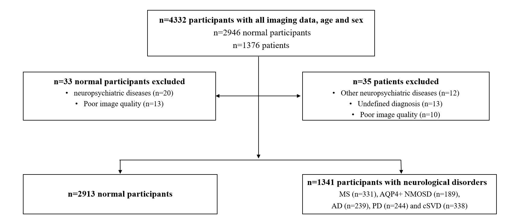
Figure 1. Study flowchart.
Abbreviations: AD, Alzheimer's disease; AQP4+ NMOSD, aquaporin-4 antibody-seropositive + neuromyelitis optica spectrum disorders; MS, multiple sclerosis; PD, Parkinson's disease; cSVD, cerebral small vessel disease.
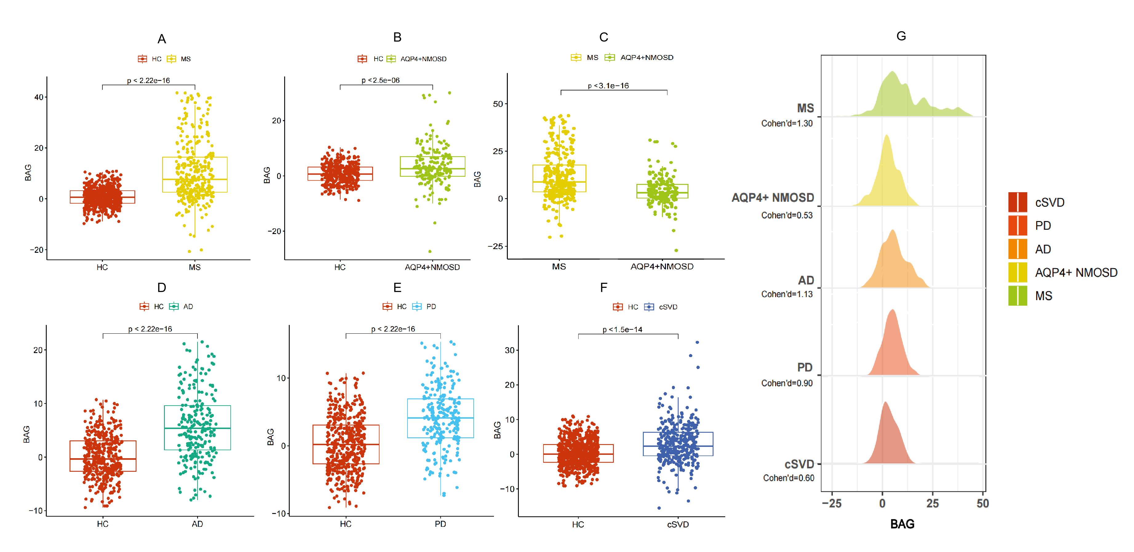
Figure 2. (A) BAG in patients with MS and HCs. (B) BAG in patients with AQP4+NMOSD and HCs. (C) BAG was greater in MS patients than AQP4+NMOSD patients. (D) BAG in patients with AD and HCs. (E) BAG in patients with PD and HCs. (F) BAG in patients with cSVD and HCs. (G) Distribution of BAG and Cohen’s d effect sizes according to neurological disorders.
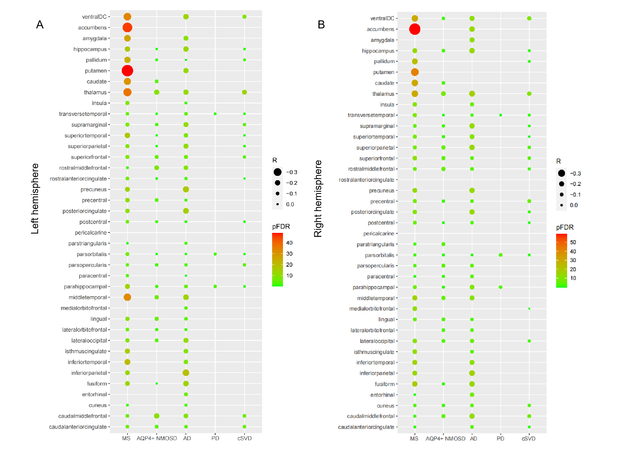
Figure 3. Correlations between BAG and brain atrophy according to neurological disorder and brain regions in the left (A) and right (B) hemispheres.
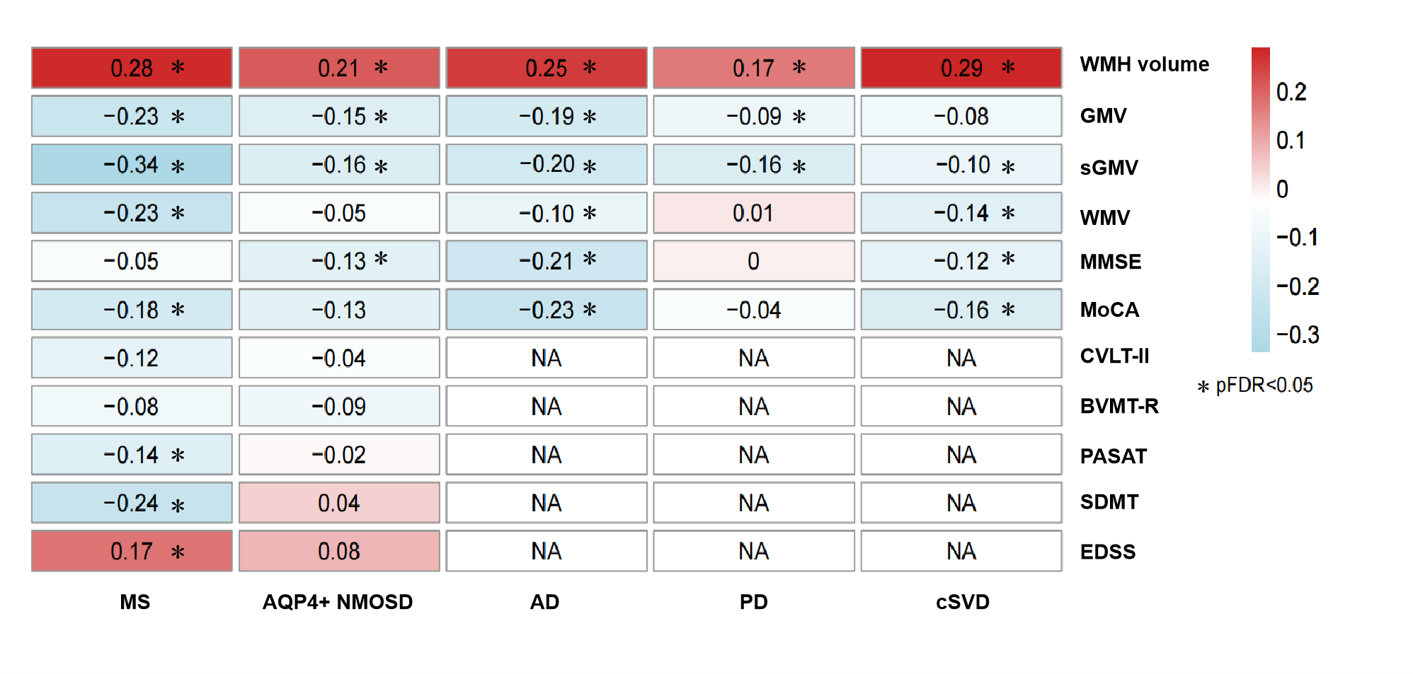
Figure 4. Correlations among BAG, magnetic resonance imaging measurements and clinical scores in patients with neurological disorders.
Abbreviations: BVMT-R, brief visuospatial memory test-revised; CVLT-II, California verbal learning test-second edition; EDSS, expanded disability status scale; MoCA, Montreal cognitive assessment; MMSE, mini-mental state examination; PASAT, paced auditory serial addition task; SDMT, symbol digit modalities test.
*P value after false discovery rate correction <0.05.