3871
Linking Myelin Integrity to Longitudinal Cognitive Processing Speed Decline in Normative Aging1National Institute on Aging, Baltimore, MD, United States
Synopsis
Keywords: Aging, Aging
Motivation: This study probes the specific impact of white matter myelin integrity on processing speed in the aging brain, responding to the need for deeper insights into cognitive decline mechanisms.
Goal(s): Our primary objective is to elucidate the relationship between myelin integrity and longitudinal changes in processing speed.
Approach: Utilizing quantitative MRI, we performed a retrospective longitudinal analysis correlating myelin water fraction (MWF) values with processing speed measurements.
Results: Significant correlations were found between decreased myelin integrity and faster decline in processing speed over the study period, affirming myelin integrity as a key factor in cognitive aging.
Impact: This research spotlights the pivotal role of myelin integrity in cognitive aging, potentially shifting existing neuroprotective strategies. Clinicians may now consider myelin preservation in cognitive health assessments, while researchers explore myelin restoration as a viable intervention for age-related cognitive decline.
INTRODUCTION
Myelin, the insulating sheath around neuronal axons, is fundamental for rapid neural signal transmission and crucial for cognitive processes. Previous studies illustrate that consistent myelination across brain regions is vital for maintaining the precise timing of neural communication 1, a factor that underpins cognitive efficiency. This relationship between myelin integrity and processing speed is evident throughout the human lifespan and has been observed in conditions ranging from healthy development 2-4 to neurodegenerative diseases 5,6. Despite insights from various studies using diverse imaging techniques, a comprehensive longitudinal understanding of how myelin content influences processing speed changes, particularly during normal aging, remains elusive. Our study employs advanced quantitative MRI 7-10 to dissect this relationship in a cohort of cognitively healthy adults. By delving into the complex interplay between myelination and cognitive aging, we aim to enhance our understanding of brain function across the lifespan and establish a basis for potential interventions and therapies targeting myelin deterioration to preserve cognitive health.METHODS
ParticipantsOur study utilized 121 cognitively normal participants from the Baltimore Longitudinal Study of Aging (BLSA) and the Genetic and Epigenetic Signatures of Translational Aging Laboratory Testing (GESTALT). Processing speed (PS) was assessed using the Digit Symbol Substitution Test (DSST) 11. Given concerns about the specificity of sole DSST, we have also adopted a PS composite score 3 by combining scores from the Trail Making Test Part A (TMT A) 12 and DSST.
MRI acquisition of in vivo myelin content
MRI scans were conducted using a 3 T Philips MRI system with a whole-brain BMC-mcDESPOT imaging protocol 13-15, following IRB approval. For each participant, MWF maps were registered to the Montreal Neurological Institute (MNI) template. Our regions of interest (ROIs) included the whole brain, frontal, parietal, temporal, occipital, and cerebellum of WM defined using MNI in FSL.
Statistical analysis
Linear mixed-effects models evaluated the relationship between MWF and PS/PS composite changes in six WM ROIs and on the voxel-wise level. The explicit regression model is given by:
$$\mathrm{PS}_{ij}=\beta_0+\beta_{\mathrm{age}}\times{\mathrm{age}}_i+\beta_{\mathrm{sex}}\times{\mathrm{sex}}_i+\beta_{\mathrm{race}}\times{\mathrm{race}}_i+\beta_{\mathrm{EDY}}\times{\mathrm{EDY}}_i\\
+\beta_{\mathrm{time}}\times{\mathrm{time}}_{ij}+\beta_{\mathrm{MWF}}\times{\mathrm{MWF}}_i+\beta_{\mathrm{time\ \times\ MWF}}\times{\mathrm{MWF}}_i\times{\mathrm{time}}_{ij}+\epsilon_{ij}+b_i$$
We note that the interaction term $$$\beta_{\mathrm{time\ \times\ MWF}}$$$ is of interest, reflecting the expectation of the difference in the longitudinal change in processing speed per unit difference in MWF. Regional analysis significance was set at uncorrected P < 0.05 and voxel-wise at P < 0.01 with a minimum cluster size of 400 voxels.
RESULTS
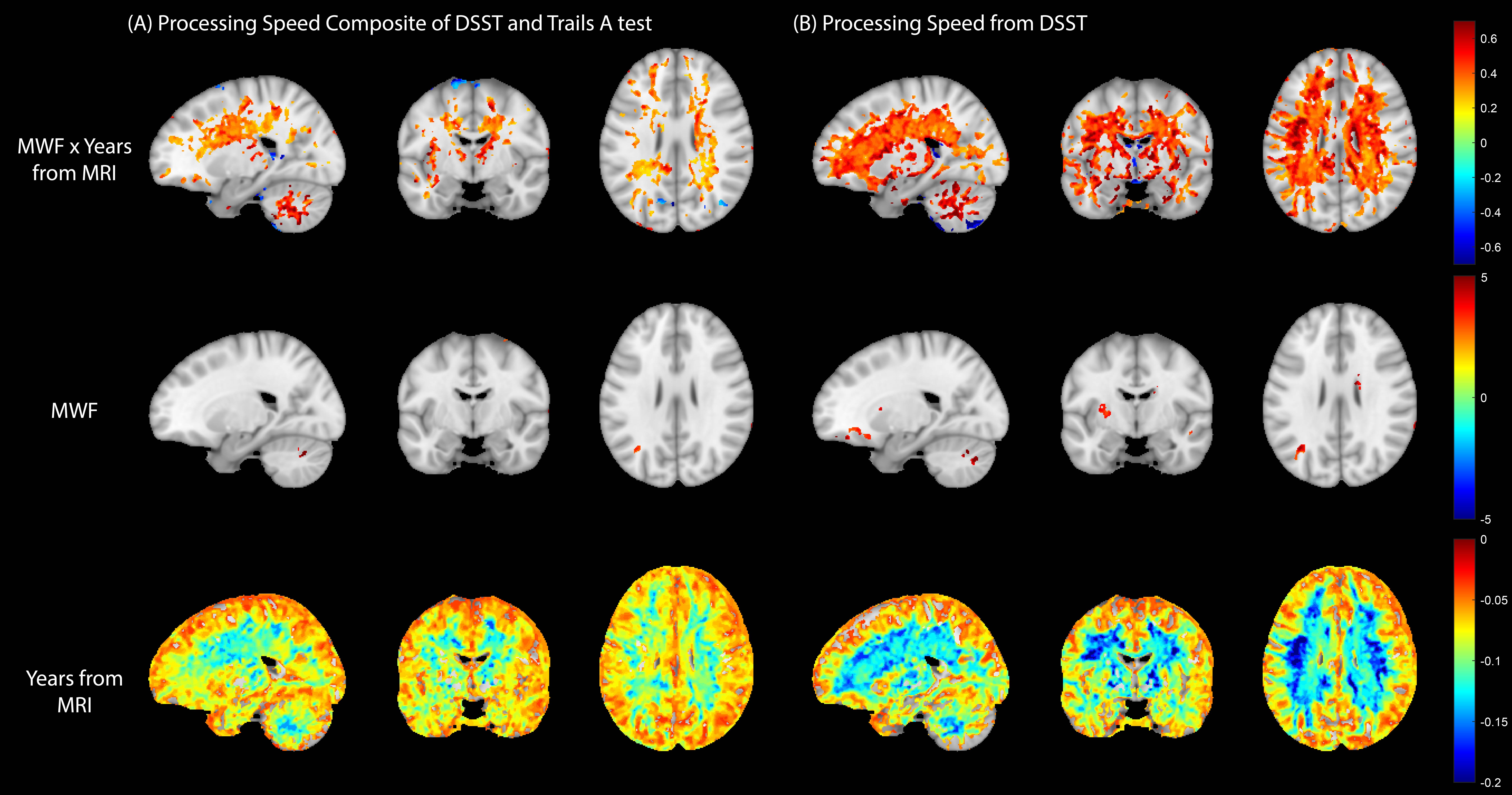
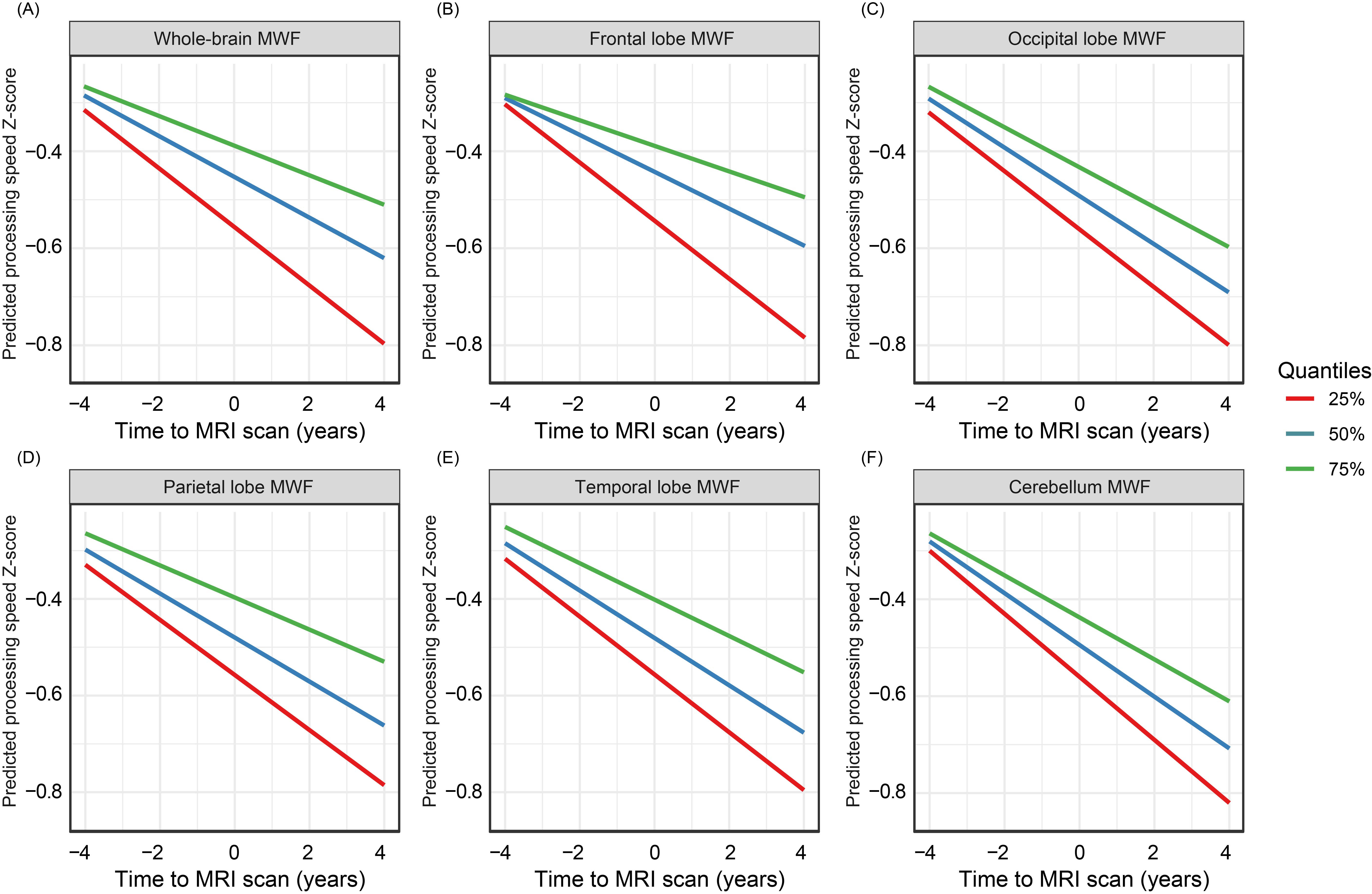
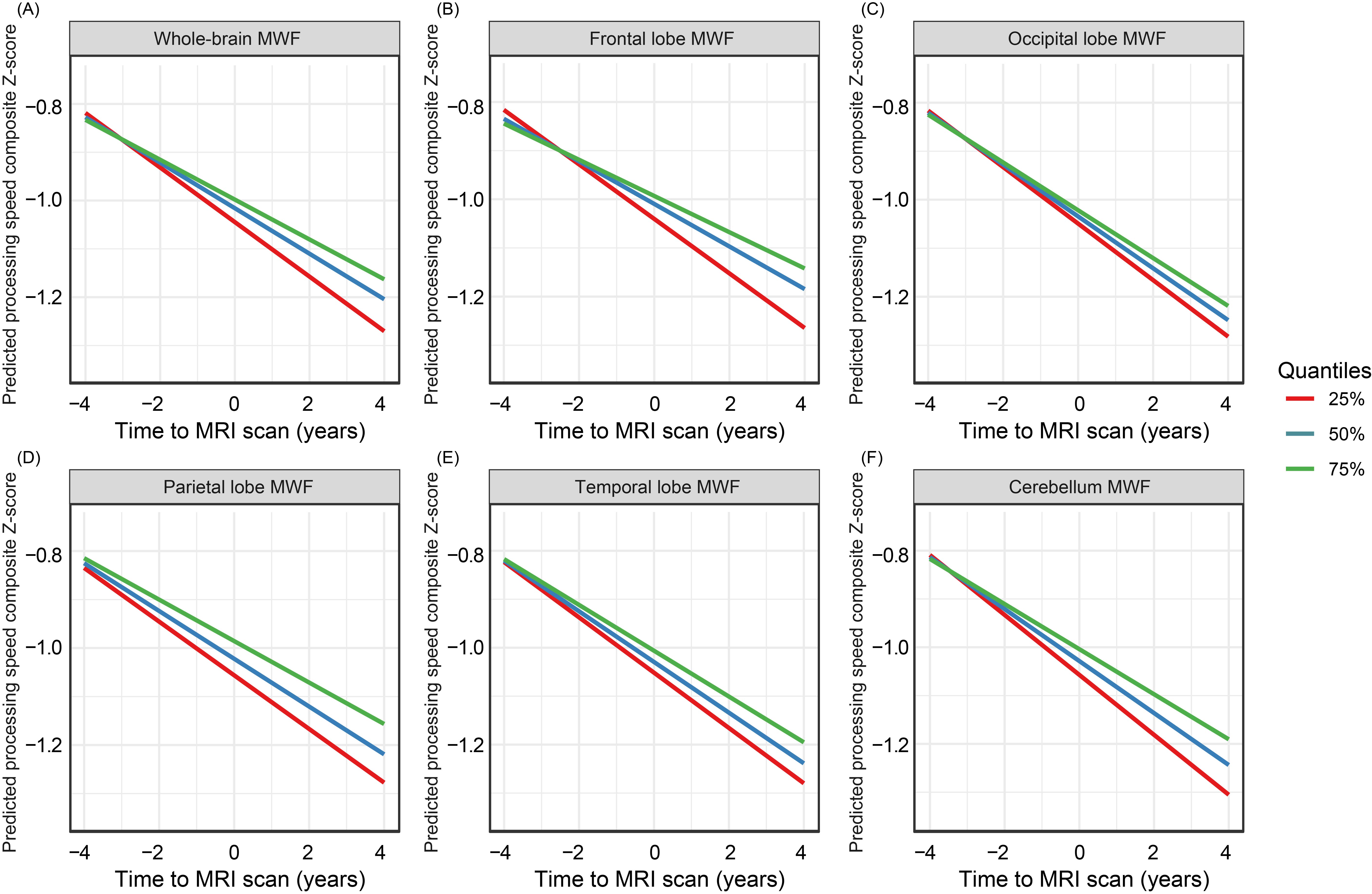
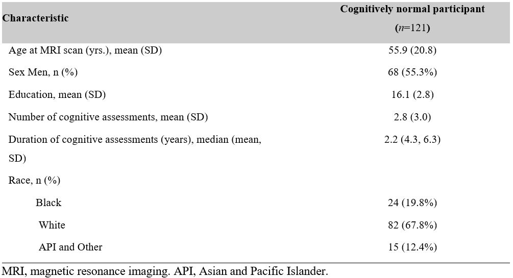
Table 1 presents the demographics of our participants. For voxel-wise analysis, Figure 1 demonstrates that cross-sectional myelin water fraction (MWF) had minimal significant associations with processing speed. However, a longer interval between initial cognitive assessment and baseline MRI scan was linked to decreased processing speed, a trend evident across nearly all examined brain regions. The interaction term highlighted positive correlations between MWF and the time from MRI across various white matter areas for both PS and PS composite scores. Regional analyses of six ROIs in Figures 2 and 3 showcase how processing speed declines vary by MWF levels. These relationships confirm that lower MWF correlates with more rapid declines in PS for all ROIs and in PS composite within most ROIs.DISCUSSION
Our study underscores the growing focus on the relationship between white matter integrity, particularly myelin, and cognitive function, given the pivotal role of white matter in neural signal conduction. Deterioration of myelin due to aging or disease disrupts neural signaling, leading to cognitive decline. With advanced neuroimaging like quantitative MRI, we can now intricately assess myelin integrity and its association with cognitive performance.In this longitudinal analysis, we established significant correlations between myelin integrity and the rate of decline in processing speed, with consistent findings across vital brain regions. This significance is anticipated considering the intricate nature of cognitive processing.
The association between MWF and processing speed was notably stronger when assessed with DSST alone compared to the PS composite score that included TMT A. This suggests DSST's heightened sensitivity to myelin's influence across neural networks. However, the weaker association of the composite measure in occipital and temporal regions implies that myelin content in these areas may not significantly impact the processing speeds evaluated by DSST and TMT A, highlighting the complexity of cognitive networks and the need for further understanding of different cognitive tests.
Our findings suggest MWF as a potential indicator of processing speed decline and call for more nuanced research to disentangle the complex relationship between white matter integrity and cognitive processing, especially in aging.
CONCLUSIONS
The study confirms that myelin integrity is crucial for cognitive processing speed and suggests myelination as a therapeutical target to maintain cognitive health and combat neurodegeneration.Acknowledgements
This work was supported by the Intramural Research Program of the National Institute on Aging of the National Institutes of Health.References
1. Salami, M.; Itami, C.; Tsumoto, T.; Kimura, F., Change of conduction velocity by regional myelination yields constant latency irrespective of distance between thalamus and cortex. Proceedings of the National Academy of Sciences 2003, 100 (10), 6174-6179.
2. Chevalier, N.; Kurth, S.; Doucette, M. R.; Wiseheart, M.; Deoni, S. C.; Dean III, D. C.; O’Muircheartaigh, J.; Blackwell, K. A.; Munakata, Y.; LeBourgeois, M. K., Myelination is associated with processing speed in early childhood: preliminary insights. PloS one 2015, 10 (10), e0139897.
3. Lu, P. H.; Lee, G. J.; Raven, E. P.; Tingus, K.; Khoo, T.; Thompson, P. M.; Bartzokis, G., Age-related slowing in cognitive processing speed is associated with myelin integrity in a very healthy elderly sample. J. Clin. Exp. Neuropsychol. 2011, 33 (10), 1059-1068.
4. Chopra, S.; Shaw, M.; Shaw, T.; Sachdev, P. S.; Anstey, K. J.; Cherbuin, N., More highly myelinated white matter tracts are associated with faster processing speed in healthy adults. Neuroimage 2018, 171, 332-340.
5. Shafer, A. T.; Williams, O. A.; Perez, E.; An, Y.; Landman, B. A.; Ferrucci, L.; Resnick, S. M., Accelerated decline in white matter microstructure in subsequently impaired older adults and its relationship with cognitive decline. Brain communications 2022, 4 (2), fcac051.
6. Abel, S.; Vavasour, I.; Lee, L. E.; Johnson, P.; Ackermans, N.; Chan, J.; Dvorak, A.; Schabas, A.; Wiggermann, V.; Tam, R., Myelin damage in normal appearing white matter contributes to impaired cognitive processing speed in multiple sclerosis. Journal of Neuroimaging 2020, 30 (2), 205-211.
7. Bouhrara, M.; Rejimon, A. C.; Cortina, L. E.; Khattar, N.; Bergeron, C. M.; Ferrucci, L.; Resnick, S. M.; Spencer, R. G., Adult brain aging investigated using BMC-mcDESPOT based myelin water fraction imaging. Neurobiology of Aging 2020, 85, 131-139.
8. Bouhrara, M.; Spencer, R. G., Rapid simultaneous high-resolution mapping of myelin water fraction and relaxation times in human brain using BMC-mcDESPOT. Neuroimage 2017, 147, 800-811.
9. Bouhrara, M.; Spencer, R. G., Improved determination of the myelin water fraction in human brain using magnetic resonance imaging through Bayesian analysis of mcDESPOT. Neuroimage 2016, 127, 456-471.
10. Bouhrara, M.; Spencer, R. G., Incorporation of nonzero echo times in the SPGR and bSSFP signal models used in mcDESPOT. Magn Reson Med 2015, 74 (5), 1227-35.
11. Chelune, G. J.; Bornstein, R. A.; Prifitera, A., The Wechsler Memory Scale—Revised. In Advances in Psychological Assessment: Volume 7, McReynolds, P.; Rosen, J. C.; Chelune, G. J., Eds. Springer US: Boston, MA, 1990; pp 65-99.
12. Reitan, R., Trail making test: Manual for administration and scoring: Reitan Neuropsychology Laboratory. Back to cited text 1992, (48).
13. Bouhrara, M.; Reiter, D. A.; Bergeron, C. M.; Zukley, L. M.; Ferrucci, L.; Resnick, S. M.; Spencer, R. G., Evidence of demyelination in mild cognitive impairment and dementia using a direct and specific magnetic resonance imaging measure of myelin content. Alzheimers Dement 2018, 14 (8), 998-1004.
14. Bouhrara, M.; Rejimon, A. C.; Cortina, L. E.; Khattar, N.; Bergeron, C. M.; Ferrucci, L.; Resnick, S. M.; Spencer, R. G., Adult brain aging investigated using BMC-mcDESPOT–based myelin water fraction imaging. Neurobiology of aging 2020, 85, 131-139.
15. Kiely, M.; Triebswetter, C.; Cortina, L. E.; Gong, Z.; Alsameen, M. H.; Spencer, R. G.; Bouhrara, M., Insights into human cerebral white matter maturation and degeneration across the adult lifespan. NeuroImage 2022, 247, 118727.
Figures