3866
Sexually dimorphic interactive effects of age and hypertension on subcortical volumes and asymmetries in normal adult brains1Canon Medical Systems Corporation, Ellicott City, MD, United States, 2Johns Hopkins University, Baltimore, MD, United States, 3Canon Medical Systems Corporation, Tochigi, Japan, 4University of Wisconsin, Madison, WI, United States
Synopsis
Keywords: Aging, Aging, thalamus
Motivation: Brain shrinkage does not happen to all areas uniformly and could be sex dependent.
Goal(s): We aimed to assess cross-sectionally the interactive effects of age and hypertension on the subcortical volumes and asymmetries in normal adult brains.
Approach: Brain structural images were acquired in 147 normal volunteers at a 3T scanner.
Results: All volumes demonstrated negative correlation with age but only the thalamic volume in both sexes, and the putamen volume in women reached the threshold of statistical significance of P=0.007. The thalamic asymmetry was the only measure that showed positive correlation to hypertension in women.
Impact: Among seven subcortical structures examined, the thalamic volume demonstrated a negative association with age in normal adult brains. Further research is needed to correlate this finding with cognition.
Introduction
Age-associated brain atrophy has been well studied. However, brain shrinkage does not happen to all areas uniformly and could be sex dependent. Many factors contribute to brain volume loss including genetic and environmental surrounding, socioeconomic status and cardiovascular risk factors but the degree of impact is less understood. The two hemispheres of the human brain are asymmetric functionally and structurally. Brain structural asymmetries might provide important clues to the neuroanatomical basis of lateralized brain functions. Characterizing subcortical asymmetries besides the absolute volume can increase sensitivity to pathological changes in neurological disease. However, subcortical asymmetries are subtle and requires accurate parcellation. Indeed, inconsistent results have been reported, and there is no consensus of biological normal ranges of subcortical asymmetries. There are also open questions of how variability in brain asymmetry relates to biological (e.g. age, sex) or modifiable risk variables (e.g. hypertension, smoking, diabetes, etc.). In this study, we aimed to assess cross-sectionally the interactive effects of age and hypertension on the subcortical volumes and asymmetries in normal adult brains.Methods
All participants were imaged using a 3T MRI systems (Galan, Canon Medical Systems, Japan). Brain structural images were acquired in sagittal orientation using a 3D magnetization-prepared rapid-acquisition gradient echo with 1-mm isotropic resolution. Segmentation and parcellation of the cortical mantle were processed using default parameters in FreeSurfer v7.2. Seven subcortical brain structures including thalamus, caudate, putamen, globus pallidus, hippocampus, amygdala, and nucleus accumbens were segmented. Asymmetry = (Left – Right)/(Left + Right). Sex difference were examined by Mann-Whitney U test for continuous variables, and Fisher’s exact test for categorial variables. Data were stratified by sex. Wilcoxon signed-rank test was used for comparison between right and left subcortical volumes. Multivariable regression was conducted for the relationship between subcortical geometry (i.e. volume and asymmetry) and age or hypertension co-adjusted for BMI and diabetes. The significance threshold was Bonferroni-adjusted to 0.007 for the analysis of seven subcortical structures.Results
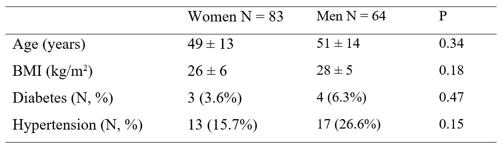
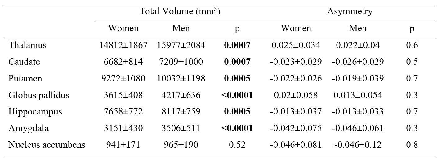
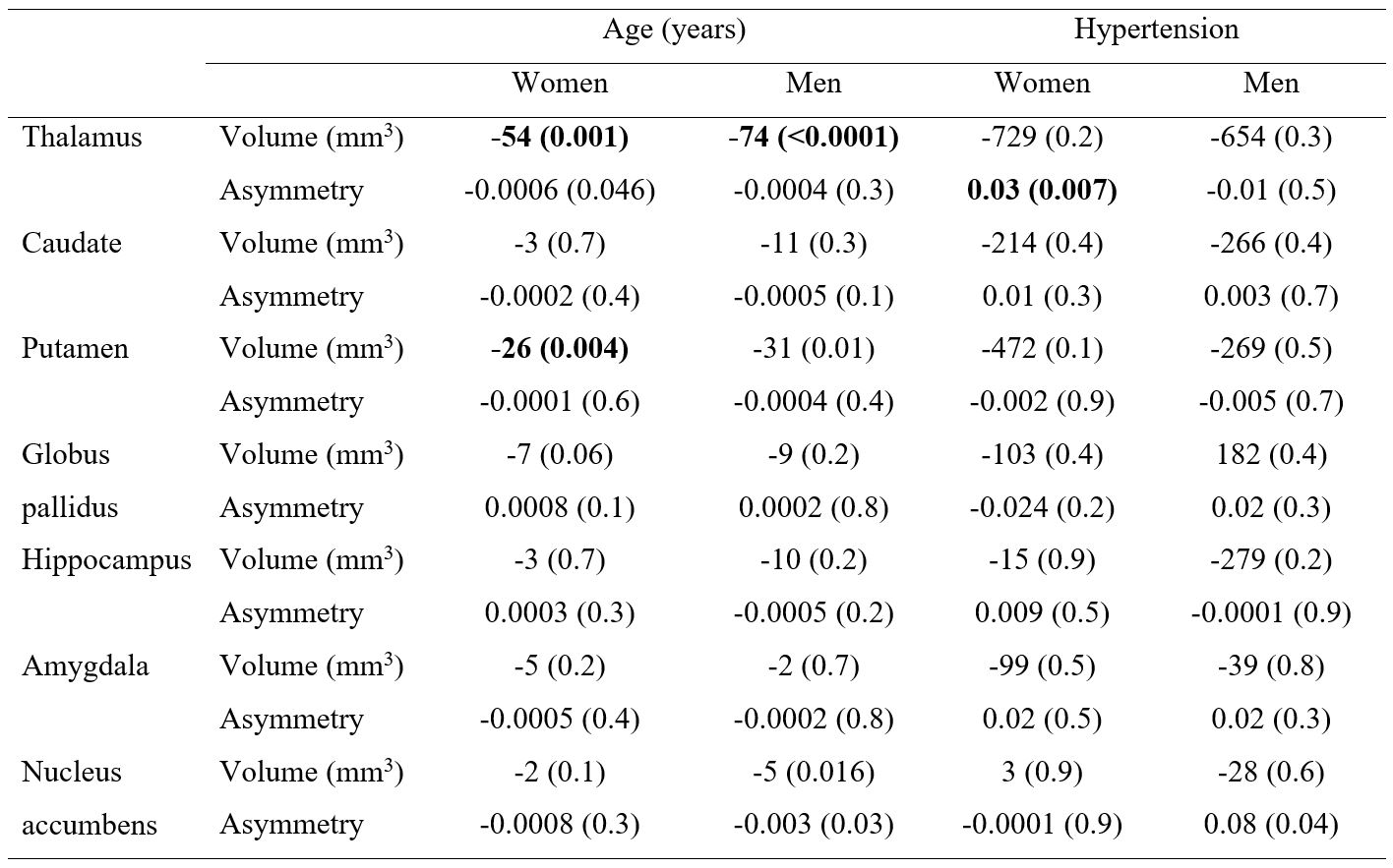
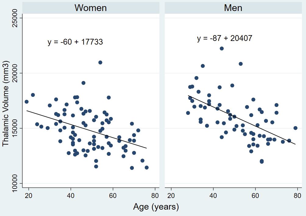
One hundred and forty-seven adult participants (mean age 50±13, 56% women) with no history of any neurological or psychiatric disorder were included in the study (Table 1). All subcortical volumes showed difference between left and right (P<0.001) except globus pallidus in men (P=0.1). In both men and women, volumes of the thalamus and the globus pallidus were larger on the left side. All other structures were larger on the right side. Results were similar between women and men. Total volume (right+left) and asymmetry of each subcortical brain structure stratified by sex is displayed in Table 2. In all segments (with the exception of nucleus accumbens, P=0.52), men demonstrated larger subcortical volumes than women (all P≤0.007). However, there was no difference between sex in the asymmetries of all subcortical structures. Association of subcortical geometries with age and hypertension stratified by sex are presented in Table 3. The thalamic volume in both sexes (P<0.007) and the putamen volume in women (P=0.004) were negatively associated with age. Figure 1 shows the relationship of thalamic volume to age. There was no relationship between asymmetry and age. Thalamic asymmetry was the only measure that showed positive correlation to hypertension in women (P=0.007).Discussion
The present study analyzed the sexual dimorphism in subcortical geometry and the association between each subcortical geometry and age/hypertension in normal adult brains. Differences by sex were present in the volumes of subcortical structures but not to the asymmetries. Age-related atrophy was demonstrated in the thalamus, while lower putamen volume with older age was only observed in women, and was generally absent in the other subcortical structures. We found greater thalamic asymmetry in women with hypertension, otherwise hypertension has no impact in the subcortical structures. It is known that anatomical features of the brain are lateralized but these findings have not been consistent across studies[1, 2]. Our results are generally in agreement with the meta-analysis conducted by Guadalupe et al. in 15,847 people from 52 studies worldwide[1]. Although we did not assess the handedness of our participants, handedness was previously found to have no association with subcortical asymmetry[1]. The effect of hypertension to the subcortical structures is less studied. We found greater thalamic asymmetry in women with hypertension, otherwise hypertension has no impact on subcortical structures of the brain.Conclusions
Among all subcortical segments, we confirmed that the thalamus demonstrated age-related shrinkage across the adult lifespan, but the asymmetry was preserved.Acknowledgements
No acknowledgement found.References
[1] Guadalupe T, Mathias SR, vanErp TGM, Whelan CD, Zwiers MP, Abe Y, et al. Human subcortical brain asymmetries in 15,847 people worldwide reveal effects of age and sex. Brain Imaging Behav 2017;11(5):1497-514.
[2] Kong XZ, Postema MC, Guadalupe T, de Kovel C, Boedhoe PSW, Hoogman M, et al. Mapping brain asymmetry in health and disease through the ENIGMA consortium. Hum Brain Mapp 2022;43(1):167-81.
Figures