3865
Tracking Age-Related Changes using NOEMTR in Brain Subregions at 3T1Radiology, University of Pennsylvania, Philadelphia, PA, United States
Synopsis
Keywords: Aging, Aging, CEST, NOE
Motivation: Demyelination and lipid degeneration occurs as the human brain ages. Our motivation was to investigate the age-dependent variations of these lipids changes in subcortical gray matter regions using NOEMTR in the brains of 15 subjects.
Goal(s): To determine the correlation of the human brain NOEMTR metric with age.
Approach: We performed NOE MRI experiments on multiple subjects from ages (24Y-76Y) using NOEMTR at 3T.
Results: The Spearman’s correlation coefficient indicates that NOEMTR declines in several brain subregions with aging.
Impact: NOEMTR can be used to track lipid changes in white and gray matter subregions of the aging brain.
Introduction
The aging brain undergoes intricate changes throughout one's life. Within the complex landscape of the human brain, various neural structures play essential roles in cognition and behavior, including the thalamus, caudate nucleus, hippocampus, pallidum, and putamen, influencing sensory perception, motor control, and memory functions1. Notably, the brain is the body's most lipid-rich organ, with lipids constituting 50% of its dry weight2. Predominantly, myelin-associated lipids like cholesterol, phospholipids, and glycolipids are fundamental components of cellular membranes, including synapses and myelin sheaths, regulating diverse biological processes3. A recent longitudinal study examining multiple "omics" data types revealed that around 40% of age-related molecular changes were linked to lipids, supporting the concept of a "LipidClock" for predicting biological aging4.Therefore, noninvasive imaging biomarkers for brain lipids hold clinical significance in studying lipid imbalances during aging and neurodegeneration. The Nuclear Overhauser Effect Magnetization Transfer Ratio (NOEMTR) is an emerging technique for studying mobile macromolecules such as lipids and proteins in the brain. This technique generates an NOE-weighted signal via the cross-relaxation of aliphatic protons from macromolecules (lipids and proteins) with bulk water, appearing at 3.5 ppm up-field from bulk water. It may also be influenced by chemical exchange saturation transfer (CEST) effects of labile protons from endogenous metabolites5. While amide CEST contrast has shown an inverse correlation with age in gray and white matter, it hasn't been extended to other gray matter sub-regions6. This study evaluates NOEMTR in 15 participants aged 24 to 76, focusing on various sub-regions, shedding light on age-related lipid metabolism in specific brain regions.Methods
All human studies were conducted under an approved University of Pennsylvania Institutional Review Board protocol. Fifteen healthy volunteers (13males, 2females) aged 24-to-76years old participated in the study. All imaging was performed on a 3T whole body scanner (MAGNETOM Prisma, Siemens Healthcare, Erlangen, Germany) with a body transmit/32-channel receive proton phased-array head coil. Three-repeated scans were acquired on one of the subjects to assess reproducibility. To obtain reproducible slice locations between subjects, we used a co-registration program, ImScribe 7. The 3D-NOE acquisition parameters are: number-of-slices=12, slice thickness=2 mm, in-plane resolution=1x1x2mm3, matrix-size=240x196, gradient-echo readout TR=3.5ms, TE=1.79 ms, read-out flip-angle=10°, averages=1, SHOT-TR = 8000ms, and saturation pulse of B1,rms=0.62µT with a saturation length of 3s.The z-spectra was acquired at varying saturation offset frequencies from -5 to 5ppm (relative to the water resonance) with a step-size of 0.25ppm. All imaging data was processed using in-house written MATLAB (MathWorks, Natick, MA) routines. The NOEMTR metric was computed as $$$ NOE_{MTR} (\%) = \frac{S_{0} - S_{-3.5}}{ S_{0} } $$$, where S0 is the signal intensity without saturation or saturation pulse at 100pm and S(-3.5) is the saturation at -3.5 ppm. Brain subregions were obtained via automated segmentation of an acquired MPRAGE image, with the following steps: 1) bias field correction using ANTs8; 2) brain masking using HD-BET9 ; 3) segmentation for subcortical gray matter regions using FSL FIRST10.Results
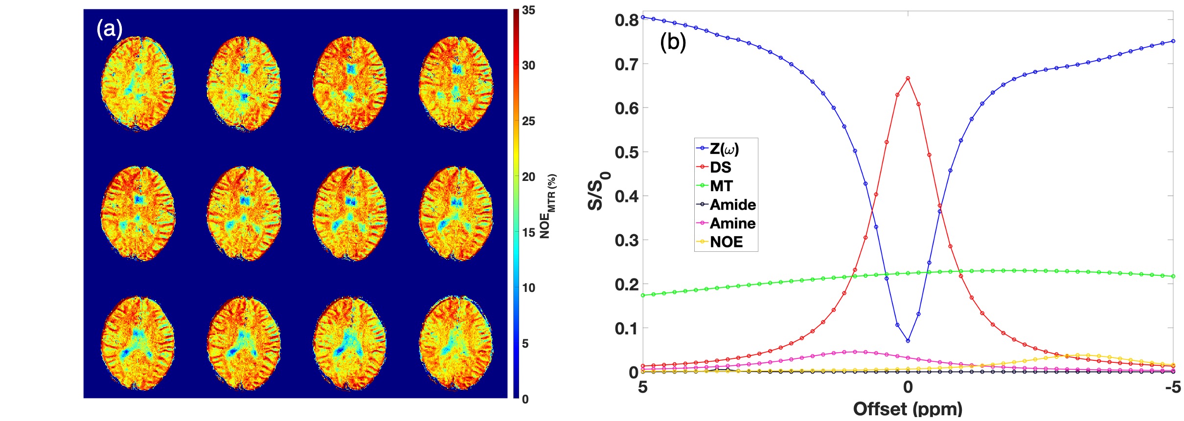
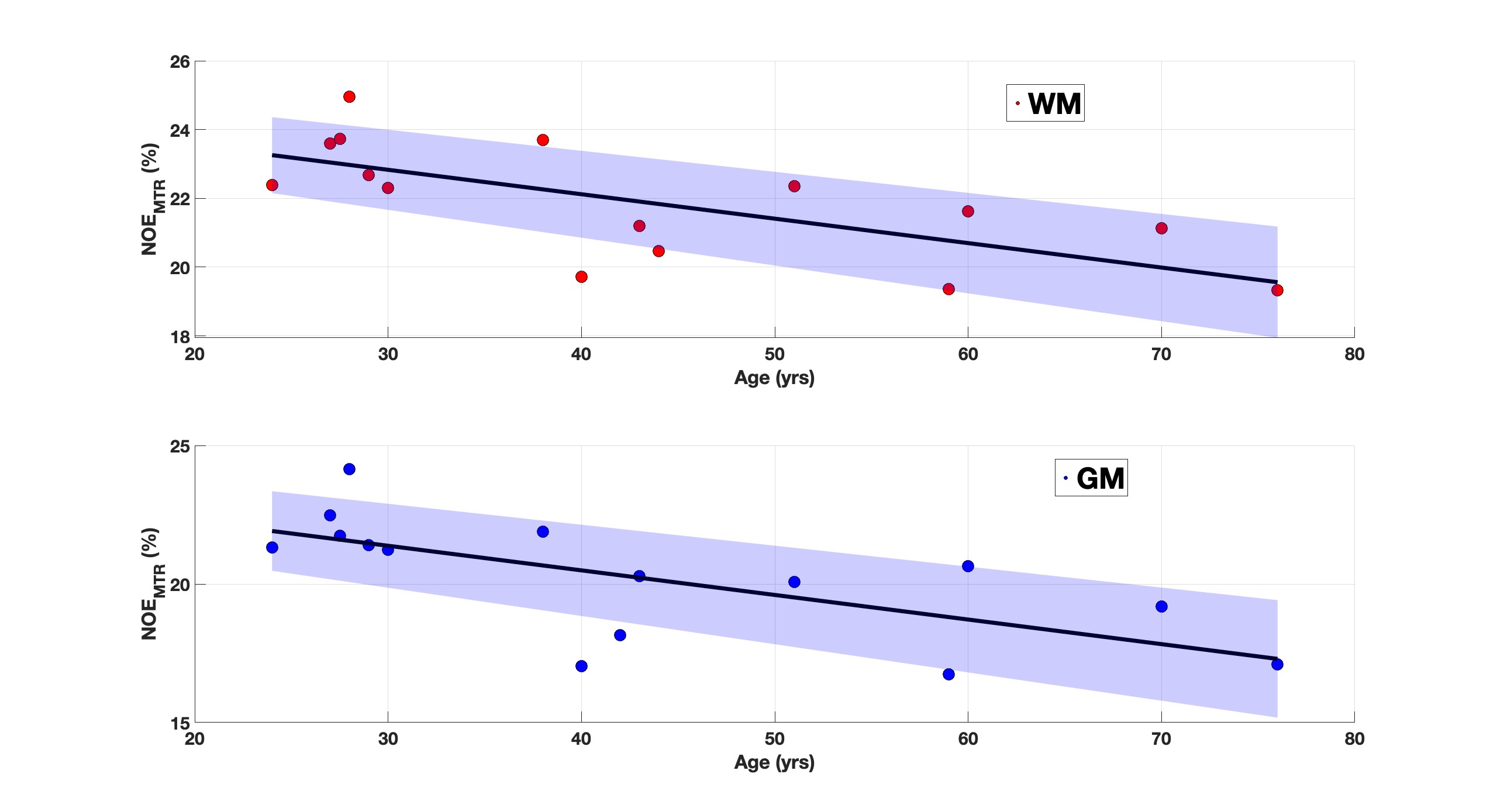
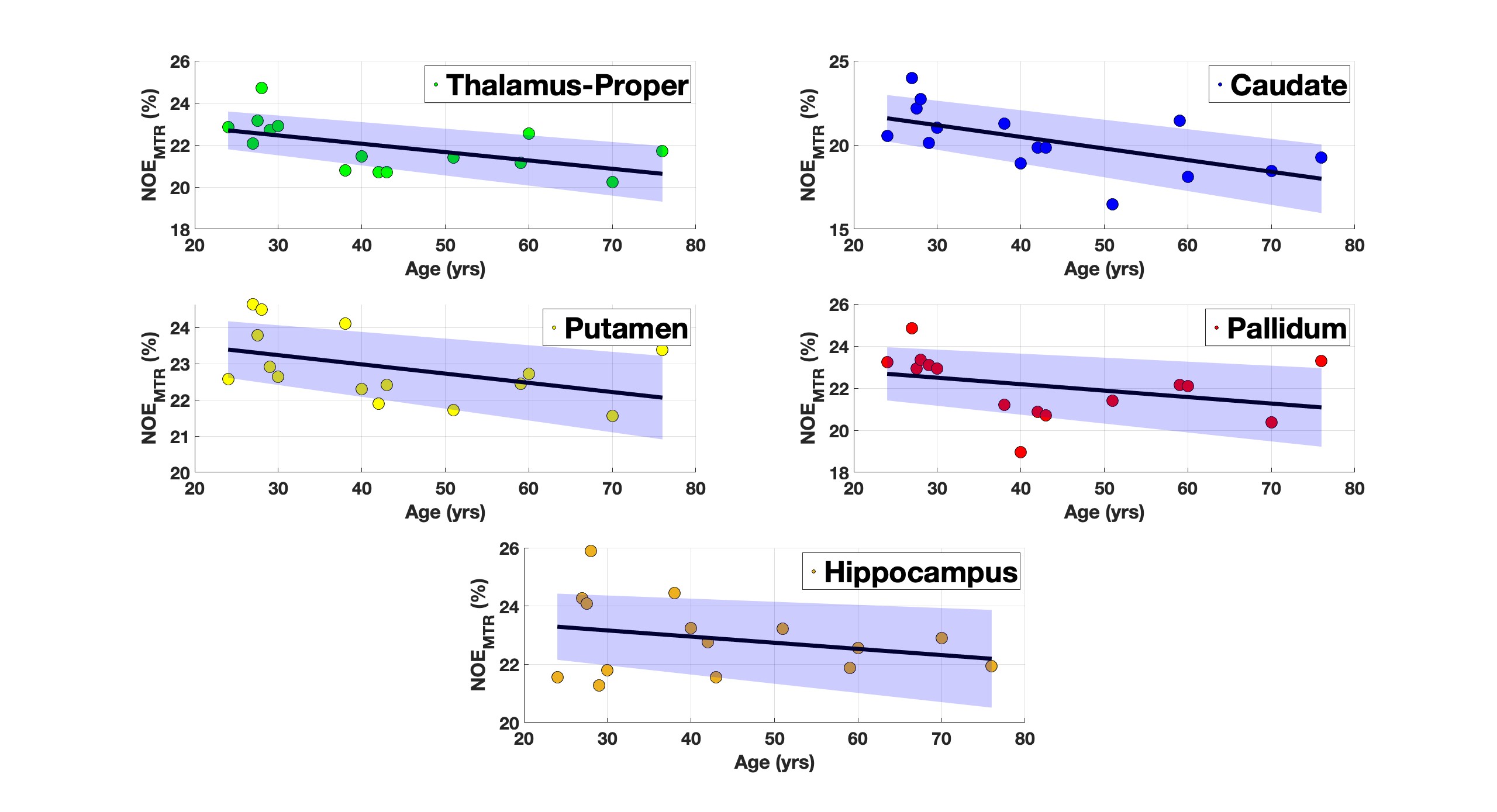
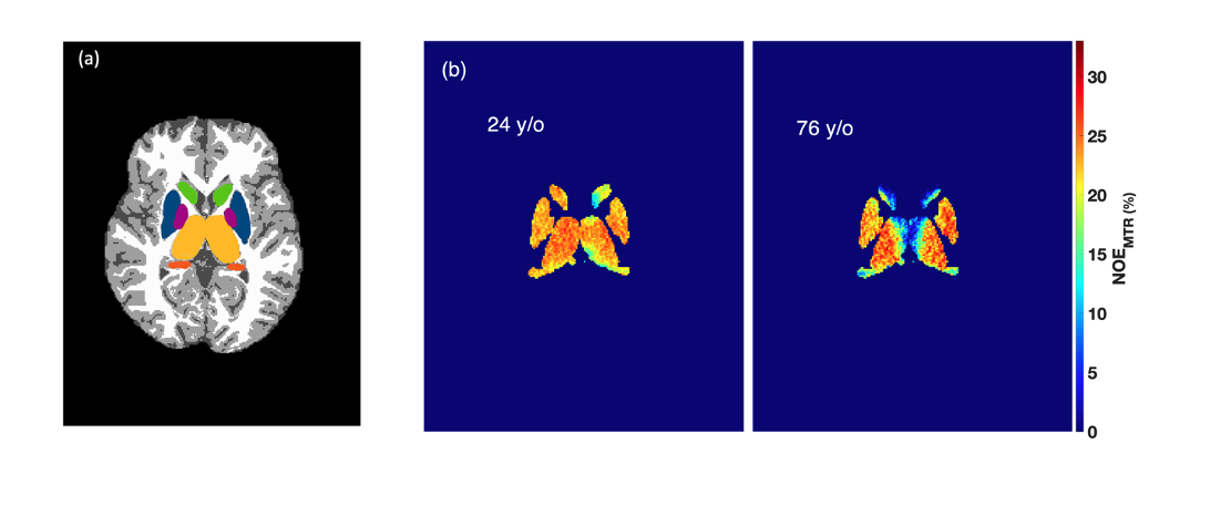
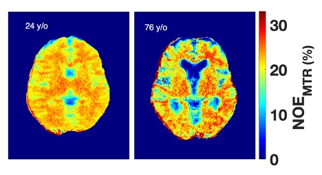
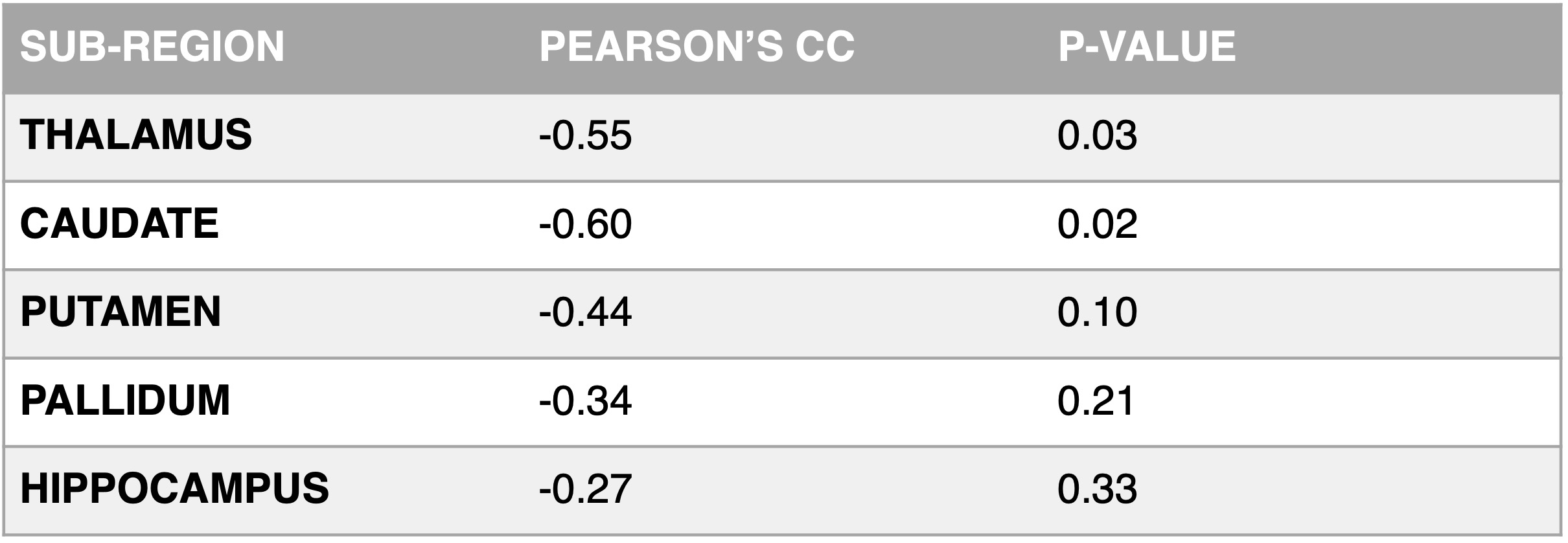
The study presents NOEMTR data and averaged Z-spectra from white matter (WM) in Figure 1, examining age-related changes in the NOEMTR metric in WM and gray matter (GM) regions (Figures 2, 3, and 4). Pearson's correlation analysis was used to assess the age dependence of the NOEMTR metric. Both WM and GM regions showed significant negative correlations with age, with Pearson's correlation coefficients (pcc) of -0.69 (p = 0.004) for WM and -0.68 (p = 0.006) for GM (Figure 2). Substructural analyses indicated significant negative age correlations in the thalamus (pcc = -0.55, p = 0.03) and caudate (pcc = -0.60, p = 0.02), suggesting a decline in NOEMTR with advancing age in these areas. The putamen displayed a negative correlation (pcc = -0.44) but didn't reach statistical significance (p = 0.10), and the pallidum exhibited a non-significant negative correlation (pcc = -0.34, p = 0.21), hinting at potential age-related changes. The hippocampus showed a weak, non-significant negative correlation with age (pcc = -0.27, p = 0.33). The study confirmed the NOEMTR metric's robustness at 3T through reproducibility assessment, with CoV values ranging from 1.94% to 4.44% for different brain subregions.Discussion & Conclusion
There are no reported age-dependent studies of NOEMTR at 3T. The only study that reported rNOE age-dependence was by Mennecke et al., [7], who demonstrated a 2% drop in the rNOE of WM and GM signals (15% to 13%) at 7T for healthy participants between ages 22Y to 73Y. In our study, we observed a 3% and 4% drop in the WM and GM regions, respectively. Results from this study collectively suggest that NOEMTR in the WM and GM subregions is significantly influenced by age.Acknowledgements
This work was supported by the National Institute of Biomedical Imaging and Bioengineering of the National Institutes of Health under award Number P41EB029460 and by the National Institute of Aging of the National Institutes of Health under award numbers R01AG071725 and R01AG063869.References
1. Stein T, Moritz C, Quigley M, et al. Functional connectivity in the thalamus and hippocampus studied with functional MR imaging. AJNR Am J Neuroradiol. 2000 Sep;21(8):1397-401.
2. Koenig, S.H. Cholesterol of myelin is the determinant of gray-white contrast in MRI of brain. Magn. Reson. Med. 1991, 20(2): 285-291.
3. Ahadi, S., Zhou, W., Schüssler-Fiorenza Rose, S.M. et al. Personal aging markers and ageotypes revealed by deep longitudinal profiling. Nat Med 26, 83–90 (2020).
4. Unfried, M., Ng, L. F., Cazenave-Gassiot, A., Batchu, K. C., Kennedy, B. K., Wenk, M. R., ... & Gruber, J. (2022). LipidClock: A Lipid-Based Predictor of Biological Age. Frontiers in Aging, 3, 828239.
5. Jones CK, Huang A, Xu J, Edden RA, et al. Nuclear Overhauser enhancement (NOE) imaging in the human brain at 7T. Neuroimage. 2013 Aug 15;77:114-24.
6. Mennecke, A, Khakzar, KM, German, A, et al. 7 tricks for 7 T CEST: Improving the reproducibility of multipool evaluation provides insights into the effects of age and the early stages of Parkinson's disease. NMR in Biomedicine. 2023; 36(6):e4717.
7. Wolf, D. H., Satterthwaite, T. D., Loughead, et al. Amygdala abnormalities in first-degree relatives of individuals with schizophrenia unmasked by benzodiazepine challenge. Psychopharmacology. 2011, 218(3), 503–512.
8. Avants BB, Yushkevich P, Pluta J, et al. The optimal template effect in hippocampus studies of diseased populations. Neuroimage. 2010 Feb 1;49(3):2457-66.
9. Isensee F, Schell M, Tursunova I, et al. Automated brain extraction of multi-sequence MRI using artificial neural networks.1901.11341, 2019.
10. Patenaude, B., Smith, S.M., Kennedy, D., and Jenkinson M. A Bayesian Model of Shape and Appearance for Subcortical Brain NeuroImage, 56(3):907-922, 2011.
Figures