3859
Identification of glioma IDH genotypes using time-dependent diffusion magnetic resonance imaging-based microstructural mapping<stork-writing-assistant></stork-writing-assistant><stork-writing-assistant></stork-writing-assistant>1Lanzhou University Second Hospital, lanzhou, China, 2Philips Healthcare, Xi'an, China
Synopsis
Keywords: Tumors (Pre-Treatment), Diffusion/other diffusion imaging techniques
Motivation: Time-diffusion-dependent diffusion MRI (t-dMRI)provides quantitative imaging of cellular microstructure; however, its value in diagnosing molecular subtypes of IDH in gliomas remains unknown.<stork-writing-assistant></stork-writing-assistant><stork-writing-assistant></stork-writing-assistant>
Goal(s): Molecular subtypes of IDH were diagnosed using quantitative time-dependent diffusion imaging parameters.<stork-writing-assistant></stork-writing-assistant><stork-writing-assistant></stork-writing-assistant>
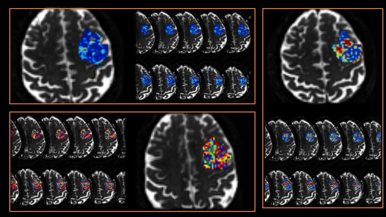
Approach: t-dMRI was acquired using OGSE and PGSE sequences, and quantitative parameters were then fitted using the Imaging Microstructural Parameters Using Limited spectrally edited diffusion(IMPULSED) method and evaluated for diagnostic potency for molecular subtypes of IDH in gliomas. <stork-writing-assistant></stork-writing-assistant><stork-writing-assistant></stork-writing-assistant>
Results: t-dMRI can identify IDH genotypes, and intracellular fraction (fin) reflects the actual state of tumor cells.<stork-writing-assistant></stork-writing-assistant><stork-writing-assistant></stork-writing-assistant>
Impact: t-dMRI can non-invasively identify IDH genotype.<stork-writing-assistant></stork-writing-assistant><stork-writing-assistant></stork-writing-assistant>
Introduction
Glioma is the most common tumor in the central nervous system1, with high lethality and poor prognosis; isocitrate dehydrogenase is an important prognostic biomarker for glioma. Previous studies have shown that the genotype of IDH correlates with overall survival and prognosis of patients, and the detection of IDH genotype still relies on invasive surgery and puncture biopsy in clinical practice; however, non-invasive detection of IDH genotype is essential for patients who refuse surgical treatment in the clinical practice.ADC values can non-invasively detect the genotype of IDH; however, they cannot reflect the actual state of the tumor cells and the surrounding cells. Recent studies2-3 have indicated that time-dependent diffusion-based imaging allows for quantitative assessment of cellular microstructure. Therefore, we aimed to use time-dependent diffusion imaging for the non-invasive identification of IDH genotypes.Methods
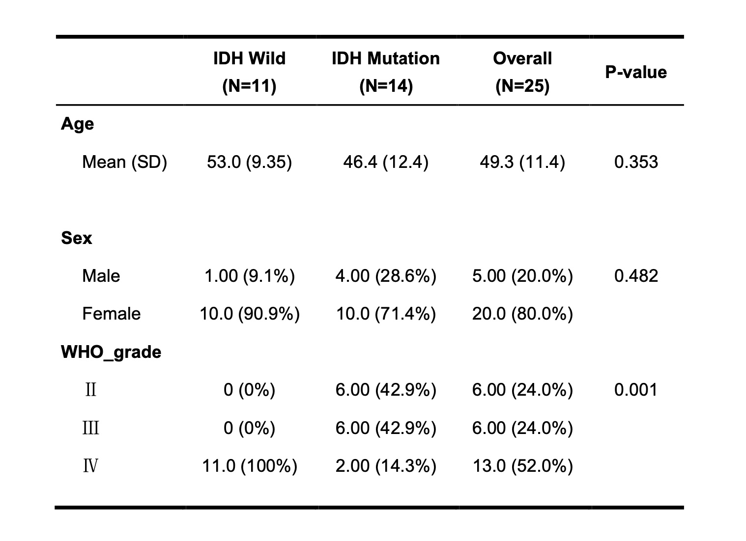
In this retrospective analysis, from June 2022 to September 2023, we collected time-dependent diffusion imaging data from 47 patients. We included 27 patients in the final based on the inclusion-exclusion criteria. All scans were performed on a 3T scanner (Ingenia CX, Phillips Healthcare, Best, The Netherlands). A 32-channel cranial coil with a maximum gradient performance of 80 mT/m and 200 mT/m/s was used. OGSE data were acquired at 22.8 Hz (b = 75/150/225/300 s/mm2), 45.6 Hz (b = 250/500/750 /1000 s/mm2), PGSE was acquired with diffusion duration/separation = 60/82.3 ms at b-value of 450/900/1350/1800 s/mm2, other parameters: Echo time/repetition time = 168/3000 ms, other parameters: Echo time/repetition time = 168/3000 ms, field of-view = 224 × 224 mm, matrix size = 160 × 160, slice thickness = 5 mm, 10slices, 1 non-diffusion-weighted image (b0), 10 diffusion directions per b value, and SENSE acceleration factor = 2. We corrected for patient head motion using FSL (https://fsl.fmrib. ox.ac.uk/fsl/fslwiki) and, finally, a two-compartment IMPULSED algorithm was used to fit the OGSE quantitative parameters4-5. The nuclei of the pathology images were further segmented using a deep learning algorithm, and the number of nuclei was analysed for correlation with the quantitative parameters to verify their reproducibility and consistency.Results
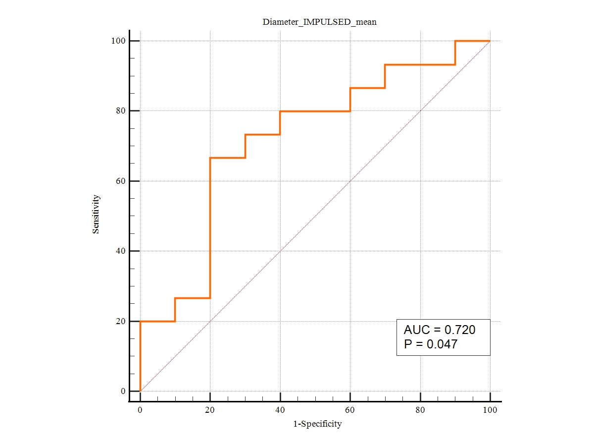
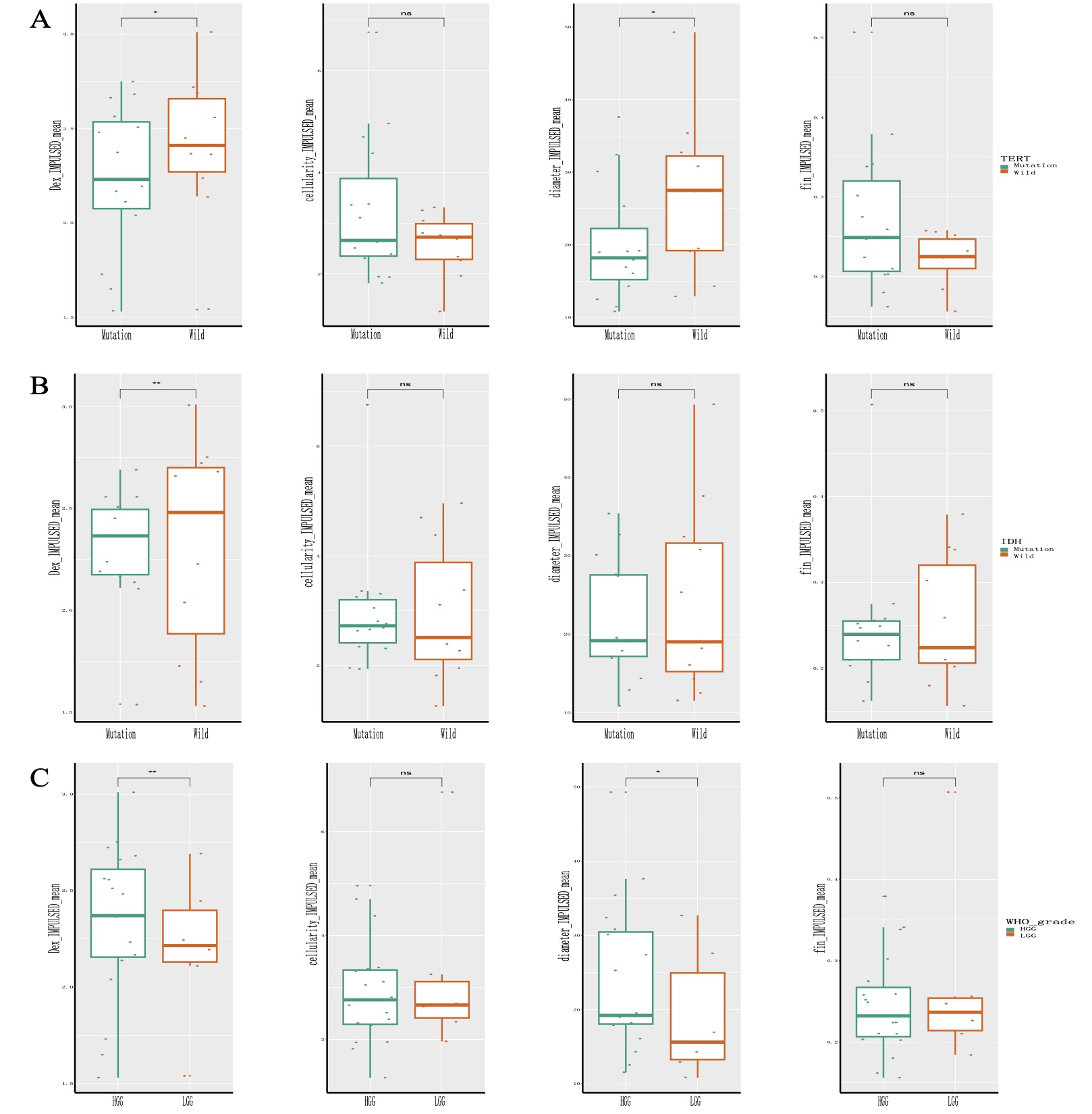
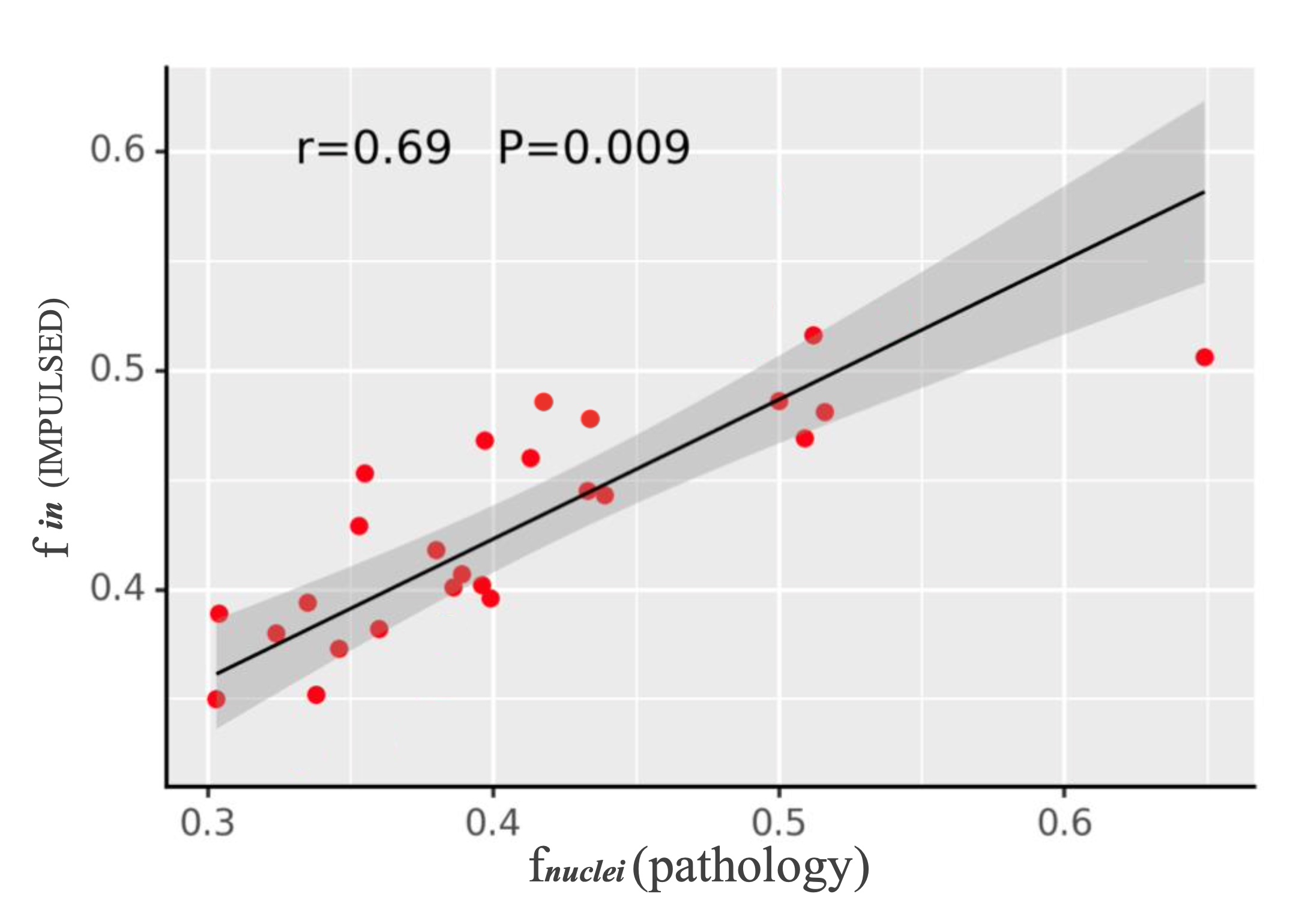
In our study, we demonstrated that cell diameter identified IDH genotype with AUC= 0.72 (95% CI:0.68-0.80), and the IMPULSED model yielded a positive correlation between f in and f nuclei in the pathological images of 25 patients (r=0.69,p<0.05). In addition, we compared the results based on the IMPULSED model in terms of IDH genotype, TERT genotype, and high and low tumor grade. The extracellular diffusivity (Dex) was higher in IDH wild-type patients than in IDH mutant patients (p<0.05); the Dex, as well as cell diameter, were higher in high-grade glioma patients than in low-grade patients (p<0.05).Discussion
Our study identified IDH genotypes based on t-dMRI cellular microstructure quantitative parameters and compared the IMPULSED model parameters across genotypes and high and low-grade gliomas. Previous studies2 utilized IMPULSED model parameters to identify the H3K27 genotype in pediatric gliomas with an AUC value of 0.91. The diagnostic performance was higher than that of our study, and we believe that it is due to the choice of the whole tumor area to be delineated when manually delineating the region of interest and that delineating the region of interest based on the parenchymal portion of the tumor may be more reflective of the cellular microstructural properties of the tumor. Our study significantly correlated fin with f-nuclei in pathological images, suggesting that the intracellular fraction may characterize the actual nuclear state. However, more extensive sample size data still needs to validate this.Conclusion
t-dMRI can identify IDH genotypes non-invasively, and each quantitative parameter is significantly different in different genotypes and high- and low-grade tumor states, with fin characterizing the actual state of the tumor cells.Acknowledgements
No acknowledgement found.References
1. Louis DN, Perry A, Wesseling P, et al. The 2021 WHO Classification of Tumors of the Central Nervous System: a summary [J]. Neuro-Oncology, 2021,23(8): 1231-1251. 2. Zhang H, Liu K, Ba R, et al. Histological and molecular classifications of pediatric glioma with time-dependent diffusion MRI-based microstructural maping [J]. Neuro-oncology, 2023, 25(6): 1146-1156. 3. Hoffmann E, Gerwing M, Niland S, et al. Profiling specific cell populations within the inflammatory tumor microenvironment by oscillating-gradient diffusion-weighted MRI[J]. Journal for Immunotherapy of Cancer, 2023, 11(3). 4. Wu D, Jiang K, Li H, et al. Time-dependent diffusion MRI for quantitative microstructural maping of prostate cancer[J]. Radiology, 2022, 303(3): 578-587. 5. Li H, Jiang X, **e J, et al. Impact of transcytolemmal water exchange on estimates of tissue microstructural properties derived from diffusion MRI[J]. Magnetic resonance in medicine, 2017, 77(6): 2239-2249.Figures