3856
The value of DCE-MRI and IVIM in predicting TERTp mutation status in glioblastoma1Department of Radiology, First Affiliated Hospital of Kunming Medical University, Kunming, Yunnan province, China, 2GE HealthCare MR Research, Beijing, China, China
Synopsis
Keywords: Tumors (Pre-Treatment), DSC & DCE Perfusion, Telomerase reverse transcriptase
Motivation: This study aims to predict the TERTp mutation status noninvasively before surgery, which is crucial for determining treatment strategies and prognosis in GBM patients.
Goal(s): Investigate the predictive value of DCE-MRI and IVIM for TERTp mutation status in GBM patients.
Approach: 68 GBM patients were enrolled according to the 2021 WHO classification, and divided into TERTp mutant group and TERTp wild group. Compare DCE-MRI and IVIM parameters between the two groups.
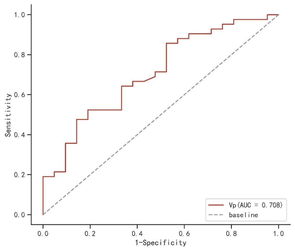
Results: The DCE-MRI parameter Vp was significantly higher in the TERTp mutation group (AUC=0.708), indicating its potential as a predictor for TERTp mutation status.
Impact: This study suggests that Vp, a DCE-MRI parameter, can predict TERTp mutation status noninvasively. This has important implications for targeted therapy and prognosis prediction in GBM patients.
Introduction
The Telomerase reverse transcriptase (TERT) promoter mutation status is not only an important criterion for glioblastoma (GBM) diagnosis according to the 2021 WHO classification1,but also makes the tumor have a dismal prognosis2.What’s more, TERTp mutation is a therapeutic target in GBM3.Therefore, noninvasive and repeatable methods for preoperative testing of TERTp mutations are necessary. Dynamic Contrast-Enhanced Magnetic Resonance Imaging (DCE-MRI) can evaluate tumor tissue perfusion, angiogenesis, and microvascular permeability4. Intravoxel incoherent motion (IVIM) can reflect cell proliferation and tumor blood flow5. This study aims to explore the application of IVIM and DCE-MRI in predicting TERTp mutation status in GBM.Methods
PatientsThe protocol for the present study was approved by the institutional review board. 68 patients diagnosed with GBM based on histopathology and genetic testing were enrolled in our study. Diagnostic criteria followed the definition of GBM in the 5th edition of the 2021 WHO classification.
MRI Acquisition
All patients underwent MR examinations using a 3.0T MR scanner (Discovery MR 750W, GE Healthcare, Milwaukee, WI) with a 32-channel head-neck unite coil. IVIM scanning parameters included: repetition time (TR)/ echo time (TE)(5200 ms/103.6 ms), slice thickness(5 mm), b values (0, 25, 50, 80, 100, 200, 400, 800, 1000s/mm2). DCE-MRI was performed using FSPGR sequence with pre- and post-contrast scans. The scanning parameters included: TR/TE (5.318 ms/ 1.196 ms), FOV (240 × 240 mm2), matrix (256 ×192) and slice thickness (5 mm). The flip angles for the pre-contrast scans were 4°, 8°, and 15°, and which for the post-contrast scan was 15°. Ten dynamic pre-contrast scans were acquired for each flip angle, followed by 180 dynamic post-contrast scans with a temporal resolution of 2 seconds. The contrast agent used was Gadoterate Meglumine, administered at an injection rate of 3 mL/sec, with a dose of 0.1 mmol/kg body weight.
Data Analysis
Post-processing was performed using the MADC program in the Functool software of GE ADW 4.6 post-processing workstation for IVIM images. The GenIQ software was used for post-processing DCE-MRI images.
Statistical analysis was conducted using independent samples t-test and Mann-Whitney U test to compare IVIM and DCE-MRI parameters between TERTp mutant and wild groups. A p-value of less than 0.05 was considered statistically significant. ROC curves were generated to evaluate the predictive efficacy of each parameter.
Results
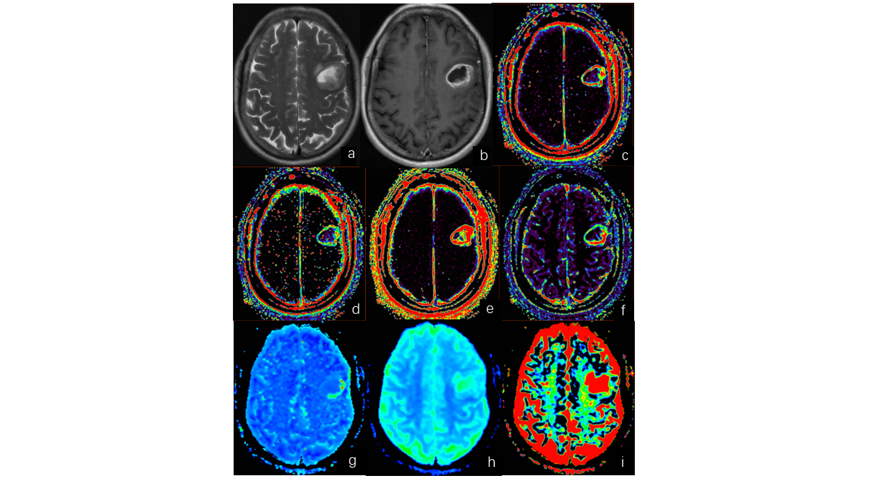
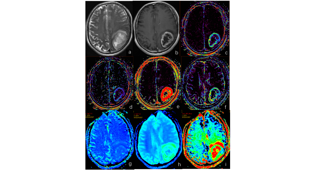
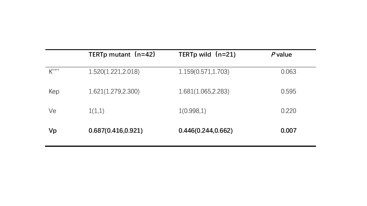
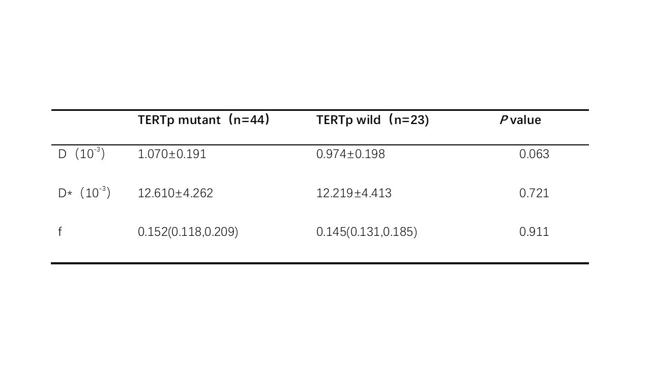
DCE-MRI parameter Vp showed a significant difference (p < 0.05) and Vp value in the TERTp mutation group (Figure 1) was significantly higher than that in the TERTp wild group (Figure 2). The AUC was 0.708 (Figure 3). Other DCE-MRI parameters, Volume transfer constant (Ktrans), rate constant (Kep) and extravascular extracellular volume fraction (Ve), did not show significant differences between the two groups (p > 0.05, Table 1).Regarding the IVIM parameters, including the true diffusion coefficient (D), pseudo-diffusion coefficient (D*), and perfusion fraction (f), there were no significant differences observed between the TERTp mutant and wild groups (p > 0.05, Table 2).
Discussion and Conclusion
The expression level of TERT is closely associated with telomerase activity, and its mRNA expression has been observed in vascular endothelial cells of the GBM with TERTp mutant6. GBM cells with TERTp mutant in vitro do induce hTERT mRNA expression, as well as telomerase enzyme activity in the endothelial cells7. Inhibition of telomerase activity in GBM endothelial cells has been shown to disrupt tumor angiogenesis8. All of these studies suggest a role for TERT mRNA expression in promoting tumor angiogenesis in TERTp mutant GBM. Our study demonstrated that Vp, which reflects the density of tumor blood vessels, has a certain predictive value for TERTp mutation status, providing strong evidence supporting the association between TERT expression and tumor angiogenesis.There were no significant differences in the DCE-MRI parameters Ktrans and Kep, which reflect tumor vascular permeability. This suggests that the tumor vascular permeability was similar between the two groups. Additionally, IVIM parameter D, which reflect tumor cell proliferation, was not significantly different between the two groups, indicating a similar degree of tumor cell proliferation. However, the parameters D* and f in IVIM, which also reflect tumor perfusion, were not statistically different between the two groups. This may be attributed to the fact that the two methods reflect different aspects of perfusion. Vp primarily reflects perfusion through the density of tumor angiogenesis, while D* and f reflect perfusion through the diffusion of water molecules in blood vessels.
Our study demonstrates that the DCE-MRI parameter Vp has predictive value for TERTp mutation status in GBM, indicating increased tumor neovascularization. Noninvasive assessment of TERTp mutation status using Vp can inform personalized treatment strategies and prognostic prediction in GBM management.
Acknowledgements
No acknowledgement found.References
1. Louis DN, Perry A, Wesseling P, Brat DJ, Cree IA, Figarella-Branger D, Hawkins C, Ng HK, Pfister SM, Reifenberger G, Soffietti R, von Deimling A, Ellison DW. The 2021 WHO Classification of Tumors of the Central Nervous System: a summary. Neuro Oncol. 2021 Aug 2;23(8):1231-1251.
2. Olympios N, Gilard V, Marguet F, Clatot F, Di Fiore F, Fontanilles M. TERT Promoter Alterations in Glioblastoma: A Systematic Review. Cancers (Basel). 2021 Mar 8;13(5):1147.
3. Aquilanti E, Kageler L, Wen PY, Meyerson M. Telomerase as a therapeutic target in glioblastoma. Neuro Oncol. 2021 Dec 1;23(12):2004-2013. doi: 10.1093/neuonc/noab203. PMID: 34473298; PMCID: PMC8643448.
4. Türkbey B, Thomasson D, Pang Y, Bernardo M, Choyke PL. The role of dynamic contrast-enhanced MRI in cancer diagnosis and treatment. Diagn Interv Radiol. 2010 Sep;16(3):186-92. doi: 10.4261/1305-3825.DIR.2537-08.1. Epub 2009 Nov 2.
5. Le Bihan D, Breton E, Lallemand D, Grenier P, Cabanis E, Laval-Jeantet M. MR imaging of intravoxel incoherent motions: application to diffusion and perfusion in neurologic disorders. Radiology. 1986 Nov;161(2):401-7. doi: 10.1148/radiology.161.2.3763909.
6. Pallini R, Pierconti F, Falchetti ML, D'Arcangelo D, Fernandez E, Maira G, D'Ambrosio E, Larocca LM. Evidence for telomerase involvement in the angiogenesis of astrocytic tumors: expression of human telomerase reverse transcriptase messenger RNA by vascular endothelial cells. J Neurosurg. 2001 Jun;94(6):961-71.
7. Falchetti ML, Pierconti F, Casalbore P, Maggiano N, Levi A, Larocca LM, Pallini R. Glioblastoma induces vascular endothelial cells to express telomerase in vitro. Cancer Res. 2003 Jul 1;63(13):3750-4.
8. Falchetti ML, Mongiardi MP, Fiorenzo P, Petrucci G, Pierconti F, D'Agnano I, D'Alessandris G, Alessandri G, Gelati M, Ricci-Vitiani L, Maira G, Larocca LM, Levi A, Pallini R. Inhibition of telomerase in the endothelial cells disrupts tumor angiogenesis in glioblastoma xenografts. Int J Cancer. 2008 Mar 15;122(6):1236-42. doi: 10.1002/ijc.23193.
Figures